Western Blotting Method: A Detailed Guide
advertisement
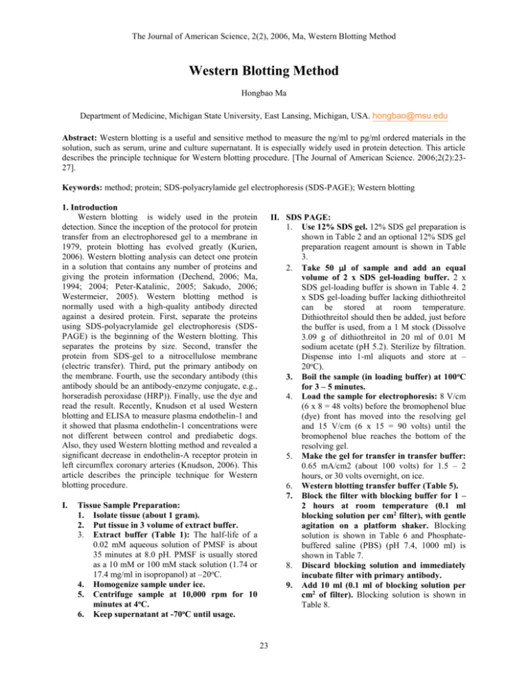
The Journal of American Science, 2(2), 2006, Ma, Western Blotting Method Western Blotting Method Hongbao Ma Department of Medicine, Michigan State University, East Lansing, Michigan, USA. hongbao@msu.edu Abstract: Western blotting is a useful and sensitive method to measure the ng/ml to pg/ml ordered materials in the solution, such as serum, urine and culture supernatant. It is especially widely used in protein detection. This article describes the principle technique for Western blotting procedure. [The Journal of American Science. 2006;2(2):2327]. Keywords: method; protein; SDS-polyacrylamide gel electrophoresis (SDS-PAGE); Western blotting 1. Introduction Western blotting is widely used in the protein detection. Since the inception of the protocol for protein transfer from an electrophoresed gel to a membrane in 1979, protein blotting has evolved greatly (Kurien, 2006). Western blotting analysis can detect one protein in a solution that contains any number of proteins and giving the protein information (Dechend, 2006; Ma, 1994; 2004; Peter-Katalinic, 2005; Sakudo, 2006; Westermeier, 2005). Western blotting method is normally used with a high-quality antibody directed against a desired protein. First, separate the proteins using SDS-polyacrylamide gel electrophoresis (SDSPAGE) is the beginning of the Western blotting. This separates the proteins by size. Second, transfer the protein from SDS-gel to a nitrocellulose membrane (electric transfer). Third, put the primary antibody on the membrane. Fourth, use the secondary antibody (this antibody should be an antibody-enzyme conjugate, e.g., horseradish peroxidase (HRP)). Finally, use the dye and read the result. Recently, Knudson et al used Western blotting and ELISA to measure plasma endothelin-1 and it showed that plasma endothelin-1 concentrations were not different between control and prediabetic dogs. Also, they used Western blotting method and revealed a significant decrease in endothelin-A receptor protein in left circumflex coronary arteries (Knudson, 2006). This article describes the principle technique for Western blotting procedure. I. II. SDS PAGE: 1. Use 12% SDS gel. 12% SDS gel preparation is shown in Table 2 and an optional 12% SDS gel preparation reagent amount is shown in Table 3. 2. Take 50 l of sample and add an equal volume of 2 x SDS gel-loading buffer. 2 x SDS gel-loading buffer is shown in Table 4. 2 x SDS gel-loading buffer lacking dithiothreitol can be stored at room temperature. Dithiothreitol should then be added, just before the buffer is used, from a 1 M stock (Dissolve 3.09 g of dithiothreitol in 20 ml of 0.01 M sodium acetate (pH 5.2). Sterilize by filtration. Dispense into 1-ml aliquots and store at – 20oC). 3. Boil the sample (in loading buffer) at 100 oC for 3 – 5 minutes. 4. Load the sample for electrophoresis: 8 V/cm (6 x 8 = 48 volts) before the bromophenol blue (dye) front has moved into the resolving gel and 15 V/cm (6 x 15 = 90 volts) until the bromophenol blue reaches the bottom of the resolving gel. 5. Make the gel for transfer in transfer buffer: 0.65 mA/cm2 (about 100 volts) for 1.5 – 2 hours, or 30 volts overnight, on ice. 6. Western blotting transfer buffer (Table 5). 7. Block the filter with blocking buffer for 1 – 2 hours at room temperature (0.1 ml blocking solution per cm2 filter), with gentle agitation on a platform shaker. Blocking solution is shown in Table 6 and Phosphatebuffered saline (PBS) (pH 7.4, 1000 ml) is shown in Table 7. 8. Discard blocking solution and immediately incubate filter with primary antibody. 9. Add 10 ml (0.1 ml of blocking solution per cm2 of filter). Blocking solution is shown in Table 8. Tissue Sample Preparation: 1. Isolate tissue (about 1 gram). 2. Put tissue in 3 volume of extract buffer. 3. Extract buffer (Table 1): The half-life of a 0.02 mM aqueous solution of PMSF is about 35 minutes at 8.0 pH. PMSF is usually stored as a 10 mM or 100 mM stack solution (1.74 or 17.4 mg/ml in isopropanol) at –20oC. 4. Homogenize sample under ice. 5. Centrifuge sample at 10,000 rpm for 10 minutes at 4oC. 6. Keep supernatant at -70oC until usage. 23 The Journal of American Science, 2(2), 2006, Ma, Western Blotting Method 10. Add 0.005 ml of primary antibody (1:2000) in to blocking solution. 11. Incubate at 4oC for 2 hours or overnight with gentle agitation on a platform shaker. 12. Discard blocking solution and wash filter 3 times (10 minutes each time) with 250 ml of PBS. 13. Incubate the filter with 150 mM NaCl, 50 mM Tris-HCl (pH 7.5) (phosphate-free, azide-free blocking solution) for 3 times for 10 minutes each time. 14. Immediately incubate the filter with secondary antibody. 15. Add 10 ml of phosphate-free, azide-free solution (150 mM NaCl, 50 mM Tris-HCl, 5% nonfat dry milk pH 7.5). Phosphate-free, azide-free blocking solution (pH 7.5, 1000 ml) is shown in Table 9. 16. Add 0.005 ml of secondary antibody solution (1:2000). 17. Incubate 1 – 2 hours at room temperature with gentle agitation. 18. Discard secondary and wash with 150 mM NaCl, 50 mM Tris-HCl (pH 7.5) (phosphatefree, azide-free solution) for 3 times for 10 minutes each time. III. Alkaline phosphatase stain: 1. Add 5 ml of the substrate 5-brono-4chloro3-indolyl phosphate/nitro blue tetrazolium (BCIP/NBT) solution (Sigma). 2. Observe the filter for the blue color on the filter (about 20 minutes). 3. Discard BCIP/NBT solution when the bands are clear (about 20 minutes). 4. Immediately stop the enzymatic reaction by add water. 5. Cover the filter with plastic membrane and keep the filter. 6. Analyze the blue bands and compare the color. The half-life of a 0.02 mM aqueous solution of PMSF is about 35 minutes, at 8.0 pH. PMSF is usually stored as a 10 mM or 100 mM stock solution (1.74 or 17.4 mg/ml in isopropanol) at –20oC. 1X SDS gel-loading buffer lacking dithiothreitol can be stored at room temperature. Dithiothreitol should then be added, just before the buffer is used, from a 1 M stock (Dissolve 3.09 g of dithiothreitol in 20 ml of 0.01 M sodium acetate (pH 5.2). Sterilize by filtration. Dispense into 1-ml aliquots and store at –20oC). Table 1. Extract buffer for Western blotting 50 mM Tris-HCl (pH 8.0) or 50 mM HEPES (pH 7.0) 150 mM NaCl 0.02% sodium azide 0.1% SDS 0.1 mg/ml phenylmethylsulfonyl fluoride (PMSF) 0.001 mg/ml aprotinin 1% Nonidet P-40 (NP-40) or 1% Triton X-100. Table 2. 12% SDS gel preparation reagent amount (l) Separating gel (%12) Stacking gel (4%) Water 1672 3020 Tris-HCl 1250 (1.5 M, pH 8.8) 1250 (0.5 M, pH 6.8) SDS (10%) 50 50 Acr-Bis (30%) 2000 650 AP 25 25 TEMED 3 5 Sum 5000 5000 (AP is ammonium persulfate) 24 The Journal of American Science, 2(2), 2006, Ma, Western Blotting Method Table 3. Optional 12% SDS gel preparation reagent amount (l) Separating gel (%12) Stacking gel (4%) Water 3344 3020 Tris-HCl 2500 (1.5 M, pH 8.8) 1250 (0.5 M, pH 6.8) SDS (10%) 100 50 Acr-Bis (30%) 4000 650 AP 50 25 TEMED 6 5 Sum 10000 5000 (AP is Ammonium persulfate) Table 4. 2 x SDS gel-loading buffer for Western blotting 100 mM Tris-HCl (pH 6.8) 200 mM dithiothreitol 4% SDS 0.2% bromophenol blue 0.2% glycerol Table 5. Western blotting transfer buffer (1000 ml) Tris 48 mM 5.814 g Glycine 39 mM 2.928 g SDS 0.037% 0.37 g Methanol 20% 200 ml SDS: 3.7 ml of 10% SDS Table 6. Western blotting blocking solution Blocking solution, 100 ml, in PBS Nonfat dried milk 5% Antifoam A 0.01% Sodium azide 0.02% Sodium azide: 1 ml of 2% solution Table 7. Phosphate-buffered saline (PBS), pH 7.4, 1000 ml NaCl 8g KCl 0.2 g Na2HPO4 1.44 g KH2PO4 0.24 g Adjust to pH 7.4 with HCl Table 8. Blocking solution Blocking solution, 10 ml, in PBS (pH 7.4) Nonfat dried milk 5% Antifoam A 0.01% Sodium azide 0.02% Table 9. Phosphate-free, azide-free blocking solution (pH 7.5, 1000 ml) NaCl 150 mM 8.766 g Tris-HCl (pH 7.5) 50 mM 6.057 g 12 N HCl about 3.35 ml Nonfat dried milk 5% (w/v) 25 The Journal of American Science, 2(2), 2006, Ma, Western Blotting Method IV. Overall of Western blotting solutions (Table 10) Table 10. Overall table of Western blotting solutions Tissue Extract buffer, 100 ml 50 mM Tris-HCl (pH 8.0), 0.6 g (Or 50 mM HEPES (pH 7.0), 1.19 g) 150 mM NaCl, 0.88 g 0.02% sodium azide, 0.02 g 0.1% SDS, 0.1 g 0.1 mg/ml phenylmethylsulfonyl fluoride (PMSF), 0.01 g 0.001 mg/ml aprotinin, 0.1 mg 1% Nonidet P-40 (NP-40), 1 ml (Or 1% Triton X-100, 1 ml) 2 X SDS gel-loading buffer, 100 ml 100 mM Tris-HCl (pH 6.8), 1.21 g 200 mM dithiothreitol 4% SDS, 0.4 g 0.2% bromophenol blue, 0.2 g 20% glycerol, 20 ml 1.5 M Tris-HCl, pH 8.8, 300 ml Tris, 54.5 g HCl, 12 N, 6.375 ml 0.5 M Tris-HCl, pH 6.8, 300 ml Tris, 18.17 g HCl, 12 N, 11.5 ml SDS 5x Running Buffer, pH 8.3, 1000 ml Tris, 125 mM, 15.14 g Glycine, 1.25 M, 93.84 g SDS, 0.50%, 5 g Western Transfer Buffer, 1000 ml Tris, 48 mM, 5.814 g Glycine, 39 mM, 2.928 g SDS, 0.04%, 0.37 g, 3.7 ml of 10% SDS Methanol, 20%, 200 ml Blocking solution, 100 ml, in 100 ml phosphate-buffered saline (PBS, pH 7.4) Nonfat dried milk, 5%, 5 g Antifoam A, 0.01%, 10 ml Sodium azide, 0.02%, 20 mg Phosphate-buffered saline (PBS), pH 7.4, 1000 ml (adjust to pH 7.4 with HCl) NaCl, 8 g KCl, 0.2 g Na2HPO4, 1.44 g KH2PO4, 0.24 g Phosphate-free, azide-free blocking solution, 1000 ml (adjust pH with 12 N HCl about 3.35 ml) 150 mM NaCl, 8.766 g 50 mM Tris-HCl (pH 7.5), 6.057 g 5% (w/v) nonfat dried milk 26 The Journal of American Science, 2(2), 2006, Ma, Western Blotting Method Correspondence to: Hongbao Ma 138 Service Road B410 Clinical Center Michigan State University East Lansing, MI 48824 Telephone: 517-432-0623 Email: hongbao@msu.edu 3. 4. 5. 6. References 1. 2. 7. Dechend R, Homuth V, Wallukat G, Muller DN, Krause M, Dudenhausen J, Haller H, Luft FC. Agonistic antibodies directed at the angiotensin II, AT1 receptor in preeclampsia. J Soc Gynecol Investig. 2006;13(2):79-86. Knudson JD, Rogers PA, Dincer UD, Bratz IN, Araiza AG, Dick GM, Tune JD. Coronary Vasomotor Reactivity to Endothelin-1 8. 27 in the Prediabetic Metabolic Syndrome. Microcirculation. 2006;13(3):209-218. Kurien BT, Scofield RH. Western blotting. Methods. 2006;38(4):283-93. Ma H. The Structure and Function of Metallothionein. Ph.D. Dissertation of Peking University. 1994:37-46. Ma H, Ruiping Huang, George S. Abela. Heat shock protein 70 expression is reduced in rat myocardium following transmyocardial laser revascularization. Journal of Investigative Medicine 2004;52:36. Peter-Katalinic J. Methods in enzymology: O-glycosylation of proteins. Methods Enzymol. 2005;405:139-71. Sakudo A, Suganuma Y, Kobayashi T, Onodera T, Ikuta K. Near-infrared spectroscopy: promising diagnostic tool for viral infections. Biochem Biophys Res Commun. 2006;341(2):27984. Westermeier R, Marouga R. Protein detection methods in proteomics research. Biosci Rep. 2005;25(1-2):19-32.
