Self-assembly of Deinococcus radiodurans S
advertisement

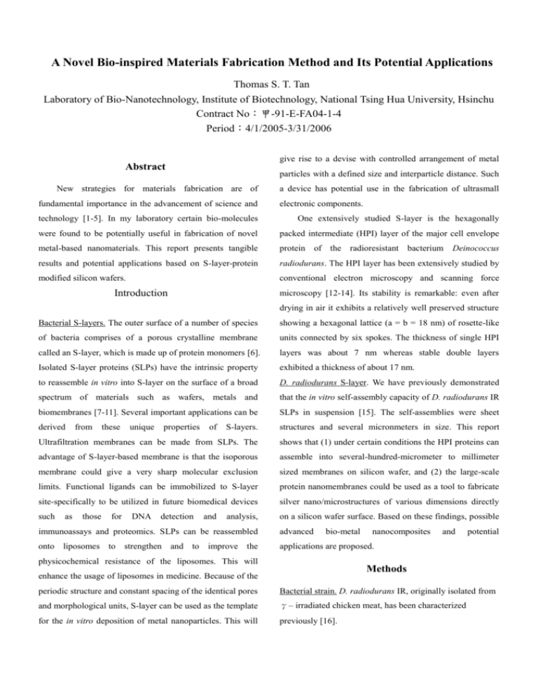
A Novel Bio-inspired Materials Fabrication Method and Its Potential Applications Thomas S. T. Tan Laboratory of Bio-Nanotechnology, Institute of Biotechnology, National Tsing Hua University, Hsinchu Contract No:甲-91-E-FA04-1-4 Period:4/1/2005-3/31/2006 give rise to a devise with controlled arrangement of metal Abstract particles with a defined size and interparticle distance. Such New strategies for materials fabrication are of fundamental importance in the advancement of science and a device has potential use in the fabrication of ultrasmall electronic components. technology [1-5]. In my laboratory certain bio-molecules One extensively studied S-layer is the hexagonally were found to be potentially useful in fabrication of novel packed intermediate (HPI) layer of the major cell envelope metal-based nanomaterials. This report presents tangible protein of the radioresistant bacterium results and potential applications based on S-layer-protein radiodurans. The HPI layer has been extensively studied by modified silicon wafers. conventional electron microscopy and scanning force Introduction Deinococcus microscopy [12-14]. Its stability is remarkable: even after drying in air it exhibits a relatively well preserved structure Bacterial S-layers. The outer surface of a number of species showing a hexagonal lattice (a = b = 18 nm) of rosette-like of bacteria comprises of a porous crystalline membrane units connected by six spokes. The thickness of single HPI called an S-layer, which is made up of protein monomers [6]. layers was about 7 nm whereas stable double layers Isolated S-layer proteins (SLPs) have the intrinsic property exhibited a thickness of about 17 nm. to reassemble in vitro into S-layer on the surface of a broad D. radiodurans S-layer. We have previously demonstrated spectrum of materials such as wafers, metals and that the in vitro self-assembly capacity of D. radiodurans IR biomembranes [7-11]. Several important applications can be SLPs in suspension [15]. The self-assemblies were sheet derived S-layers. structures and several micronmeters in size. This report Ultrafiltration membranes can be made from SLPs. The shows that (1) under certain conditions the HPI proteins can advantage of S-layer-based membrane is that the isoporous assemble into several-hundred-micrometer to millimeter membrane could give a very sharp molecular exclusion sized membranes on silicon wafer, and (2) the large-scale limits. Functional ligands can be immobilized to S-layer protein nanomembranes could be used as a tool to fabricate site-specifically to be utilized in future biomedical devices silver nano/microstructures of various dimensions directly such on a silicon wafer surface. Based on these findings, possible as from those these for unique DNA properties detection of and analysis, immunoassays and proteomics. SLPs can be reassembled advanced bio-metal onto liposomes to strengthen and to improve the applications are proposed. physicochemical resistance of the liposomes. This will nanocomposites and potential Methods enhance the usage of liposomes in medicine. Because of the periodic structure and constant spacing of the identical pores Bacterial strain. D. radiodurans IR, originally isolated from and morphological units, S-layer can be used as the template γ– irradiated chicken meat, has been characterized for the in vitro deposition of metal nanoparticles. This will previously [16]. Isolation of SLPs. It is well documented that SLPs have the further exploration of integration and functionality As intrinsic tendency to reassemble into two-dimensional summarized in Fig, 6, possible advanced nanostrutures may arrays after removal of the disrupting agent, such as include interconnects, switch, array, and living electric guanidine hydrochloride (GHCl) and urea, used in the circuit (i.e., living cells are interconnected). S-layer dissolution procedure. Cells of D. radiodurans IR, Applications. washed and suspended in phosphate buffer, were broken by method may find potential industrial applications in sonication. production of The cell-wall fraction, collected by centrifugation, washed and suspended in Tris-HCl buffer The novel bio-inspired nanofabrication (1) ultrasensitive sensors, (2) miniturizd devices, and (3) smart drug delivery systems. Conclusions (50 mM, pH 7.4), was treated with deoxyribonuclease I (1 mg/ml), ribonuclease A (0.5 mg/ml), and Triton X-100 We have established novel methods using bacterial (0.5%). The partially purified S-layer preparation was S-layer proteins to functionalize wafer surface in large areas. suspended in Tris-HCl buffer for further use. We have successfully established that our S-wafer products have potential uses in the synthesis of not only Microscopy. The S-layer preparation was treated with 5 M zero-dimensional but also one-dimensional silver GHCl to dissolve the SLPs. The GHCl extract was stored at 4℃ before use. Samples were observed with confocal laser nanostructures. For examples, one of the products was capable of transforming silver ions into silver nanocubes. scanning microscopy, atomic force microscopy, and electron Another product can even help fabricate silver nanowires microscopy when appropriate. under certain conditions. Results and Discussion Atomic force microscopy. The atomic force micrograph of the HPI-protein-based self-assemblies on a silicon wafer (S-wafer) suggested a nonrandom arrangement of the HPI-protein-based nanoparticles (with a morphological unit sized around 50 – 100 nm) (Fig. 1). Confocal laser scanning microscopy. By using confocal laser scanning microscopy, we noted that in some area on the wafer several self-assembly sheets further formed multi-layered structures (Fig. 2). A layer appeared to serve as the template for the next one with smaller size, thus forming a layered cake-like structure. Fabication of silver nanostructures. We have produced a number of S-layer modified wafers by different procedures. One of the products was found to effectively transform silver ions into silver nanocubes under certain reaction conditions (Fig. 3). Use of another protocol resulted in the synthesis and simultaneous immobilization of silver nanowires directly on wafer surfaces (Fig. 4 & 5). Possible advanced nanostructures. More advanced fabrication work is currently being conducted with a view to Fig. 1. Atomic force micrograph of a S-wafer. Fig. 4. SEM micrograph showing silver nanowires on an S-wafer. Fig. 2. Scanning laser confocal micrograph showing multi-layered self-assembly. Bar = 100 μm. Fig. 5. A silver nanobridge on an S-wafer. Fig. 3. Silver nanocubes on an S-wafer. Fig. 6. Possible advanced bio-metal nanocomposites. References [1] A. M. Morales and C. M. Lieber. Science 279, 209, 1998. [2] H. M. Huang et al. Science 292, 1897, 2001. [3] Z. W. Pan et al. Science 291, 1947, 2001. [4] X. F. Duan et al. Nature 409, 66, 2001. [5] Y. Huang et al. Science 291, 630, 2001. [6] T. J. Beverage and L. L. Graham. Microbiol. Rev 55, 684, 1991. [7] U. B. Sletr and P. Messner. “Electro Microscopy of Subcellular Dynamics ed H Plattner,” CRC Press, 1989. [8] D. Pum and U. B. Sleytr. Superamol. Sci. 2, 193, 1995 [9] S. Kupcil et al. Biochim. Biophys. Acta. 1235, 263, 1995. [10] D. Pum et al. Microelectron. Engr. 35, 297, 1997 [11] U. B. Sleytr et al. Amgew Chemie Int. Ed. 38, 1034, 1999. [12] D. Pum and U. B. Sleytr. Trends Biotech. 17, 8, 1999. [13] U. B. Sleytr et al. “(Supramolecular Polymerization) ed A Ciferri,” Marcel Dekker, 2000. [14] W. Baumeister et al. J. Mol. Biol. 187, 241, 1986. [15] J. Lin and T.S.T. Tan. Techn. Proc. 2003 Nanotech. Conf, Trade Show. 3, 39, 2003. [16] F. I. Chou and S. T. Tan. J. Bacteriol. 173, 3184, 1991.


