ORIGINAL ARTICLE A CLINICAL STUDY ON OLIGOHYDRAMNIOS
advertisement

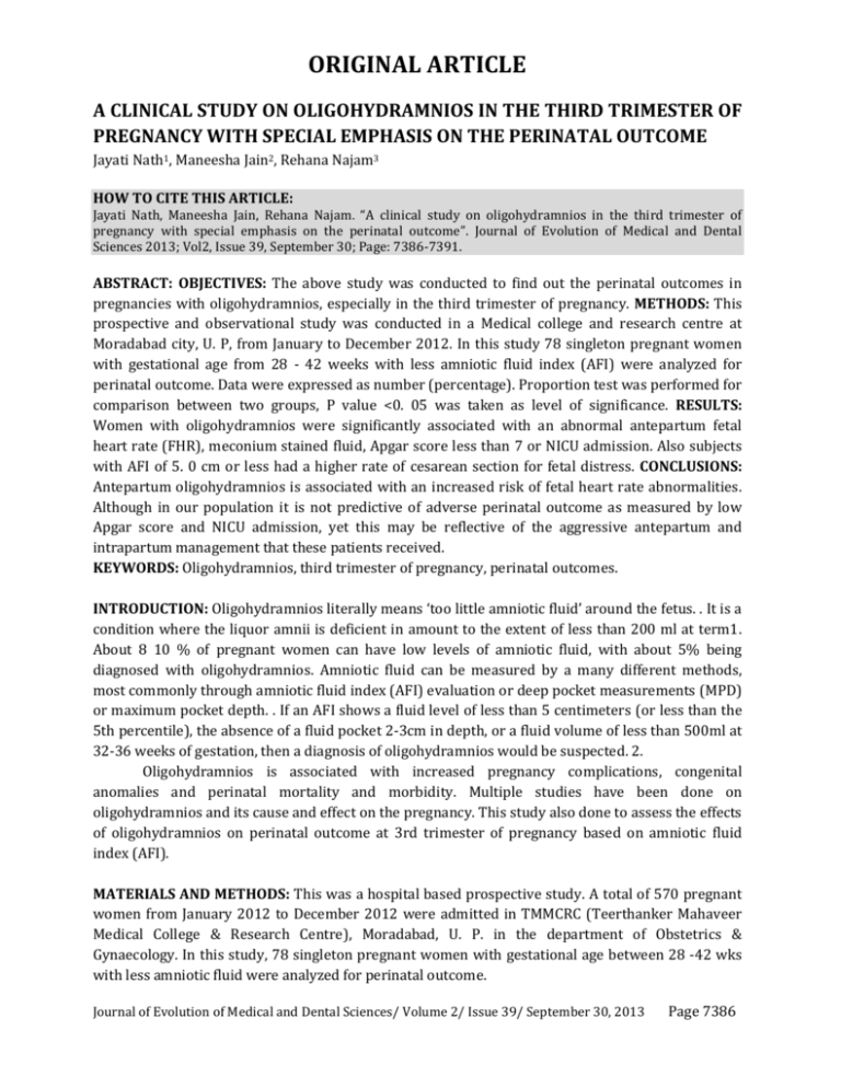
ORIGINAL ARTICLE A CLINICAL STUDY ON OLIGOHYDRAMNIOS IN THE THIRD TRIMESTER OF PREGNANCY WITH SPECIAL EMPHASIS ON THE PERINATAL OUTCOME Jayati Nath1, Maneesha Jain2, Rehana Najam3 HOW TO CITE THIS ARTICLE: Jayati Nath, Maneesha Jain, Rehana Najam. “A clinical study on oligohydramnios in the third trimester of pregnancy with special emphasis on the perinatal outcome”. Journal of Evolution of Medical and Dental Sciences 2013; Vol2, Issue 39, September 30; Page: 7386-7391. ABSTRACT: OBJECTIVES: The above study was conducted to find out the perinatal outcomes in pregnancies with oligohydramnios, especially in the third trimester of pregnancy. METHODS: This prospective and observational study was conducted in a Medical college and research centre at Moradabad city, U. P, from January to December 2012. In this study 78 singleton pregnant women with gestational age from 28 - 42 weeks with less amniotic fluid index (AFI) were analyzed for perinatal outcome. Data were expressed as number (percentage). Proportion test was performed for comparison between two groups, P value <0. 05 was taken as level of significance. RESULTS: Women with oligohydramnios were significantly associated with an abnormal antepartum fetal heart rate (FHR), meconium stained fluid, Apgar score less than 7 or NICU admission. Also subjects with AFI of 5. 0 cm or less had a higher rate of cesarean section for fetal distress. CONCLUSIONS: Antepartum oligohydramnios is associated with an increased risk of fetal heart rate abnormalities. Although in our population it is not predictive of adverse perinatal outcome as measured by low Apgar score and NICU admission, yet this may be reflective of the aggressive antepartum and intrapartum management that these patients received. KEYWORDS: Oligohydramnios, third trimester of pregnancy, perinatal outcomes. INTRODUCTION: Oligohydramnios literally means ‘too little amniotic fluid’ around the fetus. . It is a condition where the liquor amnii is deficient in amount to the extent of less than 200 ml at term1. About 8 10 % of pregnant women can have low levels of amniotic fluid, with about 5% being diagnosed with oligohydramnios. Amniotic fluid can be measured by a many different methods, most commonly through amniotic fluid index (AFI) evaluation or deep pocket measurements (MPD) or maximum pocket depth. . If an AFI shows a fluid level of less than 5 centimeters (or less than the 5th percentile), the absence of a fluid pocket 2-3cm in depth, or a fluid volume of less than 500ml at 32-36 weeks of gestation, then a diagnosis of oligohydramnios would be suspected. 2. Oligohydramnios is associated with increased pregnancy complications, congenital anomalies and perinatal mortality and morbidity. Multiple studies have been done on oligohydramnios and its cause and effect on the pregnancy. This study also done to assess the effects of oligohydramnios on perinatal outcome at 3rd trimester of pregnancy based on amniotic fluid index (AFI). MATERIALS AND METHODS: This was a hospital based prospective study. A total of 570 pregnant women from January 2012 to December 2012 were admitted in TMMCRC (Teerthanker Mahaveer Medical College & Research Centre), Moradabad, U. P. in the department of Obstetrics & Gynaecology. In this study, 78 singleton pregnant women with gestational age between 28 -42 wks with less amniotic fluid were analyzed for perinatal outcome. Journal of Evolution of Medical and Dental Sciences/ Volume 2/ Issue 39/ September 30, 2013 Page 7386 ORIGINAL ARTICLE For the purpose of this study oligohydramnios was defined as when clinically an amniotic fluid was suspected to be reduced and sonographically AFI was less than 8 cm. Pregnant women having normal amniotic fluid volume with medical complications like gestational diabetes mellitus, hypertension, heart disease or any obstetric complications like preeclampsia, eclampsia, multiple pregnancy, antepartum hemorrhage etc were excluded from this study. A written informed consent was taken from each of the patient included in the study & the study was passed by the ethics society of the university. Entry and baseline demography, past obstetrics and medical history were recorded in a data sheet, clinically suspected cases of oligohydramnios were sonographically confirmed by measuring AFI. Previous sonographic report if available, were also recorded. By transabdominal ultrasonography AFI (amniotic fluid index) was measured by four-quadrant technique by dividing the uterus into four quadrants. The transducer was placed on the maternal abdomen along the longitudinal axis. The vertical diameter of the largest amniotic fluid pocket in each quadrant was measured with the transducer head held perpendicular to the foot. These measurements were summed in centimeter and the result was recorded as the amniotic fluid index (AFI). On admission, fetal surveillance was done by BPP, which included foetal cardiotocography (CTG) and ultrasonography. Fetal heart rate was monitored by CTG. It was done for 20 minutes. Baseline FHR, beat to beat variability acceleration and decelerations were observed. Variable deceleration or late deceleration or prolonged bradycardia was taken as indicators of foetal distress and these had influenced the pattern of management towards caesarean section. Gestational age at the time of delivery was recorded. Liquor was assessed (volume, colour etc) at the time of rupture of the membranes, during labour and at the time of lower segment caesarean section (LSCS). Mode of delivery, either normal or assisted vaginal delivery or caesarean section was recorded. APGAR score and neonatal birth weight were also recorded. All relevant information recorded were appropriately analysed by SPSS methods. RESULTS: A total of 570 pregnant women were admitted during this 12 months period. Among them 78 were diagnosed as oligohydramnios. Table -I shows characteristics of pregnant mother including age, parity, educational status, AFI on admission, estimated age of delivery and mode of delivery. Mean age of the patients was 24. 583. 99 SD and of all these 46. 15% were between 21-25 years. 36% patients were nulliparous and 64% patients were multiparous. Among 78 pregnant women borderline oligohydramnios was 74% and severe oligohydramnios was 25%. About 68% patients were delivered at less than 37 completed weeks i. e. preterm delivery. In most of the cases (72%) delivery was by caesarean section and 51% caesarian sections were because of fetal distress (table -IV). Table -II shows 33% normal CTG and 66% abnormal CTG on admission. Table-III indicates colour of the liquor. At the time of membrane rupture colour of the liquor was found normal in 69% cases and 31 % cases was meconium stained. Table -V shows that caesarean section was significantly higher in severe oligohydramnios group than in borderline oligohydramnios group. Table – 6 shows perinatal outcome including birth weight, Apgar score, meconium aspiration syndrome and NICU admission. Among 78 babies low birth weight baby was 65%. Apgar score <7 at 5 minute was found in 21 babies. Among 78 babies Journal of Evolution of Medical and Dental Sciences/ Volume 2/ Issue 39/ September 30, 2013 Page 7387 ORIGINAL ARTICLE 15% suffered from respiratory distress and 10% from meconium aspiration syndrome. 15 neonates were admitted in neonatal ward with these complications. DISCUSSION: It is well established that oligohydramnios is associated with high risk adverse perinatal outcomes. On the other hand, oligohydramnios is a poor predictor for adverse outcomes. 4. But oligohydramnios is often used as an indicator for delivery. So assessment of amniotic fluid volume in antenatal period is a helpful tool in determining who is at risk for potentially adverse perinatal outcome. In our study, maximum number of women (n=36) were in the age group 21-25 years (46%). Sixty four percent women were multigravida, 38% women presented at gestational age 34-36 wks. Studies done by Cosey et al3, Chauhan4, Magann5 et al. there was no significant relation of age and parity with oligohydramnios. In our study only 24 patients had meconium stained liquor which was 30. 76%. In Coseys3 study among 147 oligohydramnios patients meconium stained liquor was found only in 9 patients, which was only 6%. He stated that meconium stained liquor less often complicated the pregnancy with oligohydramnios. This study showed no obvious relation between meconium stained liquor and oligohydramnios. Elective caesarian section was done in 12%, 28% women had normal vaginal delivery and 71% underwent caesarian section, out of which 51% had fetal distress. Chauhan et al found that AFI <5cm was associated with an increased incidence of caesarian section delivery for fetal distress. Anna et al 6 found that 15. 2% caesarian section delivery among 341 oligohydramnios patients. Voxman 7 also found increased rate caesarian section (14. 7%) for fetal distress in oligohydramnios group. In Annaet al study and voxmans study caesarian section rate was high in oligohydramnios patients but not significantly higher as it is found in this study. Probably due to less facilities for fetal monitoring well being during antepartum and intrapartum period. So for the avoidance of adverse effects on perinatal outcome in most cases caesarian section was done. Meconium stained liquor was seen in 44% of women in our study, while youseff et al8 indentified it is 40% of females. This suggests that there is high incidence of meconium stained liquor and poor placental reserve in oligohydramnios patient. Sarno et al 9 noted a significantly higher rate of fetal distress and low Apgar score in women with AFI 5 cm. This is reported to be due to head and cord compression. Golam et al 10 reported a low Apgar score at 5 minutes in 4. 6% babies, in contrast to a figure of 21%noted by us. This difference in rates observed is because of better intrapartum fetal assessment facilities available in developed countries. They concluded that liberal use of amnioinfusion in women diagnosed with oligohydramnios might have resulted in improved outcomes which were not seen in previous Oligohydramnios at third trimester and perinatal outcome studies. Casey et al 3 found respiratory distress in 3. 4% of neonates at birth in contrast to 15. 3% as noted by us. The incidence of NICU admission was found to be 19% by Garmel et al11 which is in accordance to our results (15%). Oligohydramnios has been recognized as a clinical hallmark of impending severe perinatal compromise. We have found 2. 4% perinatal deaths (1 still birth and 1 neonatal death) where as Casey et al reported 6. 4% perinatal deaths. Ja -young et al12 in a recent study have concluded that in the borderline AFI group, the presence of abnormal dorsal velocimetry measurement was related to adverse perinatal outcomes and mandates closer antenatal surveillance. Journal of Evolution of Medical and Dental Sciences/ Volume 2/ Issue 39/ September 30, 2013 Page 7388 ORIGINAL ARTICLE CONCLUSION: Oligohydramnios is associated with a high rate of pregnancy complications and increased perinatal morbidity and mortality. AFI measurement in antepartum or intrapartum period can help to identify women who need increased antepartum surveillance for avoidance of pregnancy complications and such women should be managed in a special unit to combat the complications effectively so that perinatal outcomes can be better. Thus ensuring a healthy mother delivering a healthy baby the ultimate aim of every obstetrician. REFERENCE: 1. D. C. Dutta, A text book of Obstetrics including perinatology & contraception. 8th Edition, s 218 2. Boys Rl. Polyhydramnios and oligohydramnios. Available at: URL: http://www.E medicine. com/PED/TOPIC854htm. 3. Casey BM, Mclntire DD, Bloom SL et al. Pregnancy outcomes after antepartum diagnosis of oligohydramnios at or beyond 34 weeks of gestation. Am J Obstet Gynecol 2000; 182: 909-12 http://dx. doi. org/10. 1016/S0002-9378(00)70345-0 4. Chauhan SP, Sanderson M, Hendrix NW et al. Perinatal outcome and amniotic fluid index in the antepartum and intrapartum periods: A meta-analysis. Am J Obstet Gynecol 1999; 181: 1473-78 http://dx. doi. Org/10. 1016/S0002-9378(99)70393-5 5. Magann EF Kinsella JM, Chauhan SP McNamanra MF, Gehring BW and Morison JC. Does an amniotic fluid index of <5 necessitate delivery in high risk pregnancies? A case control study. Am J Obstet Gynecol 1999; 181: 1473-8 PMid:10601931 6. Localeli A vergani P, Pezzullo JC, Toso L and Verderio M. Perinatal outcome associated with oligohydramnios in uncomplicated pregnancies. Am J Obstet Gynecol 2004; 269: 130-3. 7. Voxman EG, Train S and Wing BA. Low amniotic fluid index as a predictor of adverse perinatal outcome. Journal of Perinatology02; 22: 282-85 8. Youssef AA, Abdullah SD, Sayed EH et al. Superiority of amniotic fluid pocket measurement for predicting bad fetal outcomes. Southern Medical Journal 1993; 86: 426-29 http://dx. doi. org/10. 1097/00007611-199304000- 00011 PMid:8465220 9. Sarno AP Jr, Ahn MO, Brar HS et al. Intrapartum Doppler velocimetry, amniotic fluid volume and fetal heart rate as prediction of subsequent fetal distress. Am J Obstet Gynecol 1989; 161: 1508-14 PMid:2690625 10. Golan A Lin G, Evron S et al. Oligohydramnios: maternal complication and fetal outcome in 145 cases. Gynaecol Obstet Invest 1994:37:91-95 http://dx. doi. org/10. 1159/000292532 PMid:8150377 11. Garmel SH, Chelmow D Sha et al. Oligohydramnios and appropriately grown fetus. Am J Perinatol 1997; 14: 359-63 http://dx. doi. org/10. 1055/s-2007-994161 PMid:9217959 12. Ja-Y-K, Han-s k, Young-H K et al. Abnormal Doppler Velocimetry is related to adverse perinatal outcomes for borderline amniotic fluid index during third trimester. J Obstet Gynecol Res 2006: 32: 545-49http://dx. doi. Org/10. 1111/j. 1447-0756. 2006. 00459. X PMid: 17100815. Journal of Evolution of Medical and Dental Sciences/ Volume 2/ Issue 39/ September 30, 2013 Page 7389 ORIGINAL ARTICLE Number Percentage Age group 18 – 20 15 19. 23 21 – 25 36 46. 15 26 – 30 18 23. 07 >31 9 11. 53 Parity Nulliparous 28 35. 89 Multiparous 50 64. 10 Education Upto class XII 35 44. 87 Class XII 43 55. 12 AFI on admission 5. 1 – 8 cm(borderline oligo) 58 74. 35 <5 cm (severe oligohydramnios) 20 25. 64 GA at delivery (weeks) < 37 22 28. 20 >37 56 71. 79 Table-1: Demographic characteristics of the patients CTG Number Percentage (%) Normal CTG 26 33. 33 Abnormal CTG 52 66. 66 Table – 2: (CTG on admission) N= 78 Colour of liquor Clear/ normal Meconium stained liquor Number 54 24 Percentage (%) 69 30. 76 Table-3: Colour of liquor at the time of rupture of membranes Indication Number Percentage Fetal distress 46 58. 97 Elective CS 10 12. 82 Table-4: Indication of C-Section (N=56) Oligohydramnios Borderline Oligohydramnios(N=58) Severe Oligohydramnios(N=20) CS 36 20 NVD 22 0 X2 = ‘P’ value 11. 67; P< 0. 001 Table-5: Comparison between C-Section between borderline & severe oligohydramnios Journal of Evolution of Medical and Dental Sciences/ Volume 2/ Issue 39/ September 30, 2013 Page 7390 ORIGINAL ARTICLE Disease Number Conservative treatment Admission Birth weight <2500 gm 51 (65. 38%) >2500 gm 27 (34. 61%) APGAR Score <7 at 5 minutes 21 (26. 9%) Birth asphyxia 12 (15. 38%) 7 (8. 9%) 6 (7. 6 %) Meconium aspiration syndrome(MAS) 8 (10. 2%) 8 (10. 2 %) Early Neonatal Death 1 (1. 2 %) 1 (1. 2 %) Stillbirth 1 (1. 2 %) Table-6: Perinatal Outcomes (N=78) AUTHORS: 1. Jayati Nath 2. Maneesha Jain 3. Rehana Najam PARTICULARS OF CONTRIBUTORS: 1. Associate Professor, Department of Obstetrics & Gynaecology, Teerthanker Mahaveer Medical College & Research Centre, Moradabad, U.P. 2. Assistant Professor, Department of Obstetrics & Gynaecology, Teerthanker Mahaveer Medical College & Research Centre, Moradabad, U.P. 3. Associate Professor, Department of Obstetrics & Gynaecology, Teerthanker Mahaveer Medical College & Research Centre, Moradabad, U.P. NAME ADDRESS EMAIL ID OF THE CORRESPONDING AUTHOR: Dr. Jayati Nath, Associate Professor, Department of Obstetrics & Gynaecology, Teerthanker Mahaveer Medical College & Research Centre, TMU Campus, Delhi Road, Moradabad, U.P. Email- jayati.nath@yahoo.com Date of Submission: 03/09/2013. Date of Peer Review: 04/09/2013. Date of Acceptance: 18/09/2013. Date of Publishing: 24/09/2013 Journal of Evolution of Medical and Dental Sciences/ Volume 2/ Issue 39/ September 30, 2013 Page 7391


