Template for Communications for the Journal of the - HAL
advertisement
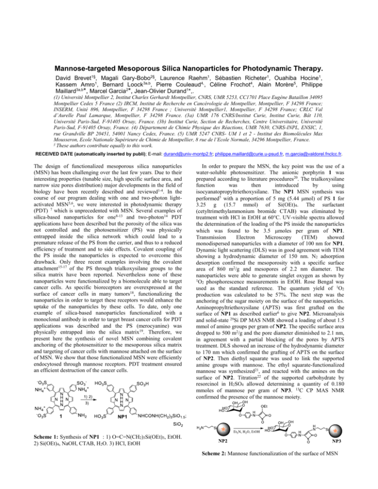
Mannose-targeted Mesoporous Silica Nanoparticles for Photodynamic Therapy. David Brevet1§, Magali Gary-Bobo2§, Laurence Raehm1, Sébastien Richeter1, Ouahiba Hocine1, Kassem Amro1, Bernard Loock3a,b, Pierre Couleaud4,, Céline Frochot4, Alain Morère5, Philippe Maillard3a,b*, Marcel Garcia2*, Jean-Olivier Durand1*,. (1) Université Montpellier 2, Institut Charles Gerhardt Montpellier, CNRS, UMR 5253, CC1701 Place Eugène Bataillon 34095 Montpellier Cedex 5 France (2) IRCM, Institut de Recherche en Cancérologie de Montpellier, Montpellier, F 34298 France; INSERM, Unité 896, Montpellier, F 34298 France ; Université Montpellier1, Montpellier, F 34298 France; CRLC Val d’Aurelle Paul Lamarque, Montpellier, F 34298 France. (3a) UMR 176 CNRS/Institut Curie, Institut Curie, Bât 110, Université Paris-Sud, F-91405 Orsay, France. (3b) Institut Curie, Section de Recherches, Centre Universitaire, Université Paris-Sud, F-91405 Orsay, France. (4) Département de Chimie Physique des Réactions, UMR 7630, CNRS-INPL, ENSIC, 1, rue Grandville BP 20451, 54001 Nancy Cedex, France. (5) UMR 5247 CNRS- UM 1 et 2 - Institut des Biomolécules Max Mousseron, Ecole Nationale Supérieure de Chimie de Montpellier, 8 rue de l’Ecole Normale, 34296 Montpellier, France. § These authors contribute equally to this work. RECEIVED DATE (automatically inserted by publi); E-mail: durand@univ-montp2.fr; philippe.maillard@curie.u-psud.fr, m.garcia@valdorel.fnclcc.fr. The design of functionalized mesoporous silica nanoparticles (MSN) has been challenging over the last few years. Due to their interesting properties (tunable size, high specific surface area, and narrow size pores distribution) major developments in the field of biology have been recently described and reviewed1-4. In the course of our program dealing with one and two-photon lightactivated MSN5,6, we were interested in photodynamic therapy (PDT) 7 which is unprecedented with MSN. Several examples of silica-based nanoparticles for one8-13 and two-photon14 PDT applications have been described but the porosity of the silica was not controlled and the photosensitizer (PS) was physically entrapped inside the silica network which could lead to a premature release of the PS from the carrier, and thus to a reduced efficiency of treatment and to side effects. Covalent coupling of the PS inside the nanoparticles is expected to overcome this drawback. Only three recent examples involving the covalent attachment15-17 of the PS through trialkoxysilane groups to the silica matrix have been reported. Nevertheless none of these nanoparticles were functionalized by a biomolecule able to target cancer cells. As specific bioreceptors are overexpressed at the surface of cancer cells in many tumors18, functionalizing the nanoparticles in order to target these receptors would enhance the uptake of the nanoparticles by these cells. To date, only one example of silica-based nanoparticles functionalized with a monoclonal antibody in order to target breast cancer cells for PDT applications was described and the PS (merocyanine) was physically entrapped into the silica matrix19. Therefore, we present here the synthesis of novel MSN combining covalent anchoring of the photosensitizer to the mesoporous silica matrix and targeting of cancer cells with mannose attached on the surface of MSN. We show that those functionalized MSN were efficiently endocytosed through mannose receptors. PDT treatment ensured an efficient destruction of the cancer cells. In order to prepare the MSN, the key point was the use of a water-soluble photosensitizer. The anionic porphyrin 1 was prepared according to literature procedures20. The trialkoxysilane function was then introduced by using isocyanatopropyltriethoxysilane. The NP1 MSN synthesis was performed5 with a proportion of 5 mg (5.44 mol) of PS 1 for 3.25 g (15.7 mmol) of Si(OEt)4. The surfactant (cetyltrimethylammonium bromide CTAB) was eliminated by treatment with HCl in EtOH at 60°C. UV-visible spectra allowed the determination of the loading of the PS inside the nanoparticles which was found to be 3.5 moles per gram of NP1. Transmission Electron Microscopy (TEM) showed monodispersed nanoparticles with a diameter of 100 nm for NP1. Dynamic light scattering (DLS) was in good agreement with TEM showing a hydrodynamic diameter of 150 nm. N2 adsorption desorption confirmed the mesoporosity with a specific surface area of 860 m2/g and mesopores of 2.2 nm diameter. The nanoparticles were able to generate singlet oxygen as shown by 1O phosphorescence measurements in EtOH. Rose Bengal was 2 used as the standard reference. The quantum yield of 1O2 production was calculated to be 57%. The next step was the anchoring of the sugar moiety on the surface of the nanoparticles. Aminopropyltriethoxysilane (APTS) was first grafted on the surface of NP1 as described earlier6 to give NP2. Microanalysis and solid-state 29Si DP MAS NMR showed a loading of about 1.5 mmol of amino groups per gram of NP2. The specific surface area dropped to 500 m2/g and the pore diameter diminished to 2.1 nm, in agreement with a partial blocking of the pores by APTS treatment. DLS showed an increase of the hydrodynamic diameter to 170 nm which confirmed the grafting of APTS on the surface of NP2. Then diethyl squarate was used to link the supported amine groups with mannose. The ethyl squarate-functionalized mannose was synthesized21, and reacted with the amines on the surface of NP2. Titration22 of the supported carbohydrate by resorcinol in H2SO4 allowed determining a quantity of 0.180 mmoles of mannose per gram of NP3. 13C CP MAS NMR confirmed the presence of the mannose moiety. Scheme 1: Synthesis of NP1 : 1) O=C=N(CH2)3Si(OEt)3, EtOH. 2) Si(OEt)4, NaOH, CTAB, H2O. 3) HCl, EtOH Scheme 2: Mannose functionalization of the surface of MSN In order to study the efficiency of these MSN for PDT, human breast cancer cells (MDA-MB-231) were incubated at different times with or without 20 µg/ml of MSN in the presence or absence of 10 mM mannose and then submitted to monophotonic irradiation (630-680 nm; 6 mW / cm²) for 40 min. A 3-(4,5-dimethylthiazol-2-yl)-2,5-diphenyltetrazolium bromide (MTT) assay15 was performed two days after irradiation, to establish the cytotoxicity of MSN. First, as showed in Figure 1, the irradiation of cancer cells alone did not induce any toxicity. We then compared the cytotoxic efficiency of NP1 and NP3 on MDA-MB-231 cancer cells incubated for 24 h with MSN. NP1 incubated with cancer cells for 24 h and not submitted to irradiation, induced 7% cytotoxicity whereas their irradiation induced 45% cell death. In the same conditions, mannose-functionalized NP3 induced 19% cell death without irradiation and 99% cell death when irradiated. The higher efficiency of mannose-functionalized MSN must be due to an active endocytosis via unidentified mannose receptors23. NP1 120 Living cells (%) Notes and references : PM and CF are members of GDR 3049 “Medicaments Photoactivables-Photochimiothérapie (PHOTOMED)”. (1) (2) (3) (4) (5) NP3 100 (6) 80 * * 60 (7) 40 (8) 20 ** 0 MSN (24 h) Irradiation - + + - + + - + + - (9) + + Figure 1 : PDT-induced cytotoxicities of NP1 and NP3. Means ± SD of 3 experiments. * p< 0.05; ** p< 0.01 Student’s t test. To prove this, cancer cells were incubated for 1 h in a serum free medium with MSN, in presence or in absence of 10 mM mannose. Table 1 shows that cell irradiation without MSN pre-incubation did not induce any significant toxicity and neither did cell incubation with NP1 or NP3 without irradiation. By contrast, cell treatment with NP3 and submission to irradiation as already described, generated a significant cell death of 30%. This effect was totally reversed (no significant cell death occurred) by incubating NP3 in the presence of mannose. This indicates that mannose acts as a competitor of NP3. Therefore, mannose receptors are involved in the active endocytosis of NP3, which thus have a higher therapeutic efficiency than NP1. MSN (1 h) - NP3 Mannose - Irradiation % cell viability Acknowledgments: Financial support by ANR PNANO. 07-102 is gratefully acknowledged. We thank Dr Corine Gérardin for DLS Measurements. NP3 NP3 NP3 NP1 NP1 + - - + - - - - - + + - + 100 99 ±1 101 ±2 70 * ±6 97 ±5 99 ±2 98 ±2 Table 1 : MSN cytotoxicity mediated through mannose receptors. Means ± SD of 3 experiments. * p< 0.05 Student’s t test. In conclusion, a new class of MSN was elaborated by covalent incorporation of a water-soluble PS and by covering the external surface with mannose residues. We have proved that these MSN presented a much higher in vitro photoefficiency in MDA-MB-231 cancer cells through mannose-dependent endocytosis than non functionalized nanoparticles. Studies are in progress to adapt these nanotools to in vivo PDT applications. (10) (11) (12) (13) (14) (15) (16) (17) (18) (19) (20) (21) (22) (23) Slowing, I. I.; Vivero-Escoto, J. L.; Wu, C.-W.; Lin, V. S. Y. Adv. Drug. Deliv. Rev. 2008, 60, 1278-1288. Trewyn, B. G.; Giri, S.; Slowing, I. I.; Lin, V. S. Y. Chem. Com. 2007, 3236-3245. Slowing, I. I.; Trewyn, B. G.; Giri, S.; Lin, V. S. Y. Adv. Func. Mater. 2007, 17, 1225-1236. Johansson, E.; Choi, E.; Angelos, S.; Liong, M.; Zink, J. I. J. Sol-Gel Sci. Technol. 2008, 46, 313-322. Lebret, V.; Raehm, L.; Durand, J. O.; Smaihi, M.; Gerardin, C.; Nerambourg, N.; Werts, M. H. V.; Blanchard-Desce, M. Chem. Mater. 2008, 20, 2174-2183. Lebret, V.; Raehm, L.; Durand, J. O.; Smaihi, M.; Werts, M. H. V.; Blanchard-Desce, M.; Methy-Gonnod, D.; Dubernet, C. J. Sol-Gel Sci. Technol. 2008, 48, 32-39. Bechet, D.; Couleaud, P.; Frochot, C.; Viriot, M.-L.; Guillemin, F.; Barberi-Heyob, M. Trends in Biotechnology, In Press. Liu, F.; Zhou, X.; Chen, Z.; Huang, P.; Wang, X.; Zhou, Y. Mater. Lett. 2008, 62, 2844-2847. Tada, D. B.; Vono, L. L. R.; Duarte, E. L.; Itri, R.; Kiyohara, P. K.; Baptista, M. S.; Rossi, L. M. Langmuir 2007, 23, 8194-8199. Tang, W.; Xu, H.; Kopelman, R.; Philbert, M. A. Photochem. Photobiol. 2005, 81, 242-249. Yan, F.; Kopelman, R. Photochem. Photobiol. 2003, 78, 587-591. Zhou, J.; Zhou, L.; Dong, C.; Feng, Y.; Wei, S.; Shen, J.; Wang, X. Mater. Lett. 2008, 62, 2910-2913. Roy, I.; Ohulchanskyy, T. Y.; Pudavar, H. E.; Bergey, E. J.; Oseroff, A. R.; Morgan, J.; Dougherty, T. J.; Prasad, P. N. J. Am. Chem. Soc. 2003, 125, 7860-7865. Kim, S.; Ohulchanskyy, T. Y.; Pudavar, H. E.; Pandey, R. K.; Prasad, P. N. J. Am. Chem. Soc. 2007, 129, 26692675. Ohulchanskyy, T. Y.; Roy, I.; Goswami, L. N.; Chen, Y.; Bergey, E. J.; Pandey, R. K.; Oseroff, A. R.; Prasad, P. N. Nano Lett. 2007, 7, 2835-2842. Lai, C.-W.; Wang, Y.-H.; Lai, C.-H.; Yang, M.-J.; Chen, C.-Y.; Chou, P.-T.; Chan, C.-S.; Chi, Y.; Chen, Y.-C.; Hsiao, J.-K. Small 2008, 4, 218-224. Rossi, L. M.; Silva, P. R.; Vono, L. L. R.; Fernandes, A. U.; Tada, D. B.; Baptista, M. S. Langmuir 2008, 24, 12534-12538. Low, P. S.; Henne, W. A.; Doorneweerd, D. D. Acc. Chem. Res. 2008, 41, 120-129. Zhang, P.; Steelant, W.; Kumar, M.; Scholfield, M. J. Am. Chem. Soc. 2007, 129, 4526-4527. Chen, Y.; Parr, T.; Holmes, A. E.; Nakanishi, K. Bioconjug. Chem. 2008, 19, 5-9. Sperling, O.; Fuchs, A.; Lindhorst, T. K. Org. Biomol. Chem. 2006, 4, 3913-3922. Park, I. Y.; Kim, I. Y.; Yoo, M. K.; Choi, Y. J.; Cho, M.H.; Cho, C. S. Int. J. Pharma. 2008, 359, 280-287. East, L.; Isacke, C. M. Biochem. Biophys. Acta 2002, 1572, 364-386. Graphical abstract An efficient method for the synthesis of mesoporous silica nanoparticles with a covalent attachment of a photosensitizer inside the silica matrix for PDT applications is described. The surface of the nanoparticle was functionalized with mannose and the mannose-derivatized particles were much more efficient than non functionalized ones for the treatment of cancer cells.

