ELBOW JOINT & ANASTOMOSIS AROUND ELBOW JOINT
advertisement
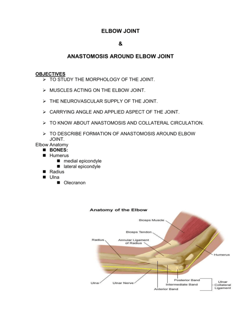
ELBOW JOINT & ANASTOMOSIS AROUND ELBOW JOINT OBJECTIVES TO STUDY THE MORPHOLOGY OF THE JOINT. MUSCLES ACTING ON THE ELBOW JOINT. THE NEUROVASCULAR SUPPLY OF THE JOINT. CARRYING ANGLE AND APPLIED ASPECT OF THE JOINT. TO KNOW ABOUT ANASTOMOSIS AND COLLATERAL CIRCULATION. TO DESCRIBE FORMATION OF ANASTOMOSIS AROUND ELBOW JOINT. Elbow Anatomy BONES: Humerus medial epicondyle lateral epicondyle Radius Ulna Olecranon Elbow Joint Articulation - Elbow consists of 3 articulations: Humeroulnar (elbow flexion/extension) Humeroradial (forearm pronation/supination) Radioulnar (forearm pronation/supination) INTRODUCTION Type: hinge joint, compound synovial type Articulation: between lower end of humerus and upper end of radius and ulna Flexion and Rotation: biceps muscles in the arm. Ligaments located at the front, back, and sides of the elbow help stabilize the joint. CAPSULAR LIGAMENT Superiorly it is attached the lower end of the humerus in such a way that it covers the capitulum, trochlea, radial fossa, coronoid fossa and the olecranon fossa. Inferiomedially it is attached to the margin of the trochlear notch of ulna. Inferiolaterally it is attached to the annular ligament of the superior radioulnar joint. Medial Ligamentous Structures: Medial/Ulnar Collateral Ligament Anterior bundle – most discrete segment Posterior bundle – thickening of posterior capsule Transverse bundle – spans medial border of semilunar notch, little/no contribution to elbow stability lateral liagamentous structures: Lateral medial / ulnar collateral ligament: – present in approximately 50% of population. Accessory lateral/radial collateral ligament: - tight only during various stress maneuvers and assists annular ligament when stress applied to elbow synovium and bursa: A synovial membrane envelops the elbow and superior radioulnar articulations and lubricates the deeper structures of the two joints. The two main bursae are the bicipital bursae which cushions the tendon when the forearm is pronated and the olecranon bursae which forms a liquid cushion. MUSCLES ACTING ON JOINT Flexion:biceps brachii, brachialis, brachioradialis, pronator teres. Extension: triceps brachii and anconeus. Supination: biceps brachii, supinator, brachioradialis. Pronation:pronator teres, pronator quadratus, anconeus, brachioradialis. Elbow Anatomy - muscles Flexors (3 B’s) Bicepslong (bicipital groove) and short head (coracoid) Brachioradialis Brachialis Extensors Triceps Anconeus Elbow Flexors: Biceps Brachii Origin: long head - supraglenoid tubercle short head - coracoid process Insertion: radial tuberosity Action: elbow flexion forearm supination Elbow Flexors: Brachialis Origin: Anterior surface of distal humerus Insertion: ulnar tuberosity Action: elbow flexion Elbow Flexors Brachioradialis Origin: lateral supracondylar ridge of humerus Insertion: radial styloid process Action: elbow flexion Elbow Extenders Triceps brachii Origin: long head - infraglenoid tubercle lateral - posterior humerus(above spiral groove) medial - posterior humerus(below spiral groove) Insertion: olecranon process Action: elbow extension NEUROVASCULAR SUPPLY AND BLOOD SUPPLY: brachial artery and it’s branches. NERVE SUPPLY: musculocutaneous, radial, and median nerves. Carrying Angle/Cubitus Valgus Formed by long axis of humerus and midline of forearm COMMON ELBOW PROBLEMS Male norms – 11-14 degrees Female norms – 13-16 degrees Larger angles are considered abnormal Arthritis: Common forms of arthritis that can affect the elbow include osteoarthritis, rheumatoid arthritis, infectious arthritis. Bursitis: Bursitis of the elbow, also called olecranon bursitis, occurs as a result of injury or constant pressure on the elbow (for example, when leaning on a hard surface). Fractures: Falling on an outstretched hand or directly on the tip of the elbow can result in dislocation and/or several types of fractures, depending on the fall. Injury: Repetitive strain on the elbow can cause inflammation Elbow Pathology Tendonitis (epicondylitis) Medial (golfer’s elbow) flexor mass Lateral (tennis elbow) extensor mass Grip size Pressure point Elbow Pathology: Sprains Capsular MCL/LCL Dislocations Rare due to stability Almost always posterior Bursitis Result of direct trauma Pad and protect Anastomosis around elbow joint The vessels engaged in this anastomosis divided into those situated in front of and those behind the medial and lateral epicondyles of the humerus. The branches anastomosing in front of the medial epicondyle are : The anterior branch of the inferior ulnar collateral, the anterior ulnar recurrent (branch of ulnar artery), and the anterior branch of the superior ulnar collateral Those behind the medial epicondyle are : The inferior ulnar collateral, the posterior ulnar recurrent, and the posterior branch of the superior ulnar collateral. The branches anastomosing in front of the lateral epicondyle are : The radial recurrent (branch of radial artery) and the anterior descending (radial collateral) branch of the profunda brachii. Those behind the medial epicondyle are : The inferior ulnar collateral, the posterior ulnar recurrent, and the posterior branch of the superior ulnar collateral. The branches anastomosing in front of the lateral epicondyle are : The radial recurrent (branch of radial artery) and the anterior descending (radial collateral) branch of the profunda brachii. Those behind the lateral epicondyle are: The inferior ulnar collateral, the interosseous recurrent, and the radial collateral branch of the profunda brachii
