X ray imaging worksheet
advertisement
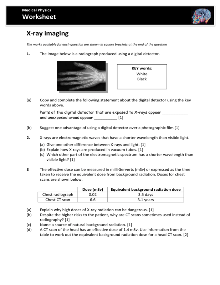
Medical Physics Worksheet X-ray imaging The marks available for each question are shown in square brackets at the end of the question 1. The image below is a radiograph produced using a digital detector. KEY words: White Black (a) Copy and complete the following statement about the digital detector using the key words above. Parts of the digital detector that are exposed to X-rays appear __________ and unexposed areas appear _________ [1] (b) Suggest one advantage of using a digital detector over a photographic film [1] 2. X-rays are electromagnetic waves that have a shorter wavelength than visible light. (a) Give one other difference between X-rays and light. [1] (b) Explain how X-rays are produced in vacuum tubes. [1] (c) Which other part of the electromagnetic spectrum has a shorter wavelength than visible light? [1] 3 The effective dose can be measured in milli-Serverts (mSv) or expressed as the time taken to receive the equivalent dose from background radiation. Doses for chest scans are shown below. Chest radiograph Chest CT scan (a) (b) (c) (d) Dose (mSv) 0.02 6.6 Equivalent background radiation dose 3.5 days 3.1 years Explain why high doses of X-ray radiation can be dangerous. [1] Despite the higher risks to the patient, why are CT scans sometimes used instead of radiography? [1] Name a source of natural background radiation. [1] A CT scan of the head has an effective dose of 1.4 mSv. Use information from the table to work out the equivalent background radiation dose for a head CT scan. [2]
