2009 exam 4
advertisement
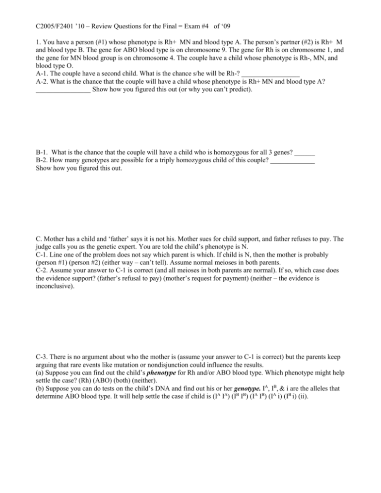
C2005/F2401 ’10 – Review Questions for the Final = Exam #4 of ‘09 1. You have a person (#1) whose phenotype is Rh+ MN and blood type A. The person’s partner (#2) is Rh+ M and blood type B. The gene for ABO blood type is on chromosome 9. The gene for Rh is on chromosome 1, and the gene for MN blood group is on chromosome 4. The couple have a child whose phenotype is Rh-, MN, and blood type O. A-1. The couple have a second child. What is the chance s/he will be Rh-? _________________ A-2. What is the chance that the couple will have a child whose phenotype is Rh+ MN and blood type A? ________________ Show how you figured this out (or why you can’t predict). B-1. What is the chance that the couple will have a child who is homozygous for all 3 genes? ______ B-2. How many genotypes are possible for a triply homozygous child of this couple? _____________ Show how you figured this out. C. Mother has a child and ‘father’ says it is not his. Mother sues for child support, and father refuses to pay. The judge calls you as the genetic expert. You are told the child’s phenotype is N. C-1. Line one of the problem does not say which parent is which. If child is N, then the mother is probably (person #1) (person #2) (either way – can’t tell). Assume normal meioses in both parents. C-2. Assume your answer to C-1 is correct (and all meioses in both parents are normal). If so, which case does the evidence support? (father’s refusal to pay) (mother’s request for payment) (neither – the evidence is inconclusive). C-3. There is no argument about who the mother is (assume your answer to C-1 is correct) but the parents keep arguing that rare events like mutation or nondisjunction could influence the results. (a) Suppose you can find out the child’s phenotype for Rh and/or ABO blood type. Which phenotype might help settle the case? (Rh) (ABO) (both) (neither). (b) Suppose you can do tests on the child’s DNA and find out his or her genotype. IA, IB, & i are the alleles that determine ABO blood type. It will help settle the case if child is (IA IA) (IB IB) (IA IB) (IA i) (IB i) (ii). C2005/F2401 ’09 Exam #4 12/22/09 ID #: ___________________________________ 2. Suppose you are examining the incidence of trisomy 16. It is the most common trisomy in humans, but is incompatible with postnatal survival. Trisomy 16 is found in about 5% of spontaneous abortions. You have a polymorphic site very close to the centromere of chromosome 16. The site is so close that there is no recombination between the site and the centromere. At the polymorphic site there are a variable number (6-23) of short repeats. All possible haplotypes (numbers of repeats) are about equally common. A. To figure out the number of repeats, you do PCR on DNA from an individual, using primers that bracket the polymorphic section with the repeats. You take the PCR products and run them on a gel. A-1. If you take DNA from a trisomic individual (from a miscarriage), the maximum number of bands you should see on the gel would be (1) (2) (3) (4) (5) (6) ( >6). A-2. Suppose you take DNA samples from many different unrelated individuals and do the same procedure for each sample. The max. number of different bands on the entire gel (including all lanes) would probably be (1) (2) (3) (4) (5) (6) ( >6) (depends on whether individuals are normal or trisomic). Explain briefly. B. You have two normal parents. The mother’s haplotype at the polymorphic site is 12/10. (12 repeats on one chromosome; 10 on the other.) The father’s haplotype is 6/6. Which of the following haplotypes are likely in a normal child? (6/6) (12/10) (6/10) (6/12) (12/12) (10/10) (none of these). Explain how you know. C. Parents are as in B for all parts of C. C-1. Suppose nondisjunction (ND) occurred in the father to produce a trisomic child. How many bands should be observed on the gel of PCR products from the child? (1) (1 or 2) (2) (2 or 3) (3) (3 or 4) (4) (5) (6) ( >6). Explain how the gel would (or would not) look different from that from a normal child. C-2. Suppose the parents have a trisomic child. Using PCR, you could tell the difference between ND at first division and at second division in (the father) (the mother) (both) (neither). Explain as specifically as possible how you could (or couldn’t) tell. D. For humans, N = 23. D-1. When a trisomic cell finishes mitosis, each daughter cell should have ___________ chromosomes and _________________ chromatids. No explanation required for this one. D-2. Suppose a human cell were triploid, and in G-2. What would the DNA content be? (c) (2c) (3c) (4c) (6c) (12c) (23) (46) (92) (69) (138). Explain D-2. C2005/F2401 ’09 Exam #4 12/22/09 ID #: ___________________________________ 3. You have three markers (polymorphic sites) on chromosome 7, A, B and D. Haplotypes, distances, etc. are given on the last page. You can tear that page off – you don’t have to flip back and forth. A. Consider the following 4 gametes: #1. (A-1 B-1 D-1) #2. (A-1 B-2 D-1) #3. (A-1 B-2 D-2) #4. (A-1 B-1 D-2) #5. (A-1 B-3 D-3). A-1. List the gametes in order of expected frequency. Indicate which is lowest and which is highest, and explain your ranking. A-2. Gamete #3 should be about the same frequency as (A-2 B-1 D-1) (A-4 B-3 D-3) (both) (neither). Explain briefly. B. You measure the RF between A and D by the standard procedure. Which of the following is the best estimate for the expected value of RF? (0%) (about 3%) (about 7%) (>7% but < 25%) (25%) (>25% but <37%) (37%) (>37% but <50%) (50%) (100%) (beats me). Explain your estimate. 4. Look at the pedigree on the last page, and answer the questions below. A-1. CGH is (dominant) (recessive) (co-dominant) (dominant or recessive) (can’t tell). Explain briefly. A-2. The gene responsible for CGH is probably on (an autosome) (the X) (the Y) (the X or the Y). (an autosome or the X) (can’t tell). Explain how you made your choice. B. Suppose that IV-11 marries has many children. Given your answers above, what do you expect? B-1. What proportion of his daughters should have CGH? (0) (some, but not all) (all) (depends on his partner). B-2. What proportion of his sons should have CGH? (0) (some, but not all) (all) (depends on his partner). Write out the genotypes of IV-11 and his children, and show how you get the predicted pattern of inheritance. If you said above that there were multiple possibilities, just pick one and go with it. C2005/F2401 ’09 Exam #4 12/22/09 ID #: ___________________________________ 5. Bowen-Conradi Syndrome (BCS) is found almost exclusively in Hutterite communities. All individuals with BCS have the same mutation, which is presumed to be inherited from one of the founders of the Hutterites. BCS is autosomal recessive, affects development of many tissues, and is usually lethal during the first year of life. The prevalence of BCS (among the Hutterites) is estimated at about 1/400. A. If you assume genetic equilibrium, what is the carrier frequency for BCS? The proportion of the Hutterite community who are carriers is about (>1/4) (¼) (1/2) (1/3) (1/5) (2/5) (3/5) (1/10) (1/20) (1/25) (1/40) (1/50) (1/100) (1/400) (<1/400). B. The mutation in BCS is a change of one base in the gene for the EMG1 protein. The protein is necessary for production of the small ribosomal subunit. The mutation changes AA #86 (amino acid #86) in the protein from asp to gly. In normal protein, the asp interacts with arg # 84. In individuals with BCS, the EMG1 protein function is defective. The amount of mRNA for EMG1 is normal in the individuals with BCS. For both parts of B, pick the best answer and explain your choice briefly. (You don’t have to rule out all the other choices.) B-1. EMG1 probably does not function properly in BCS because of a problem with (folding of EMG1 protein) (transcription of the EMG1 gene) (translation of EMG1 mRNA) (splicing of transcript of EMG1gene). B-2. Why does a defect in EMG1 protein affect many tissues, and have an (ultimately) lethal effect? The most likely explanation is (lack of proper splicing of multiple mRNAs) (low levels of synthesis of many proteins) (complete failure to make ribosomes) (low levels of transcription of many mRNAs). C. All eukaryotic organisms checked make the EMG1 protein, and all have asp at position #86. This AA is the same in plants, yeast, flies, slime molds & worms. Other AAs in the protein, such as #25, are much more variable from one organism to another. Why is position #25 variable and #86 isn’t? C-1. The region of the DNA encoding AA #25 probably has (a lower mutation rate) (a higher mutation rate) (about the same mutation rate) as the DNA encoding AA #86. C-2. Variations in the region of the protein containing AA #25 probably have (a bigger effect on function) (a smaller effect on function) (about the same effect on function) as variations in the region of the protein containing AA #86. Explain why position #25 varies more from species to species than position #86. Information for problem 3. The three markers (polymorphic sites) on chromosome 7 are in the order A – B – D. A and B are 15cM apart. B and D are 22 cM apart. Each marker or locus has multiple haplotypes we will call A-1, A-2, A-3 or D-1, D-2…. Etc. Both parents are heterozygous for all 3 loci. Mother is A-1 B-1 D-1/A-2 B-2 D-2. Father is A-3 B-3 D-3/A-4 B-4 D-4. Pedigree for Problem 4 This is a pedigree for a very rare condition – congenital generalized hypertrichosis (wolfman syndrome) or CGH. Affected individuals have excessive hairiness due to the extreme growth of hair follicles. The hairiness is not affected by hormones, and is so bad that some of the affected males end up in side shows. In males, the excessive hairiness affects all areas of the skin; in females it is restricted to patches. (The key will contain a link to some gross pictures in case you like that sort of thing.) Note there are numbers (2 or 3) inside some of the symbols. That means there were 2 or 3 individuals with the same sex and phenotype. The very small filled circles represent a miscarriage. Phenotype & sex unknown. They are not counted in the pedigree. A slash through a symbol indicates that the person is deceased. Important: Some of the people are numbered or have other symbols next to the standard ones. You can ignore those symbols – they indicate which individuals were tested for various markers to try and determine linkage. Not all spouses are shown here, and several women in generation 3 had multiple partners. ↑ IV-11 is this one
