Studying the brain
advertisement

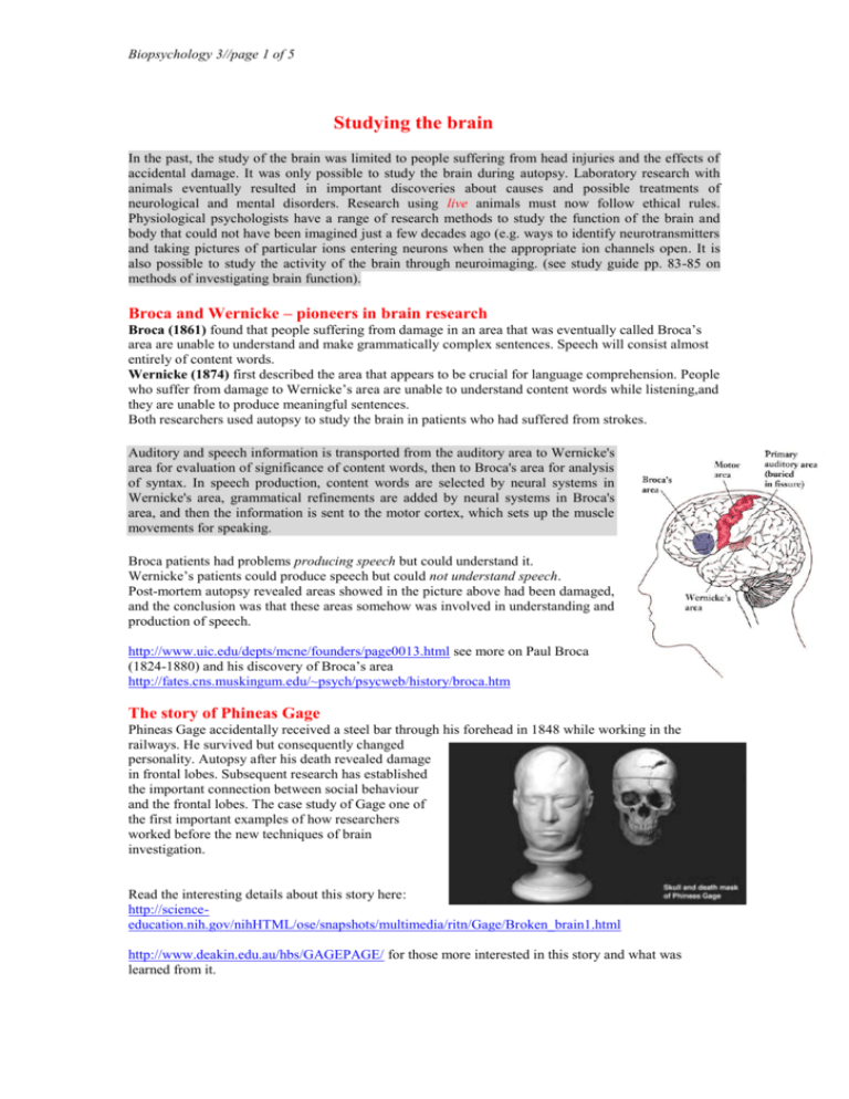
Biopsychology 3//page 1 of 5 Studying the brain In the past, the study of the brain was limited to people suffering from head injuries and the effects of accidental damage. It was only possible to study the brain during autopsy. Laboratory research with animals eventually resulted in important discoveries about causes and possible treatments of neurological and mental disorders. Research using live animals must now follow ethical rules. Physiological psychologists have a range of research methods to study the function of the brain and body that could not have been imagined just a few decades ago (e.g. ways to identify neurotransmitters and taking pictures of particular ions entering neurons when the appropriate ion channels open. It is also possible to study the activity of the brain through neuroimaging. (see study guide pp. 83-85 on methods of investigating brain function). Broca and Wernicke – pioneers in brain research Broca (1861) found that people suffering from damage in an area that was eventually called Broca’s area are unable to understand and make grammatically complex sentences. Speech will consist almost entirely of content words. Wernicke (1874) first described the area that appears to be crucial for language comprehension. People who suffer from damage to Wernicke’s area are unable to understand content words while listening,and they are unable to produce meaningful sentences. Both researchers used autopsy to study the brain in patients who had suffered from strokes. Auditory and speech information is transported from the auditory area to Wernicke's area for evaluation of significance of content words, then to Broca's area for analysis of syntax. In speech production, content words are selected by neural systems in Wernicke's area, grammatical refinements are added by neural systems in Broca's area, and then the information is sent to the motor cortex, which sets up the muscle movements for speaking. Broca patients had problems producing speech but could understand it. Wernicke’s patients could produce speech but could not understand speech. Post-mortem autopsy revealed areas showed in the picture above had been damaged, and the conclusion was that these areas somehow was involved in understanding and production of speech. http://www.uic.edu/depts/mcne/founders/page0013.html see more on Paul Broca (1824-1880) and his discovery of Broca’s area http://fates.cns.muskingum.edu/~psych/psycweb/history/broca.htm The story of Phineas Gage Phineas Gage accidentally received a steel bar through his forehead in 1848 while working in the railways. He survived but consequently changed personality. Autopsy after his death revealed damage in frontal lobes. Subsequent research has established the important connection between social behaviour and the frontal lobes. The case study of Gage one of the first important examples of how researchers worked before the new techniques of brain investigation. Read the interesting details about this story here: http://scienceeducation.nih.gov/nihHTML/ose/snapshots/multimedia/ritn/Gage/Broken_brain1.html http://www.deakin.edu.au/hbs/GAGEPAGE/ for those more interested in this story and what was learned from it. Biopsychology 3//page 2 of 5 Research methods. The earliest research method of physiological psychology involves correlating a behavioural deficit with damage to a specific part of the nervous system. This method can be used in two ways 1. Study of the effects of brain damage on function, e.g. effect of damage to the frontal lobes on the ability to create and adhere to plans (Code et al. 1996). 2. Experimental brain lesion, i.e. an injury to a particular part of the brain (only in an animal brain!). The techniques used for observing activities of the brain called non- invasive (i.e. the technique does not involve breaking the skin with an electrode e.g.) invasive where the skin is broken, e.g. because you use lesions but even injections with radio. Invasive methods: ablation/lesion, electrical stimulation, micro-electrode recording Brain lesioning involves deliberate surgically removal of brain tissue in order to study behavioural changes. You can cut, burn with electrodes or suck part of the brain tissue away. Ablation/lesion was introduced by Pierre Flourens in 1820s. He cut slices of the cerebellum in rabbits, birds and dogs and found that animals had problems in muscular co-ordination and poor sense of balance after the lesion. Flourens correctly hypothesised that removed parts were involved in muscular coordination and balance. Advantages of ablation/lesion in animal research gives a possibility to understand how the brain functions without having to wait for naturally occurring damages) Under anaesthetic, an animal’s head can be held in a fixed position in what is termed a stereotaxic1 apparatus to insert an electrode into a particular location in the brain so that you can investigate an exact correlate of behaviour by comparing between behaviour before the brain damage and afterwards. Disadavantages of ablation/lesion in animal research: perhaps limited what such studies can tell about the human brain You cannot be absolute sure that behavioural changes are only due only to ablation There may be ethical issues in using this technique Electrical stimulation of the brain (ESB): This method implies insertion of electrodes into the brain of a living animals and sending a weak electric current into the brain to mimic a nerve impulse (i.e. a false nerve impulse will make the brain react as if real impulse from sensory receptor). In the picture of the monkey here it is demonstrated that the diameter of the pupil can be electrically controlled as if it were the diaphragm of a photographic camera lens. See constriction of the right pupil evoked by stimulation of the hypothalamus (from Delgado’s book). The other pupil is big. Delgado demonstrated that he could control a bull by sending impulses via a transmitter into the brain the term ‘stereotaxic’ refers to the ability to manipulate an object in three-dimensional space. The researcher passes an electrical current through the electrode, which produces heat that destroys a small portion of the brain around the tip of the electrode. After a few days, the animal recovers from the operation, and the researchers can assess its behaviour. 1 Biopsychology 3//page 3 of 5 Olds & Milner (1954) found that electrical stimulation of hypothalamus in rats seemed extremely pleasurable and that animals who were stimulated in this area was only interested in continuing to stimulate the area. It was suggested that this centre was perhaps a pleasure centre in the brain. They also located a different centre that the animals would avoid to activate and suggested that this was a pain centre. Using the electrical stimulation method, James Olds and Peter Milner (1954) observed that some animals seem to behave in a manner that increased the amount of intracranial stimulation that they received. Further investigation demonstrated that rats will press a lever as rapidly as 2000 times each hour to obtain electrical brain stimulation, and they will continue responding at this rate for twentyfour hours or longer. They will ignore other rewards, such as water or food, to continue working for electrical stimulation. These very powerful results led to the adoption of the intracranial selfstimulation method for investigating the "reward system" in the brain and remain up to now the principal tool. Walter Penfield performed surgery on epileptics during the 1940s and 1950s. Before surgery he tried to stimulate the cortex . He found that there were no pain receptors in the brain. The patient was conscious during the stimulation and Penfield mapped the cortex in this way. Evaluation of ESB: Provides insight into functioning of brain (e.g. argued that if stimulation in one area produces a behaviour →the site is involved in that behaviour) May be efficient in treatment of certain psychological disorders (schizophrenia mentioned) Efficient in blocking pain (e.g. due to cancer) Evaluation by Valenstein (1977) no single area of the brain is the only source of a behaviour/emotion ESB-provoked behaviour is compulsive + stereotypical (i.e. does not perfectly mimic natural behaviour) ESB effects may depend on other factors (not everybody reacts in the same way to stimulation). Non-invasive methods: CAT, MRI, PET etc. Scanning and imaging devices: neuroimaging techniques. The development of several different diagnostic machines, which can be used to investigate the brain’s structure and activity, has revolutionised neuropsychological research. Since the 1970s it has been possible to study the human brain in living individuals, e.g. using X-rays. Sophisticated techniques called neuroimaging techniques now allow researchers to visualise and obtain images of brain function and structure. These techniques include e.g. CAT, PET, MRI and fMRI. CAT (computerizes axial tomogram): the technique includes that a number of X-rays pictures are taken from different locations. The scanner sends a narrow beam of X-rays through e.g. a person’s head. The beam is moved around the patient’s head, and a computer calculates the amount of radiation that passes through it at various points along each angle. The result is a three- dimensional image of a ‘slice’ of the brain’s structures. CAT scans help determine if behavioural problems have physiological determinant and it can help surgeons to determine how to proceed in an operation, or it can establish effects of therapy, e.g. radiation on brain tumours. Since you inject a radioactive substance in the patient, it is considered to be an invasive method. Biopsychology 3//page 4 of 5 MRI (Magnetic resonance imaging) gives a three-dimensional picture of the brain structures in greater detail than the CAT scanner. It uses magnetic fields and radio waves (instead of X-rays). It exploits the fact that some substances that make up the body have intrinsic magnetic properties and respond to being in a magnetic field, rather as does a compass needle. For example, water, a major component of the body is made up of hydrogen and oxygen and the hydrogen atoms exhibit such a magnetic property. When a magnetic field is passed over the head, reverberations are produced by hydrogen molecules, and these are picked up by the scanner, which can convert the activity into a structural image (of the normal brain, structural disorders in the brain etc.). CAT vs. MRI Harmless radio waves are used in MRI (not X-rays and injected dyes like in the CAT scan), more sensitive than the CAT scan (very accurate pictures) still pictures (i.e. useful for structure but not possible to deal with function) PET2 (positron emission tomography) invasive measure of brain metabolism, glucose consumption and blood flow. The procedure: the person is injected in the arm with harmless dose of a radioactive substance (glucose) that enters the brain and goesto active parts. PET measures brain activity by examining the amount of oxygen consumed by, or blood flow travelling to, neurons. The radioactive parts of glucose emit positrons, which are detected by PET scanner and this activity is represented in the form of coloured maps. PET scan depicting the effects of Alzheimer's disease on metabolism. The arrow indicates areas of low activity in the parietotemporal cortex - a region important for processing language and memory PET can diagnose abnormalities like tumours or changes like in Alzheimer. compare brain differences in normal individuals and individuals with psychological disorders (neural activity is different in persons with schizophrenia). The greatest advantage of PET (compared to MRI) is that it can record ongoing activity in the brain, e.g. thinking Some empirical research Restak (1984) used the PET to show how the front of the brain and the part that produces movement became active when a person was asked to move the right hand. When a person is asked to think about moving the hand, only the front part is active (and not the part involved in actual movement). Martin &Brust (1985) showed that participants asked to listen to and recall a story had activity in the part of the brain responsible for processing auditory information, and also in the hippocampus when asked to recall (hippocampus involved in memory). It is possible to use MRI in a functional capacity, i.e. to examine the brain’s function as well as its structure (functional magnetic resonance imaging or fMRI). MRI and fMRI are non-invasive methods (compared to PET where radioactive substances are introduced in the body). MRI and fMRI have been used to investigate similar functions to those investigated using PET: language, attention, vision, memory etc. PET and MRI can be used in combination. They have the advantage of good spatial resolution (i.e. images and structures are seen very precisely) but the disadvantage of a poor temporal resolution (i.e. it is difficult to match the psychological and neural 2 http://www.dushkin.com/connectext/psy/ch02/pet.mhtml Biopsychology 3//page 5 of 5 event in time precisely). The reason for this is that in PET and MRI, a number of scans are taken and these are averaged in time. Assignments: 1. How did researchers study the brain before the new techniques? 2. What are the advantages of the new techniques compared to the old techniques? 3. Discuss ethical implications of using animals in neurological research: see here the APA guidelines for ethical conduct in animal research http://www.apa.org/science/anguide.html 4. Compare two different methods for investigating the brain and evaluate their strength and limitation. http://web.lemoyne.edu/~hevern/psy340/lectures/psy340.04.1.research.meth.html Some links to the discussion on ethics in animal research: http://www.simr.org.uk/pages/nobel/nobel_survey.html http://www.amprogress.org/Issues/IssuesList.cfm?c=13


