Supplementary Information (docx 573K)
advertisement
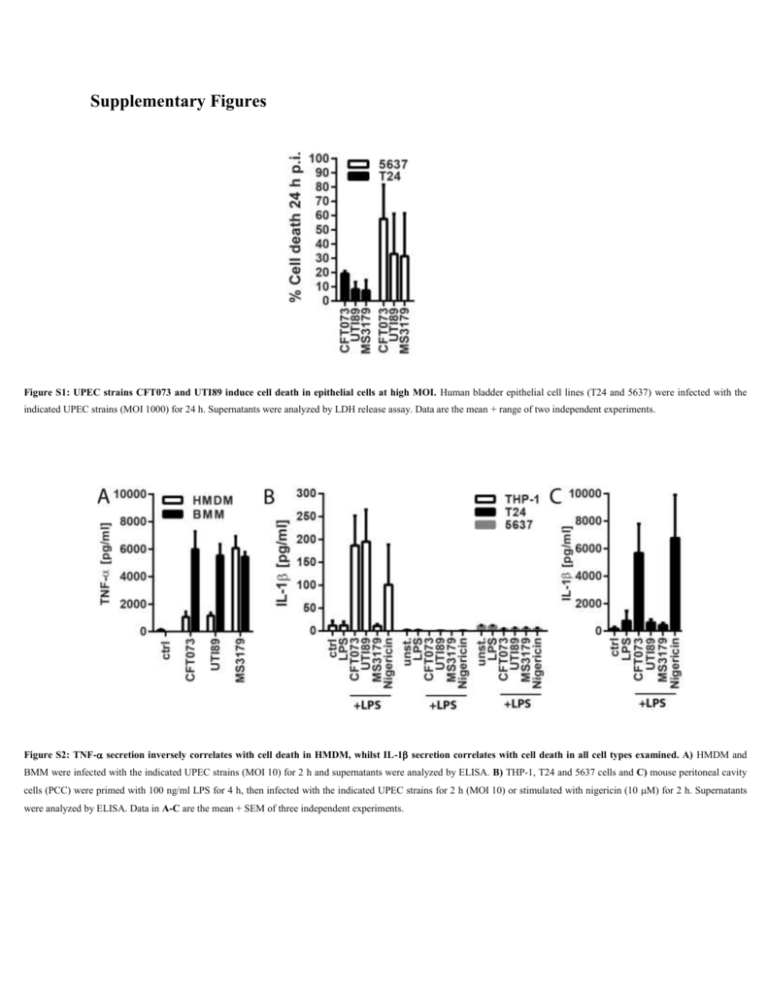
Supplementary Figures Figure S1: UPEC strains CFT073 and UTI89 induce cell death in epithelial cells at high MOI. Human bladder epithelial cell lines (T24 and 5637) were infected with the indicated UPEC strains (MOI 1000) for 24 h. Supernatants were analyzed by LDH release assay. Data are the mean + range of two independent experiments. Figure S2: TNF- secretion inversely correlates with cell death in HMDM, whilst IL-1 secretion correlates with cell death in all cell types examined. A) HMDM and BMM were infected with the indicated UPEC strains (MOI 10) for 2 h and supernatants were analyzed by ELISA. B) THP-1, T24 and 5637 cells and C) mouse peritoneal cavity cells (PCC) were primed with 100 ng/ml LPS for 4 h, then infected with the indicated UPEC strains for 2 h (MOI 10) or stimulated with nigericin (10 M) for 2 h. Supernatants were analyzed by ELISA. Data in A-C are the mean + SEM of three independent experiments. Figure S3: NLRP3 inhibition has differing effects in CFT073-infected primary human versus mouse macrophages. HMDM and BMM were primed with LPS (100 ng/ml, 4 h) or were left untreated, then pretreated for 1 h with the NLRP3 inhibitor MCC950 (10 M), and subsequently infected with the UPEC strain CFT073 (MOI 10) or stimulated with nigericin (10 M). At 2 h p.i., supernatants were collected and analyzed by A) LDH release assay and B) IL-1 ELISA. Data are the mean + SEM of three independent experiments. Figure S4: CFT073- and UTI89-triggered cell death is mediated by a large, soluble factor(s) that is heat- and protease-sensitive. A) PMA-differentiated THP-1 cells were treated with live bacteria at MOI 10, heat inactivated supernatants and bacteria (95˚C, 20 min) (HI bacteria), 10% filtered (0.45 m) supernatants from bacterial overnight cultures (S/N), or flow though from filtration with Amicon Ultra-15 centrifugal fiter units with 30 kDa cutoff (S/N < 30 kDa). Cell culture supernatants were analyzed by LDH release assay after 2 h. Similar results were obtained in two independent experiments. B) PMA-differentiated THP-1 cells were treated with bacterial culture supernatants as above (S/N) or supernatants pretreated with proteinase K (100 g/ml) for 45 min at 37˚C (S/N +Prot K). Cell viability was assessed by methylthiazolyldiphenyl-tetrazolium bromide (MTT) viability assays. Medium was replaced with medium containing 1 mg/ml MTT (Sigma-Aldrich) 2 h post-infection and incubated at 37˚C and 5% CO2 for another 2 h. Cells were lysed in isopropanol and formation of formazan was assessed by measuring absorption at 570 nm. Cell viability was calculated as % of absorption at 570 nm of untreated cells. Donor : % Cell death MCC950 LPS + LPS+CFT073 + LPS+Nig + LPS+Sal + + LPS+UTI89 Donor : IL-1 [pg/ml] MCC950 LPS + LPS+CFT073 + LPS+Nig + LPS+Sal + LPS+UTI89 + 1 2 3 4 5 6 7 8 9 0.00 0.00 0.00 0.00 0.00 0.00 0.00 0.00 0.00 0.34 0.47 1.21 2.83 -0.23 -1.16 0.00 0.00 0.00 6.93 34.49 27.71 39.05 8.08 63.04 17.32 23.25 35.83 0.51 25.89 24.66 29.14 1.18 62.69 21.88 25.61 32.43 46.68 14.72 18.63 24.28 14.73 18.89 4.60 7.91 26.25 1.06 14.98 16.85 3.19 1.89 2.63 -0.45 0.50 10.71 49.15 32.57 27.43 17.73 48.54 ND ND ND 10.95 43.51 45.27 27.51 61.45 ND ND ND 24.82 30.66 29.16 33.79 ND ND ND ND ND -0.01 22.72 24.66 20.15 ND ND ND ND 1 2 3 4 5 6 7 8 9 41.74 42.49 37.23 15.48 43.21 12.56 0.00 25.94 0.00 20.24 -1.54 ND 41.89 42.49 49.64 26.26 117.92 7.00 2.73 8.93 0.00 1877.28 232.20 2973.51 1084.81 2973.85 9706.29 685.04 2259.10 2876.68 363.65 67.74 1803.38 149.82 117.58 1093.71 501.10 1527.51 1539.38 1314.92 107.99 4872.90 1202.30 4629.54 5147.09 1497.69 2994.72 1949.71 46.94 43.10 74.98 44.01 106.71 33.27 3.47 27.50 5215.72 2708.92 7228.57 4618.81 3074.61 10308.71 ND ND ND 5701.27 2718.58 3973.76 4733.99 4182.30 ND ND ND 2235.10 238.90 4034.61 1220.35 ND ND ND ND ND 155.28 57.51 336.57 147.63 ND ND ND ND ND 6127.35 0.00 Average 0.00 0.38 28.41 24.89 19.63 4.35 31.02 34.82 29.61 16.88 Average 24.29 32.98 2740.97 795.98 2635.21 42.22 5525.89 4572.87 1932.24 174.25 Table S1: NLRP3 contributes to IL-1b release but contributes only marginally to cell death in human macrophages responding to the UPEC strains CFT073 and UTI89. HMDM were primed with LPS (100 ng/ml, 4 h), pretreated for 1 h with the NLRP3 inhibitor MCC950 (10 mM) and then subsequently infected with the UPEC strains CFT073 or UTI89 or S. Typhimurium SL1344 (MOI 10), or were stimulated with nigericin (10 mM). Supernatants were collected at 2 h p.i., and analyzed by LDH release assay and ELISA. Data are from nine (CFT073, LPS/nigericin), six (S. Typhimurium) or four (UTI89) independent experiments (different donors), respectively. ND = not determined.

