Case Scenario 1
advertisement
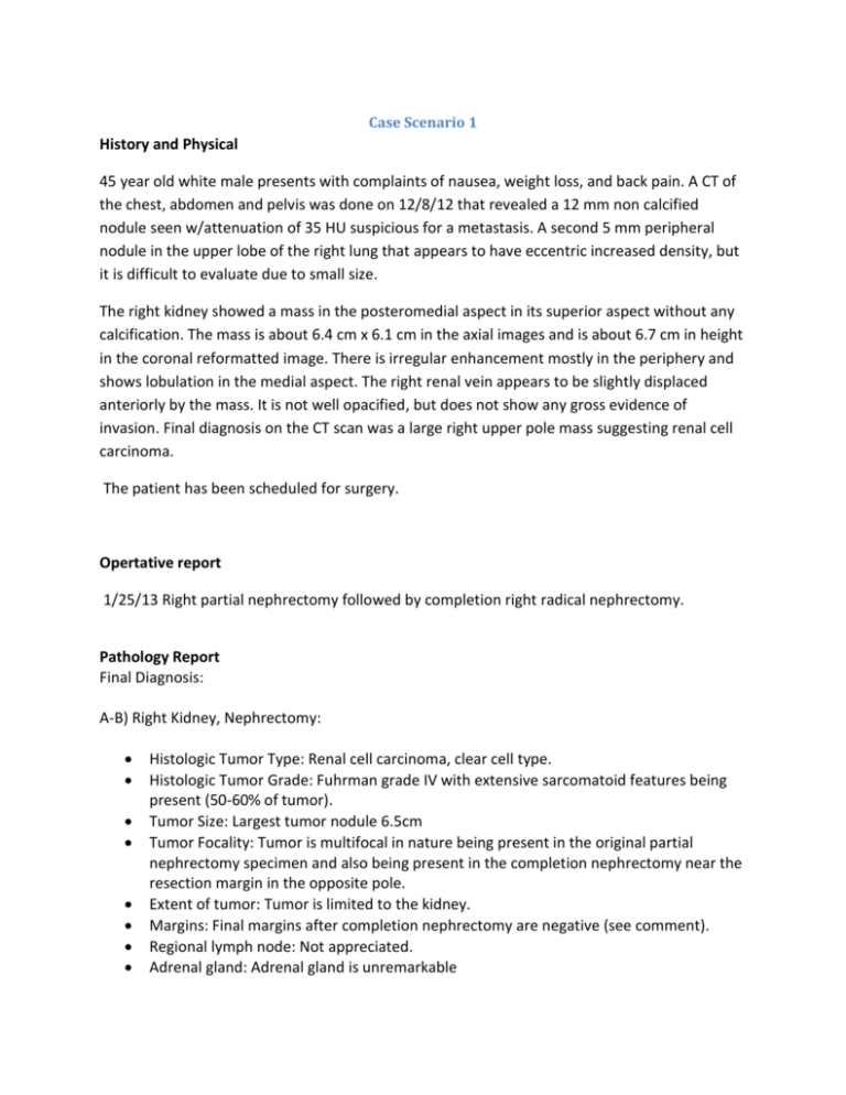
Case Scenario 1 History and Physical 45 year old white male presents with complaints of nausea, weight loss, and back pain. A CT of the chest, abdomen and pelvis was done on 12/8/12 that revealed a 12 mm non calcified nodule seen w/attenuation of 35 HU suspicious for a metastasis. A second 5 mm peripheral nodule in the upper lobe of the right lung that appears to have eccentric increased density, but it is difficult to evaluate due to small size. The right kidney showed a mass in the posteromedial aspect in its superior aspect without any calcification. The mass is about 6.4 cm x 6.1 cm in the axial images and is about 6.7 cm in height in the coronal reformatted image. There is irregular enhancement mostly in the periphery and shows lobulation in the medial aspect. The right renal vein appears to be slightly displaced anteriorly by the mass. It is not well opacified, but does not show any gross evidence of invasion. Final diagnosis on the CT scan was a large right upper pole mass suggesting renal cell carcinoma. The patient has been scheduled for surgery. Opertative report 1/25/13 Right partial nephrectomy followed by completion right radical nephrectomy. Pathology Report Final Diagnosis: A-B) Right Kidney, Nephrectomy: Histologic Tumor Type: Renal cell carcinoma, clear cell type. Histologic Tumor Grade: Fuhrman grade IV with extensive sarcomatoid features being present (50-60% of tumor). Tumor Size: Largest tumor nodule 6.5cm Tumor Focality: Tumor is multifocal in nature being present in the original partial nephrectomy specimen and also being present in the completion nephrectomy near the resection margin in the opposite pole. Extent of tumor: Tumor is limited to the kidney. Margins: Final margins after completion nephrectomy are negative (see comment). Regional lymph node: Not appreciated. Adrenal gland: Adrenal gland is unremarkable Significant Pathologic Findings in nonneoplastic kidney: There is Mild arterionephorsclerosis with limited fibrosis surrounding the tumor mass. Pathologic Stage: T1b Nx Diagnostic Comments: The first specimen received was a partial nephrectomy specimen which demonstrated a 6.5 cm. tumor which closely abutted the inked margin. It was seen to be multifocal in nature. A completion nephrectomy specimen was received which demonstrated small tumor nodules adjacent to the previous resection surface and in the opposite pole. An extensive amount of this tumor is sarcomatoid in differentiation. Follow up recommendations: 1/27/13 We will continue to monitor the metastatic bronchial lesion. At this time no adjuvant therapy has been planned. 2/4/13 CT Chest: Definite worsening in multiple pulmonary metastases when compared to 12/8. No evidence of adenopathy in the chest. 2/4/13 MRI Brain: 4 cm posterior parietal metastasis. 2/8/13 Bone Scan: Abnormal areas of increased activity of the lft humerus suspicious for bony metastasis. 2/26/13 The patient was started on Sutent and brain radiation. How many primaries are present in this case scenario? 1 per rule M11 What is the diagnosis date? 12/8/12 How would we code the histology of the primary you are currently abstracting? What is the sequence? 00 8310/3 clear cell carcinoma per rule H12 Stage/ Prognostic Factors CS Tumor Size CS Extension CS Tumor Size/Ext Eval 065 100 3 CS SSF 9 CS SSF 10 CS SSF 11 988 988 988 CS Lymph Nodes CS Lymph Nodes Eval Regional Nodes Positive Regional Nodes Examined CS Mets at Dx CS Mets Eval CS SSF 1 CS SSF 2 CS SSF 3 CS SSF 4 CS SSF 5 CS SSF 6 CS SSF 7 CS SSF 8 000 0 98 00 40 0 000 000 000 010 000 040 000 000 CS SSF 12 CS SSF 13 CS SSF 14 CS SSF 15 CS SSF 16 CS SSF 17 CS SSF 18 CS SSF 19 CS SSF 20 CS SSF 21 CS SSF 22 CS SSF 23 CS SSF 24 CS SSF 25 988 988 988 988 988 988 988 988 988 988 988 988 988 988 Treatment Diagnostic Staging Procedure Surgery Codes Surgical Procedure of Primary Site Approach-Surgery of Primary Site at this facility Scope of Regional Lymph Node Surgery Surgical Procedure/ Other Site 00 50 5 0 0 Systemic Therapy Codes Chemotherapy Hormone Therapy Immunotherapy Hematologic Transplant/Endocrine Procedure Systemic /Surgery Sequence Radiation Radiation Treatment Volume Regional Treatment Modality Reason No Radiation 00 00 00 00 0 00 00 1 Case Scenario 2 History and Physical A 63 year old white male presents with a history of left flank pain for the last month. A bone scan and CT were done on 2/22/13. The CT showed a large complex left renal mass (10 x 8 x 7.8 cm) highly suspect for renal cell carcinoma. Also noted was a left renal vein tumor thrombosis. Metastases are noted involving the bilateral adrenal glands, with a 6 cm mass in the left adrenal gland. There are multiple mildly enlarged upper abdominal lymph nodes. Also noted are bilateral pulmonary metastases and enlarged mediastinal lymph nodes, possibly metastatic. The bone scan showed a large photopenic defect in the lower pole of the left kidney corresponding to the renal mass. No intense uptake is noted to suggest osseous metastasis. Operative Report 3/11/13 Left laparoscopic conversion to open radical nephrectomy Pathology Report Final Diagnosis: Specimen: Kidney and adrenal gland, left, radical nephrectomy. Histologic Tumor Type: Sarcomatoid renal cell carcinoma Histologic Tumor Grade: Fuhrman grade 4 (of 4) Tumor Size: 9.0 X 9.0 X 8.0 CM. Tumor Focality: The tumor is unifocal within the kidney. Extent of Tumor: The tumor invades through the renal capsule into perirenal adipose tissue and abuts, but does not show definitive invasion beyond Gerota’s fascia. The tumor invades into renal sinus adipose tissue. The tumor grossly invades into the renal vein. Margins: The renal vein margin is positive for tumor. The remaining surgical margins are negative. Adrenal gland: The adrenal gland is positive for metastatic sarcomatoid renal cell carcinoma. No contiguous involvment from the kidney is seen. Regional lymph nodes: No regional lymph nodes are identified. Pathologic TNM Stage: AJCC pT3a Nx M1 PATHOLOGIC TNM STAGE: AJCC pT3a NX M1. Patient was started on preoperative Torisel chemotherapy 3/3/13. Chemotherapy was continued after surgery. How many primaries are present in this case scenario? 1 primary per rule M2 What is the diagnosis date? 2/22/13 What is the sequence? 00 How would we code the histology of the primary you are currently abstracting? 8318/3 Sarcomatoid carcinoma Stage/ Prognostic Factors CS Tumor Size CS Extension CS Tumor Size/Ext Eval 100 601 5 CS SSF 9 CS SSF 10 CS SSF 11 988 988 988 CS Lymph Nodes CS Lymph Nodes Eval Regional Nodes Positive Regional Nodes Examined CS Mets at Dx CS Mets Eval CS SSF 1 CS SSF 2 CS SSF 3 CS SSF 4 CS SSF 5 CS SSF 6 CS SSF 7 CS SSF 8 000 0 98 00 40 5 030 010 020 010 000 040 000 000 CS SSF 12 CS SSF 13 CS SSF 14 CS SSF 15 CS SSF 16 CS SSF 17 CS SSF 18 CS SSF 19 CS SSF 20 CS SSF 21 CS SSF 22 CS SSF 23 CS SSF 24 CS SSF 25 988 988 988 988 988 988 988 988 988 988 988 988 988 988 Treatment Diagnostic Staging Procedure Surgery Codes Surgical Procedure of Primary Site Approach-Surgery of Primary Site at this facility Scope of Regional Lymph Node Surgery Surgical Procedure/ Other Site 00 50 4 0 Systemic Therapy Codes Chemotherapy Hormone Therapy Immunotherapy Hematologic Transplant/Endocrine Procedure Systemic /Surgery Sequence 02 00 00 00 4 0 Radiation Radiation Treatment Volume Regional Treatment Modality Reason No Radiation 00 00 1
