05,6 PLATELETS PHYSIOLOGY AND DISORDERS
advertisement
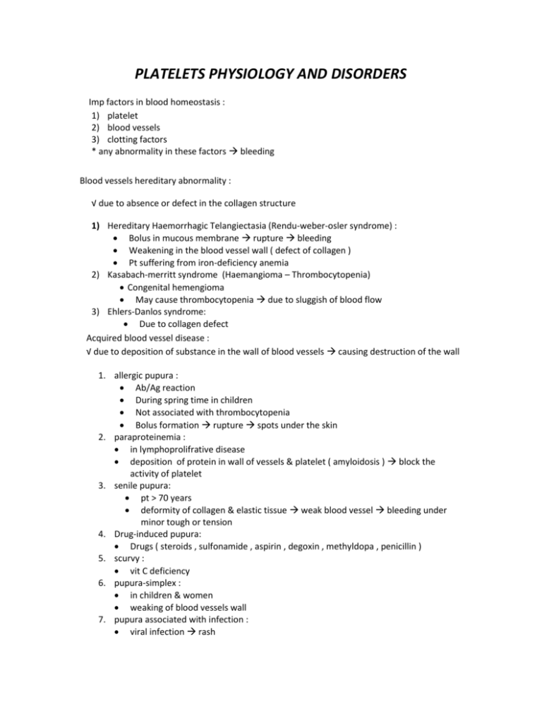
PLATELETS PHYSIOLOGY AND DISORDERS Imp factors in blood homeostasis : 1) platelet 2) blood vessels 3) clotting factors * any abnormality in these factors bleeding Blood vessels hereditary abnormality : √ due to absence or defect in the collagen structure 1) Hereditary Haemorrhagic Telangiectasia (Rendu-weber-osler syndrome) : Bolus in mucous membrane rupture bleeding Weakening in the blood vessel wall ( defect of collagen ) Pt suffering from iron-deficiency anemia 2) Kasabach-merritt syndrome (Haemangioma – Thrombocytopenia) Congenital hemengioma May cause thrombocytopenia due to sluggish of blood flow 3) Ehlers-Danlos syndrome: Due to collagen defect Acquired blood vessel disease : √ due to deposition of substance in the wall of blood vessels causing destruction of the wall 1. allergic pupura : Ab/Ag reaction During spring time in children Not associated with thrombocytopenia Bolus formation rupture spots under the skin 2. paraproteinemia : in lymphoprolifrative disease deposition of protein in wall of vessels & platelet ( amyloidosis ) block the activity of platelet 3. senile pupura: pt > 70 years deformity of collagen & elastic tissue weak blood vessel bleeding under minor tough or tension 4. Drug-induced pupura: Drugs ( steroids , sulfonamide , aspirin , degoxin , methyldopa , penicillin ) 5. scurvy : vit C deficiency 6. pupura-simplex : in children & women weaking of blood vessels wall 7. pupura associated with infection : viral infection rash PLATELETSPHYSIOLOGY: Mechanism of platelet activation : Injury to blood vessel -> activate platelet aggregation by exposure of collagen coagulation factors activation fibrinogen converted into fibrin ( insoluble ) Production: fragmentation Hematopoietic stem cell Megakaryoblasts Megakaryocytes-------------- ---- Platelets. of cytoplasm Megakaryocytes are found in the cytoplasm of the bone marrow and are released to the blood stream by a budding process Each megakaryocyte gives 4000 platelet Circulation: 1-2.5 micron in diameter Normal count 150,000 – 450,000 Normal life span 7 – 10 days. About 1 / 3 are trapped in the spleen. Young platelets are larger than old ones. Structure: Mucopolysacch. Coat: glycoprotein content which are important for interaction of platelets with each other or aggregating agents. Alpha (α) granules: " Fibronigen , Factor V, vWF, Fibronectin, β-thromboglobin, heparin antagonist (PF4), thrombospondin " Dense core:"Nucleotides (ADP), Ca+2, serotonin" Peripheral microtubule : elastic fiber , encircle the cell squeeze the platelet when activated Function: Formation of mechanical plug during normal homeostatic response to vascular injury. The main steps involved are: adhesion, release, aggregation. 1-Platelets adhesion: Adhesion of platelet to sub-endothelial collagen. Dependent on the VW factor (Von Willebrand part of fact VIII). Also dependent on glycoproteins. MCQ Note Glyco-proteins 1B, 2B, and 3A need VW factor to connect collagen; however, 1A glycoprotein does not because it has a direct connection with collagen. 1B is a receptor for vW factor bridge the gap between the subendothelial collagen & platelet 2B and 3A join platelet to each other and fibrinogen. Deficiency in 1B, 2B, 3A lead to abnormal synthesis. Deficiency in 2B, 3A is the most common and lead to Glanzman disease "congenital". Deficiency in 1B lead to Bernard Soluier's "congenital" "MCQ" Deficiency in VW factor vW disease 2-Release (secretion): Collagen exposure results in the release of granules contents (ADP, serotonin, and fibrinogen). Collagen and thrombin activate prostaglandin synthesis. cyclothrombpxane membrane phospholipids arachidonic acid ------------ --------------------- thromboxane A2 oxygenase synthetase "in platelets" Thromboxane A2: Potentiates aggregation and vasoconstrictor. Cyclooxygenase is inhibited by small dose of aspirin 3-Aggregation: Release ADP + thromboxane A2 aggregation. This is followed by more secretion secondary aggregation platelet mass or plug. Note : in epithelial cells , we have the same reaction But differ in one enzyme ( prostacyclin synthase ) instead of thromboxane synthase . prostacyclin synthase synthesize PGE2 ( which is anti-platelet ) to maintain the blood in liquide state PLATELETS DISORDERS: Platelet disorders are the most common cause of bleeding. The disorder could be decrease in number (thrombocytopenia) or defective function. A-1- DECREASED PLATELETS NUMBER OR THROMBOCYTOPENIA ( < 150,000 ) : Loss of platelets from the circulation faster than the rate of their production by the bone marrow. So Thrombocytopenia is due to: a) Failure of platelets production, most common cause. Megakaryocytes are decreased in the bone marrow e.g. drugs. b) Increase rate of removal of platelets from the circulation. i. Megakaryocytes are increased or normal in the bone marrow i.e. production is normal but platelets are destroyed e.g. by antibodies. c) Combination of A&B. Causes: Congenital * Megakaryocytic hypoplasia. * TAR Syndrome. Acquired * Immuno thrombocytopenia √ may be associated with other autoimmune disease ( e.g. SLE ) * Thrombotic thrombocytopenic purpra * DIC * Drugs( cytotoxic drug e.g. chemotherapy) * Infections * Splenomegaly. ( abnormal platelet distribution ) * Bone marrow suppression or infiltration * Aplastic anemia * HIV *Bone Marrow Suppression: Due to effect of infections (viral) or toxins or due to replacement e.g. by malignancy e.g. leukemias, metastatic tumors, or due to fibrosis of the bone marrow e.g. due to irradiation *DIC: Due to consumption of platelets "more about it in next topics ". *Drugs: Due to suppression e.g. phenylbutazone, Gold, Thiazide. Other mechanisms of action are immune, or by causing direct aggregation of platelets. May be accompanied by other sings e.g. fever, joint pain, rash, leucopenia. *Aplastic Anemia: means shut down of the bone marrow *Splenomegaly: Normally 1/3 of body platelets are in the spleen and 2/3 in the peripheral circulation. With spleen enlargement, up to 80 - 90% of body platelets will pool in the spleen decreased platelets in the peripheral circulation. This spleen enlargement could be due to many causes e.g. thalassemia, portal hypertension, gaucher's malaria, kalaazar, lymphomas, etc. Life span of the platelet is normal. NOTE one of the treatments of thrombocytopenia is by removing the spleen. But in some cases it can lead to thrombocytosis *Infection: Decreased platelets can be seen with many infections, e.g. intra–uterine infections: best examples are congenital syphilis, toxoplasmosis, rubella, cytomegalovirus (CMV), and herpes. Also seen with other infections e.g. influenza, chickenpox, rubella, infectious mononucleosis. The effect is due to suppression of bone marrow, immune mediated or due to DIC in fulminant infections. * Thrombotic thrombocytopenic purpra : Microangiopathic hemolytic anemia Fragmentation of RBCs due to hitting of them by fibrin threads in occluded microcirculation * Dilutional loss Massive transfusion of stored blood to bleeding patients Manifestations: Usually bleeding not significant until count is < 80,000. Presents as skin purpura, ecchymoses, mucosal membrane bleeding, and prolonged bleeding after trauma. The most common site of bleeding is in the retina( mirror for cerebral hemorrhage ) If the Thrombocytopenia is severe, intracranial hemorrhage can be seen. Mucoceutenous bleeding : due to platelet reduction or dysfunction Note : Skeletomuscular bleeding indicate coagulation factor defects Investigations: In addition to history and examination, the following tests are required: Platelet count Blood smear Bleeding time: To measure Platelet plug formation in vivo. Normal is 2.5-9.5 mins. Bone marrow examination. Platelet Aggregation test: - Detect aggregation using aggregation agents ADP, adrenaline, collagen, ristocetin. - Looking for 50% aggregation within 1 minute Degree of Symptoms Physical findings Thrombocytopenia Mild (>50 000/mm3) Moderate (30-50 000/mm3) Severe (10-30 000/mm3) None Bruising with minor None Scattered ecchymoses trauma at trauma site Spontaneous bruising. Petechiae and purpura, menorrhagia more prominent on extremities Marked (<10 000mm3) spontaneous brusing, Generalized purpura, mucosal bleeding, risk epistaxis, GU bleeding for CNS bleeding CNS symptoms A-2-IMMUNOTHROMBOCYTOPENAI (ITP): MCQ Autoimmune disorder characterized by platelets bound antibodies. Classification: Acute: usually in children, self limiting, and preceeded by infection usually viral. Chronic: usually in adults, more common in female. Etiology: Idiopathic , or associated with oher autoimmune disease . A- ACUTE IMMUNOTHROMBOCYTOPENIA: Self limiting usually weeks. In children. Usually preceeded by viral infection. Bone marrow shows normal or increased megakaryocytes. Due to immune complexes bound to platelets. (Complex = viral antigen –antibody complex). These complexes are removed by the reticulo-endothelial system (RE system). 5-10% can go into chronic ITP. 90% recovered. B- CHRONIC IMMUNOTHROMBOCYTOPENIA: Pathogenesis: Autoimmune. Antibodies are formed against antigens on platelet surface. Manifestations: Skin purpura, superficial bruising, epistaxis, and menorrhagia. ( mucocuetanous bleeding ) Mucosal hemorrhage is seen in severe cases and intra – cranial hemorrhage is rare. Splenomegaly: 10% of cases Pathogenesis of immunothrombocytopenia (ITP): 1. Platelets are sensitized with auto antibodies. + Anti-platelets Platelets Antibodies (usually IgG) 2. Premature removal of platelets from the circulation by macrophages of the RE system and destroyed mainly in the spleen. Laboratory findings: Thrombocytopenia with giant forms. Count usually 10 -50,000. Bone marrow shows normal or increased megakaryocytes. Platelet bound IgG is +ve . Platelet survival studies show decreased life span. ______________________ B-DEFECTIVE PLATELET FUNCTION: A defect in function is suspected if there is prolonged bleeding time with or without skin or mucosal hemorrhage in the presence of normal platelet count. Disorders: Congenital Acquired - Glanzman disease - Drugs - Bernard Soluier's - Storage granules defect -Uremia - Myeloproliferative disorders - Multiple myeloma - liver disease - valvular disease ( abnormal valve hits platelet ) - hypoglycemia ( glycogen storage disease ) - scurvy - severe burn 1)GLANZMAN'S DISEASE (THROMBASTHENIA): Autosomal recessive inheritance. Normal platelets count. and appearance. No clumps are seen on peripheral blood film (i.e. no platelets clumps). Due to decreased surface membrane glycol-proteins 11b +11a failure of primary aggregation. MCQ Platelets do not aggregate with all aggregation agents but they aggregate with ristocetin. ( in platelet aggregation test ) MCQ Bleeding time is prolonged. MCQ: in prolong bleeding time, the first test we perform is platelet aggregating test. 2) Bernard Soluier's Disease : no aggregation with Ristocetin ( in platelet aggregation test ) Deficiency in 1B glycoprotein 3) Storage pool disease : no aggregation with collagen ( in platelet aggregation test ) In Platelet Aggregation Test : Bernard-Soulier syndrome : no aggregation with Ristocetin Defective Throboxane synthesis : no aggregation with collagen & arachidonic acid(Aspirine – like syndrome ) vW disease : no aggregation with Ristocetin Storage pool deficiency : no aggregation with collagen NOTE : differential diagnosis between Bernard-Soulier syndrome & vW disease by estimation of vW factor in the blood ACQUIRED DISORDERS OF PLATELET FUNCTION: Causes: Uremia (renal failure) Uremia is the most common cause of acquired platelet function abnormality Drugs e.g. aspirin. Asprin is the best example which is the most common cause of acquired platelet function disorder. It irreversible affects the cyclooxygenase enzyme. The effect last 4 -7 days and it takes about 10 days before the platelets are replaced. Presents as increased bleeding time but purpura is unusual. Myeloproliferative disorder. Paraproteinemias e.g. multiple myeloma. Cardiopulmonary bypass. Autoimmune diseases e.g. SLE (Systemic Lupus Erythromatosis) Severe burn ( grade 4 ) Liver disease Clinical note : Expected neonate with thrombocytopenia : Can occur with alloimmune disease (maternal Ab against fetal platelet Ag ) We either do cesirian section Or give intrauterine transfusion of platelet , to prevent cerebral hemorrhage during vaginal delivery COAGULATION Among plasma factors : 4 are anticoagulant factors ( Protein C , protein S , AT-III & factor XII ) 2 are sex-linked ( Factor VIII & IX ) Hereditary coagulation disorders: Hereditary deficiencies of coagulation factors are rare. However, the following disorders are more common: Hemophilia A (Factor VIII deficiency) Hemophilia B (Christma's disease) (Factor IX deficiency) Von will brand's disease. ( mixture of platelet & coagulation factors disease ) A- HEMOPHILIA A This is the most common hereditary disorder of blood coagulation. Inheritance: X – Linked "in the long arm" Defect: The absence or low level of factor VIII MCQ Clinical features: The clinical severity of the disease correlates well with the extent of factor deficiency. Sever (Factor level= <1% of normal): - Profuse post – circumcision bleeding. - Recurrent painful hemarthroses (I.e., bleeding into joints most commonly knees) with progressive joint deformity & crippling. ( may lead to organization of the organ ( fixing ) by fibrosis ) - Muscle hematomas. - Prolonged bleeding after dental extraction. - Hematuria - Spontaneous intra-cerebral hemorrhage, uncommon. Moderate (Level of factor = <1 - 5% of normal): - Most post – traumatic bleeding. - Occasional spontaneous bleeding. Mild (Level of factor = < 5 - 20% of normal): - Post – traumatic bleeding. - Spontaneous bleeding is rare. Laboratory features: Abnormal - APTT : Prolonged MCQ - Factor VIII : Decreased Normal - PT & Bleeding time MCQ Treatment: Principles of treatment: For spontaneous bleeding: bleeding is controlled if the level of factor VIII is raised to above 20%. For major surgery or serious post – traumatic bleeding: the level should be about 100%. Complication of treatment: Disease transmission: - HIV infection - Hepatitis Development of antibodies (inhibitors to factorVIII): - Seen in 5-10% of patients - The antibodies make the patients resistant to treatment & make him require larger doses. B- VON WILLEBRAND'S DISEASE MCQ Inheritance: Autosomal dominant. Defect: 1. Reduced level of factor vWF (von Will brand's factor). This result in rapid loss of factor VIII and abnormal coagulation. 2. Defective platelet – related activity of (Von Will brand's factor). This affect platelet's adherence to sub-endothelial collagen & platelet aggregation induced by ristocetin. Clinical features: Operative & post – traumatic bleeding. Mucous membrane bleeding e.g., epistaxes, menorrhagia. MCQ Hemarthroses & muscle hematomas are rare in contrast to hemophilia A. Laboratory Features: Bleeding time: prolonged ( because it is related to platelets ) PTT: prolonged Factor Assay: low levels: VWF & VIII - Platelet aggregation: defective with ristocetin. Normal with other reagents. - Normal PT Treatment: Factor VIII concentrate - NOTE : This table is important for differential diagnosis between Hemophilia A & vW disease : C – Factor XIII deficiency : Factor XIII convert soluble fibrin ( fibrin s ) insoluble fibrin ( fibrin i ) Pt with factor XIII deficiency will have " secondary bleeding " that means pt will bleed for 4-6 hrs ( after teeth extract for example ) then will stop , in the next morning will bleed again due to fibrin instability Fibrin stabilization test : 5 molar urea conc. + blood sample if the clot is desolved Factor XIII deficiency D – Factor XII deficiency : Factor XII is called " Hugmann factor " Factor XII has fibrinolytic activity ( through plasmin pathway ) Activated by Calicrine ( chemotactic for neutrophils ) Pt with Calicrine deficiency cannot kill the bacteria due to lack the chemotactic activity of Calicrine Treatment of Factor XII deficiency : Thrombolytic drugs DISSEMINATED INTRAVASCULAR COAGULATION (DIC): Widespread intravascular deposition of fibrin with consumption of coagulation factors and platelets. This occurs as a consequence of many disorders which release pro-coagulant material into the circulation or cause widespread endothelial damage or platelet aggregation. Forms of DIC: Chronic, less server course. Fulminant hemorrhagic course. Pathology: Deposition of fibrin in the microcirculation. Formation of large amounts of fibrin monomers. Increased fibrinolysis with release of fibrin split products (FSPs) or fibrin degradation products (FDPs). These FDPs interfere with fibrin polymerization, thus causing coagulation defect. Depletion of fibrinogen and other factors. Consumption of platelets. Causes: Due to release of pro-coagulant material: e.g. in amniotic fluid embolism, premature separation of placenta, abortion , hemangioma , mucin secreting adenocarcinoma, M3 AML, falciparum malaria, hemolytic transfusion reaction, and snake bites. Widespread endothelial damage: e.g. gram negative septicemia, meningococcal septicemia. Widespread intravascular platelet aggregation: e.g. some bacterial or viral infections. Laboratory Features: Platelet count: Decreased Fibrinogen level: Decreased FDPs: Increased PT: Prolonged PTT: Prolonged ( Thrombin time) TT: Prolonged Blood film: Features of microangiopathic hemolytic anemia: there is fragmentation of RBCs when they passed through fibrous strands in small vessels. PT test : for Extrinsic & common pathways APTT test : for Intrinsic & common pathways Factor X is common factor (found in Ex. & In. pathways ) Summary of screening tests : Screening tests Defects B.T. Prolonged Platelets (↓ or dysfunction) + Von Willebrand’s disease APTT prolonged Factors: XII, XI, VIII, IX, X, V, II, I P.T. Prolonged Factors: VII, X, V, II, I T.T. Prolonged Fibrinogen (Factor I) high FDPS Reptilase time prolonged Fibrinogen (factor I) high FDPS. Not effected by Heparin therapy FDPS high ▪ D.I.C. ▪ Thrombolytic therapy ▪ Dysfibrinogenemia Platelet Count Low Thrombocytopenia Platelet Count Normal Platelet dysfunction
