Multiplexed, High Density Electrophysiology with Nanofabricated
advertisement
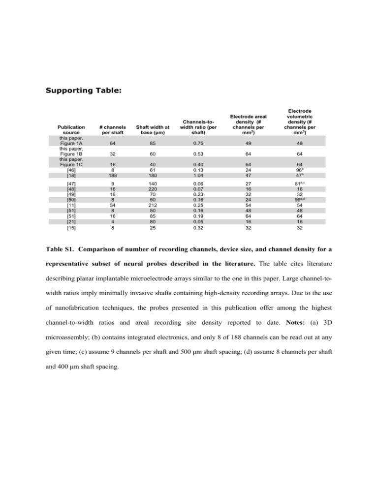
Supporting Table: Electrode volumetric density (# channels per mm3) Publication source this paper, Figure 1A this paper, Figure 1B this paper, Figure 1C [46] [18] # channels per shaft Shaft width at base (μm) Channels-towidth ratio (per shaft) Electrode areal density (# channels per mm2) 64 85 0.75 49 49 32 60 0.53 64 64 16 8 188 40 61 180 0.40 0.13 1.04 64 24 47 64 96a 47b [47] [48] [49] [50] [11] [51] [51] [21] [15] 9 16 16 8 54 8 16 4 8 140 220 70 50 212 50 85 80 25 0.06 0.07 0.23 0.16 0.25 0.16 0.19 0.05 0.32 27 16 32 24 54 48 64 16 32 81a,c 16 32 96a,d 54 48 64 16 32 Table S1. Comparison of number of recording channels, device size, and channel density for a representative subset of neural probes described in the literature. The table cites literature describing planar implantable microelectrode arrays similar to the one in this paper. Large channel-towidth ratios imply minimally invasive shafts containing high-density recording arrays. Due to the use of nanofabrication techniques, the probes presented in this publication offer among the highest channel-to-width ratios and areal recording site density reported to date. Notes: (a) 3D microassembly; (b) contains integrated electronics, and only 8 of 188 channels can be read out at any given time; (c) assume 9 channels per shaft and 500 μm shaft spacing; (d) assume 8 channels per shaft and 400 μm shaft spacing.

