Viability of dendrosomal curcumin complexes in
advertisement
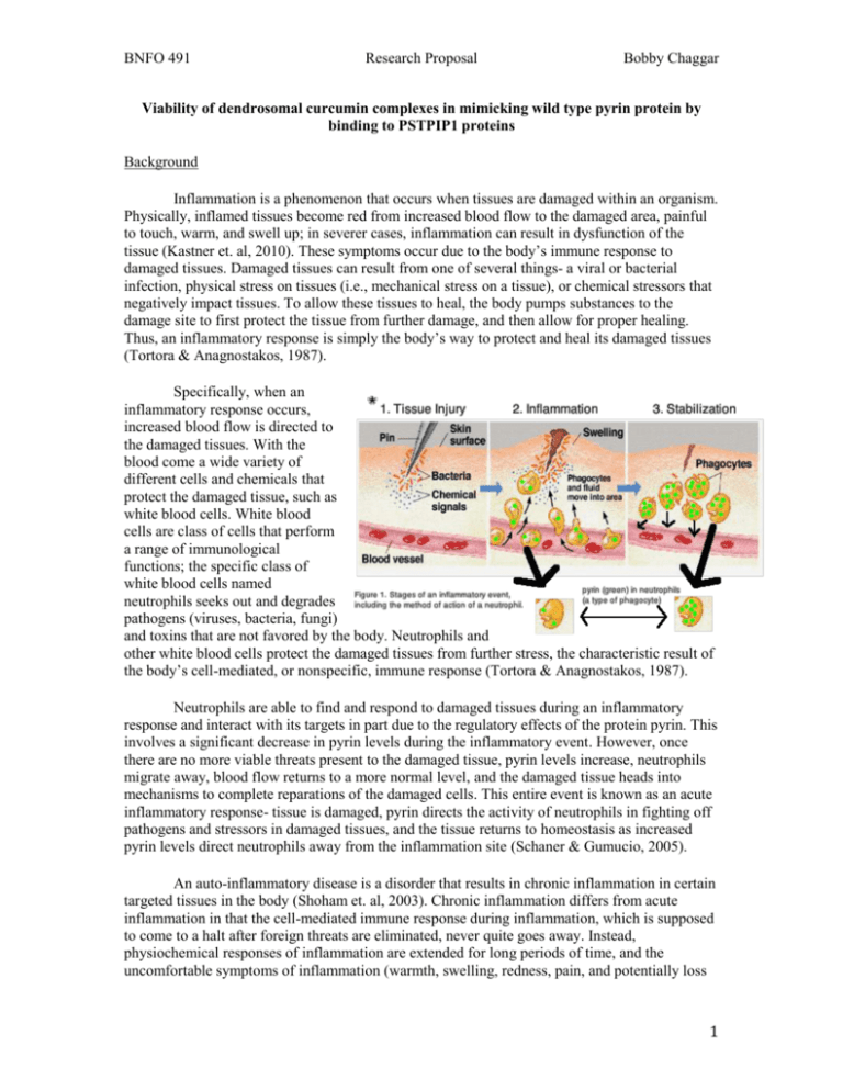
BNFO 491 Research Proposal Bobby Chaggar Viability of dendrosomal curcumin complexes in mimicking wild type pyrin protein by binding to PSTPIP1 proteins Background Inflammation is a phenomenon that occurs when tissues are damaged within an organism. Physically, inflamed tissues become red from increased blood flow to the damaged area, painful to touch, warm, and swell up; in severer cases, inflammation can result in dysfunction of the tissue (Kastner et. al, 2010). These symptoms occur due to the body’s immune response to damaged tissues. Damaged tissues can result from one of several things- a viral or bacterial infection, physical stress on tissues (i.e., mechanical stress on a tissue), or chemical stressors that negatively impact tissues. To allow these tissues to heal, the body pumps substances to the damage site to first protect the tissue from further damage, and then allow for proper healing. Thus, an inflammatory response is simply the body’s way to protect and heal its damaged tissues (Tortora & Anagnostakos, 1987). Specifically, when an inflammatory response occurs, increased blood flow is directed to the damaged tissues. With the blood come a wide variety of different cells and chemicals that protect the damaged tissue, such as white blood cells. White blood cells are class of cells that perform a range of immunological functions; the specific class of white blood cells named neutrophils seeks out and degrades pathogens (viruses, bacteria, fungi) and toxins that are not favored by the body. Neutrophils and other white blood cells protect the damaged tissues from further stress, the characteristic result of the body’s cell-mediated, or nonspecific, immune response (Tortora & Anagnostakos, 1987). Neutrophils are able to find and respond to damaged tissues during an inflammatory response and interact with its targets in part due to the regulatory effects of the protein pyrin. This involves a significant decrease in pyrin levels during the inflammatory event. However, once there are no more viable threats present to the damaged tissue, pyrin levels increase, neutrophils migrate away, blood flow returns to a more normal level, and the damaged tissue heads into mechanisms to complete reparations of the damaged cells. This entire event is known as an acute inflammatory response- tissue is damaged, pyrin directs the activity of neutrophils in fighting off pathogens and stressors in damaged tissues, and the tissue returns to homeostasis as increased pyrin levels direct neutrophils away from the inflammation site (Schaner & Gumucio, 2005). An auto-inflammatory disease is a disorder that results in chronic inflammation in certain targeted tissues in the body (Shoham et. al, 2003). Chronic inflammation differs from acute inflammation in that the cell-mediated immune response during inflammation, which is supposed to come to a halt after foreign threats are eliminated, never quite goes away. Instead, physiochemical responses of inflammation are extended for long periods of time, and the uncomfortable symptoms of inflammation (warmth, swelling, redness, pain, and potentially loss 1 BNFO 491 Research Proposal Bobby Chaggar of function) are often difficult for the body to control (Schaner & Gumucio, 2005). Familial Mediterranean Fever (FMF) is a specific example of a prevalent auto-inflammatory disease, mostly affecting people of Jewish, Armenian, Arab and Turkish ancestry (Schaner & Gumucio, 2005). FMF is caused by one or more mutations in the MEFV gene, which just so happens to encode for the pyrin protein. This aberrant pyrin protein is of concern because its loss of ability to regulate neutrophil activity during inflammatory responses plays a major role in the development of chronic inflammation in afflicted individuals (Schaner & Gumucio, 2005). Normal pyrin protein plays a variety of intracellular roles; notably, pyrin has been shown to be linked to apoptosis, cytokine secretion, and signaling via the cell cytoskeleton. In an inflammatory response, low pyrin levels indicate the need for neutrophils to act in a proinflammatory response; on the flip side, the up-regulation of pyrin indicates the end of an inflammatory response (Shoham et. al, 2003). Since the pyrin proteins translated from the mutant MEFV gene remain at low levels or do not function properly, highly levels of functional pyrin can not accumulate, perhaps explaining the inability of the tissues to prevent chronic inflammation from occurring. A clinical solution that seems to jump into one’s mind is to simply increase the body’s production of functional pyrin, or utilize a compound that can mimic the three main aforementioned molecular functions of pyrin. The latter is considered henceforth. Curcumin is a compound isolated from turmeric powder, and has ben shown in numerous studies to have a wide range of regulatory effects on cell activity in vivo, manifesting as a regulator in tumor suppression, to acting as an analgesic (pain relieving) drug, to being an antiinflammation agent, all without apparent side effects (Basnet et. al, 2011; Shing et. al, 2011). Babaei et. al found that curcumin, delivered in a dendrosome nanoparticle drug vehicle, is able to stimulate interferon-γ (IFN-γ) levels. IFN-γ is a cytokine that pyrin interacts with, and that can down-regulate the pyrin protein (Shoham et. al, 2003). Further, curcumin has been shown to be involved in another intracellular function, apoptosis, that pyrin also is integral in (Basnet et. al, 2011). Of particular interest is to find out whether or not curcumin can further act similarly to pyrin, by taking on pyrin’s third core function: cell signaling through utilization of the cytoskeleton. Namely, Shoham et. al described that a key step in pyrin’s role in a proinflammatory response is mediated through interactions with the cell’s cytoskeleton. This interaction requires the binding of pyrin to an adapter protein known as PSTPIP1, a protein heavily implicated in the reorganization of actin in the cytoskeleton (Shoham et. al, 2003). The purpose of this study is to examine whether or not curcumin, a compound that performs some of the same intracellular functions as pyrin, can also bind significantly to the protein PSTPIP1. Experimental Design Curcumin linked with a dendrosome will be the form of the compound used in drug delivery. A dendrosome is a ball-like aggregate of repeating molecular units joined with a nucleic acid. Dendrosomes have become increasingly common in targeted drug delivery to improve site bioavailability of a drug to which the dendrosome is linked (Markatou et. al 2007). Specifically, 2 BNFO 491 Research Proposal Bobby Chaggar Babaei et. al found the bioavailability of curcumin to be markedly augmented when the drug was linked into a complex with the dendrosome DenO400 (Babaei et. al, 2012). As such, DenO400 dendrosomal curcumin, or DenCur as it is known, will be prepared and utilized in this experiment, to circumvent the issue of curcumin’s poor bioavailability in its natural form. This dendrosome gradually deteriorates, and by the time the complex reaches its site of activity, the curcumin is active (Babaei et. al, 2012). To test to what extent curcumin and pyrin bind to PSTPIP1, the levels of curcuminPSTPIP1 (henceforth CUP1) and pyrin-PSTPIP1 (henceforth PYP1) must be measured from tissues that are inflamed. Levels of CUP1 and PYP1 will be measured in two groups of mice: normal mice with normal, wild type pyrin, and FMF mice with the aberrant pyrin proteins. Each main group of mice can be subdivided into two more subgroups; one of these subgroups would receive a dosage of curcumin, while the other would not. The purpose of doing this is to try and establish baseline levels of PYP1 in normal and FMF mice, and from this, gauge whether or not the level of CUP1 formation (if any) is significant. To prompt a more noticeable rate of complex formation, the mice will need to be infected with a rhinovirus, or similar stressor, to promote an initial inflammatory response throughout the mice’s tissues. To actually measure CUP1 and PYP1, the complexes have to be precipitated out of cells from inflamed tissues (if infected with a rhinovirus, then from targeted tissues from the mouth, nose, or throat). To do this, CUP1 and PYP1 will be isolated from cells following the along the lines of Shoham et. al’s methodology. Namely, the authors co-immunoprecipitated out a pyrinPSTPSP1 complex from sampled cells in their study. Co-immunoprecipitation is a technique that allows for insoluble protein-resin complexes to be separated from other proteins in samples by having a specific antibody bind to a portion of that complex, which can then be used to separate out the targeted complexes from these cells. To aid in the visualization of the separated complexes, immunofluoresence microscopy was performed by Shoham et. al in their study, to help quantify how much of the complex was present. This technique relies on flurorescent tagging of the complex, once again by antibodies developed for the protein complex. These antibodies were developed by fusing Glutathione S-transferase (GST) with a separate portion of pyrin-PSTP1P1 complexes, allowing for the formation of an antibody that would bind specifically to pyrin-PSTPIP1 complexes (Shoham et. al, 2003). In the same manner, insoluble (resin bound) CUP1 and PYP1 in this present study will be visualized with immunofluorescence microscopy, and then co-immunoprecipitated out of inflamed cells, through the use of antibodies development by fusion of GST with the constituent proteins of the protein complexes. Because both CUP1 and PYP1 share the common PSTP1P1 protein as a subunit, the antibodies need only be designed to target this protein. Once these complexes have been separated from their host cells, centrifugation will be utilized to separate CUP1, PYP1, and free PSTP1P1 into distinct phases. Confirmation of the CUP1 and PYP1 fractions can be done by taking samples from the fractions, and utilizing mass spectroscopy to determine the content in each phase, looking specifically for the presence of pyrin fragments and the curcumin molecule. From here, the amounts of each can be determined and compared using SDS-PAGE, a form of gel electrophoresis used to analyze and quantify the amounts of protein in a given sample. Expected Outcomes If curcumin is really quite similar in its molecular activity to pyrin, then it should be expected that it has some discernable amount of binding to PSTPIP1. Pyrin present in normal 3 BNFO 491 Research Proposal Bobby Chaggar mice cells ought to have a high binding fraction of pyrin present since an infection by a rhinovirus would trigger a normal inflammatory response. Mutant pyrin, however, may end up with a smaller fraction of pyrin complexed to PSTPIP1. If the hypothesis of this present study is correct, then curcumin would bind to the free, uncomplexed PSTP1P1 present in the cell. In addition, curcumin may then function in influencing the three pathways that pyrin is heavily involved in: apoptosis, cytokine secretion, and cell signaling through the cytoskeleton. While this coincidence doesn’t really mean much in itself, it could open the doors to future research in examining the potential use of curcumin in performing the functions that the aberrant pyrin should, but cannot, do in the cell. More common in life, however, is the grim expectation that things are not so easily understood. In the case where curcumin does not bind to PSTPIP1, then the idea that curcumin can be a substitute for pyrin seems to be completely unlikely. Even if, however, curcumin was found to bind with PSTPIP1, it is a far stretch to assume that it implies anything meaningful. What is not considered, for example, is whether or not curcumin’s binding actually promotes or discourages PSTPIP1 from reorganizing actin as it is supposed to do. In that sense, if curcumin binds to the protein, but inhibits its function, then it obviously is not mimicking the pro-PSTPIP1 effects of pyrin. Though, this is somewhat of a secondary step- if curcumin binds to PSTPIP1, then the next logical step would be to evaluate how this interaction affects PSTPIP1’s function. Finally, there is always the consideration of the limitations of the scope of this study. Assuming all goes well and good, and curcumin binds to PSTPIP1, and acts similarly to pyrin, these findings may or may not be able to be utilized in other instances of auto-inflammatory disorders, like Behçet’s disease or DIRA, for example. Any conclusions drawn regarding pyrin, curcumin, and PSTPIP1 can only be made in reference to FMF, because the mechanism of regulation that curcumin takes may be different in expressions of different auto-inflammatory disorders. Even though the results of this study are limited in their immediate conclusions, the potential for further research based off this topic looks rewarding. Finding compounds that mimic the function of normal, wild type proteins in these auto-inflammatory disorders is a step in helping to better understand and counteract the mechanisms of this type of disorder. Furthermore, future studies that evaluate the ability of such compounds like curcumin, and their ability to regulate an inflammatory response by binding to key proteins in signaling pathways, opens up a potential path for clinicians to develop more effective treatment options for sufferers of familial Mediterranean fever (FMF) and other auto-inflammatory disorders. 4 BNFO 491 Research Proposal Bobby Chaggar References Babaei, E., Sadeghizadeh, M., Hassan, Z. M., Feizi, M. A. H., Najafi, F., & Hashemi, S. M. (2012). Dendrosomal curcumin significantly suppresses cancer cell proliferation in vitro and in vivo. International Immunopharmacology, 12(1), 226-234. Basnet, P., & Skalko-Basnet, N. (2011). Curcumin: An anti-inflammatory molecule from a curry spice on the path to cancer treatment. Molecules, 16(6), 4567-4598. Kastner, D. L., Aksentijevich, I., & Goldbach-Mansky, R. (2010). Autoinflammatory disease reloaded: A clinical perspective. Cell, 140(6), 784-790. Markatou, E., Gionis, V., Chryssikos, G., Hatziantoniou, S., Georgopoulos, A., & Demetzos, C. (2007). Molecular interactions between dimethoxycurcumin and pamam dendrimer carriers. International Journal of Pharmaceutics, 339(1-2), 231-236. Shoham Nitza G., Centola Michael, Mansfield Elizabeth, Hull Keith M., Wood Geryl, Wise Carol A., and Kastner Daniel L.. (2003). Pyrin binds the PSTPIP1/CD2BP1 protein, defining familial Mediterranean fever and PAPA syndrome as disorders in the same pathway. PNAS 2003 100 (23) 13501-13506 Schaner, P. E., & Gumucio, D. L. (2005). Familial mediterranean fever in the post-genomic era: How an ancient disease is providing new insights into inflammatory pathways. Current Drug Targets - Inflammation & Allergy, 4(1), 67-76. 5 BNFO 491 Research Proposal Bobby Chaggar Shing, C. M., Adams, M. J., Fassett, R. G., & Coombes, J. S. (2011). Nutritional compounds influence tissue factor expression and inflammation of chronic kidney disease patients in vitro. Nutrition, 27(9), 967-972. Tortora, G. J., & Anagnostakos, N. P. (1987). The lymphatic system and immunity. In Principles of anatomy and physiology (5 ed., pp. 540-542). Zemer, D., Pras, M., Sohar, E., Modan, M., Cabill, S., & Gafni, J. (1986). Colchicine in the prevention and treatment of the amyloidosis of familial mediterranean fever. N Engl J Med, (314), 1001-1005. * Adapted from http://legacy.owensboro.kctcs.edu/gcaplan/anat2/notes/ 6
