Hemoglobin and Myoglobin Outline
advertisement
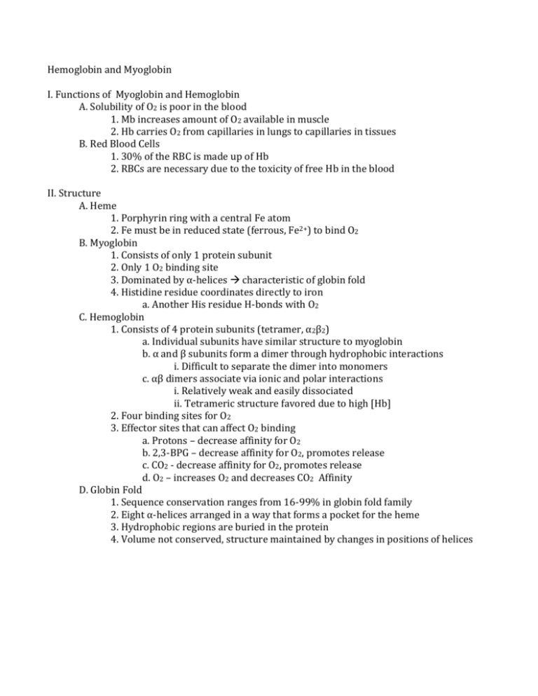
Hemoglobin and Myoglobin I. Functions of Myoglobin and Hemoglobin A. Solubility of O2 is poor in the blood 1. Mb increases amount of O2 available in muscle 2. Hb carries O2 from capillaries in lungs to capillaries in tissues B. Red Blood Cells 1. 30% of the RBC is made up of Hb 2. RBCs are necessary due to the toxicity of free Hb in the blood II. Structure A. Heme 1. Porphyrin ring with a central Fe atom 2. Fe must be in reduced state (ferrous, Fe2+) to bind O2 B. Myoglobin 1. Consists of only 1 protein subunit 2. Only 1 O2 binding site 3. Dominated by α-helices characteristic of globin fold 4. Histidine residue coordinates directly to iron a. Another His residue H-bonds with O2 C. Hemoglobin 1. Consists of 4 protein subunits (tetramer, α2β2) a. Individual subunits have similar structure to myoglobin b. α and β subunits form a dimer through hydrophobic interactions i. Difficult to separate the dimer into monomers c. αβ dimers associate via ionic and polar interactions i. Relatively weak and easily dissociated ii. Tetrameric structure favored due to high [Hb] 2. Four binding sites for O2 3. Effector sites that can affect O2 binding a. Protons – decrease affinity for O2 b. 2,3-BPG – decrease affinity for O2, promotes release c. CO2 - decrease affinity for O2, promotes release d. O2 – increases O2 and decreases CO2 Affinity D. Globin Fold 1. Sequence conservation ranges from 16-99% in globin fold family 2. Eight α-helices arranged in a way that forms a pocket for the heme 3. Hydrophobic regions are buried in the protein 4. Volume not conserved, structure maintained by changes in positions of helices III. Ligand Binding Interactions A. Binding Isotherms PL P + L PL = protein-Ligand Complex P = free protein L = free ligand Kd = [P][L]/[PL] Kd is the concentration at which 50% of the protein active sites are occupied Y = [PL]/[Ptotal] Y is the fractional saturation, or fraction of the total protein with bound ligand Y = [O2]/(Kd + [O2]) Fractional saturation for myoglobin, where O2 is the ligand When [O2] = Kd then Y=0.5, the definition of Kd When [O2] = 0, then Y=0, as nothing is bound without ligand When [O2] >> Kd, then Y ≅ 1, as a high ligand concentration will bind most sites For Myoglobin: [PL] = [Ptotal][L]/(Kd + [L]) When [L] << Kd, the graph is linear with respect to [L] When [L] >> Kd, no dependence on Kd, saturation is limited by [Ptotal] B. Determining Kd 1. Determine [PL] at several concentrations of [L] 2. Graphic or computer methods to estimate Kd 3. Several graphing methods: a. Hyperbolic saturation plot b. Linear transformations of binding equations 4. Linear or nonlinear least squares fit of function to data to get Kd estimate C. Hill Approximations For a ligand that binds to multiple sites P + nL PLn Y = [L]n/(Kn + [L]n) n = Hill Coefficient, gives measure of cooperativity When n = 1, there is no cooperativity When n >1, positive cooperativity When n < 1, negative cooperativity IV. Models for Cooperativity A. Monod (Concerted) Model 1. Two conformational states of subunits a. R – Relaxed, high affinity state b. T – Taut, low affinity state 2. Tetramer must be all R or all T 3. Equilibrium exists between the two states a. High [O2] favors R-state i. R-state binds O2 in the lungs b. Low O2 favors T-state i. T-state releases O2 to the tissues c. Binding of O2 shifts equilibrium to R-state 4. Only explains positive cooperativity B. Koshland Sequential Model 1. R and T states exist in this model 2. Without ligand, protein is in one conformation 3. Symmetry is not required a. Subunits can be a mix of R and T 4. Sequential change in conformation a. Ligand binding induces TR transition 5. Ligated subunit can influence conformation/affinity of a neighboring subunit 6. Can explain positive and negative cooperativity C. General Model of Cooperativity 1. Neither the Monod nor Koshland model adequately describes hemoglobin 2. It is actually a variety of microstates with varying oxygen affinities a. Not just T and R 3. The first O2 has an equal probability of binding to the α or β subunit a. The second O2 binds with a much higher affinity to the other dimer subunit 4. Subsequent O2 bind with increasing affinity 5. Multiple subunits are not needed for cooperativity a. Hexokinase exists in equilibrium between high and low affinity states b. Glucose binds to high affinity conformer i. Shifts the protein equilibrium to the high affinity state V. O2 Binding and Tertiary Structure A. Allosteric Effectors 1. Homotropic effectors – bind to same site as ligand (orthosteric) 2. Heterotropic effectors – bind to site that is different from ligand (allosteric) a. H+ - binds to His146, contributes to Bohr effect dec 02 affinity b. 2,3-BPG – binds to center of tetramer dec 02 affinity c. CO2 – reacts with N-termini to alter O2 heme binding dec 02 d. O2 – binds to heme and alters CO2 binding at N-termini dec affinity for CO2 B. O2 Induced Structural Changes 1. Binding of O2 alters contacts between subunits 2. Particular stabilizing interactions are lost a. Allows for transition from low affinity to high affinity state C. Bohr Effect 1. pH affects O2 binding to hemoglobin a. pH decreases, curve shifts right, lower affinity for O2 b. pH increases, curve shifts left, higher affinity for O2 2. Inverse relationship between [H+] and O2 affinity 3. Due to the protonation of His146 a. pH tends to be higher in the lungs and lower in the tissues VI. CO2 Transport A. CO2 Binding 1. CO2 binds to the N-terminus of deoxyhemoglobin α subunits a. Forms carbamino hemoglobin b. Carbamino has a (-) charge, forms a salt bridge to stabilize T-state i. Encourages the release of O2 2. Allows hemoglobin to effectively carry O2 B. Bohr Effect and CO2 1. More CO2 in the tissues, causes an increase in [H+] 2. The increase in H+ protonates Hb and stabilizes the T-state a. Promotes release of O2 in the tissues 3. Exhalation of CO2 creates a lower [CO2] and [H+] in the lungs a. Promotes binding of O2 in the lungs C. Haldane Effect 1. Deoxygenation of hemoglobin increases its basicity and affinity for CO2 HbO2 + H+ H-Hb + O2 H-Hb + CO2 H-Hb-CO2 carbamino 2. Oxygenation of hemoglobin increases its acidity and decreases affinity for CO2 a. Oxygen binding promotes release of CO2 in the lungs b. Oxygenation promotes H+ dissociation i. Shifts bicarbonate buffer equilibrium toward CO2 H-Hb + O2 Hb-O2 + H+ HCO3- + H+ CO2 + H2O 3. Carbonic anhydrase is essential for this, as it catalyzes CO2 H2CO3 a. Allows the buffer equilibrium to exist VII. Additional Effector Molecules A. 2,3-Bisphosphoglycerate (BPG) 1. Hemoglobin binds one molecules of BPG a. Decreases O2 affinity by stabilizing T-state 2. Fetal hemoglobin has low affinity for BPG a. Also has a higher affinity for O2 3. BPG is an allosteric effector B. Hemoglobin Effectors and O2 Binding C. Chloride 1. Chloride binds to deoxyhemoglobin, decreases O2 affinity 2. Forms a salt bridge that stabilizes T-state D. Temperature 1. Inverse relationship between O2 affinity and temperature a. Reduced O2 requirement for metabolism that occurs during hypothermia b. Increased O2 requirement during fever E. Carbon Monoxide 1. Binds to O2 site reversibly a. Binds tightly, but important to remember that it is reversible 2. Binds 200 times stronger than O2 a. Also increases affinity for O2, making it difficult to release it to the tissues F. Flow of Oxygen Maternal Hb Fetal Hb Fetal Mb cytochromes VIII. Sickle Cell Disease A. Point Mutation 1. HbS: Conversion GluVal 2. Results in altered hemoglobin conformation a. Val residue inserts into a hydrophobic pocket of another HbS molecule b. Allows for polymer formation i. Aggregation restricted to deoxy form c. Aggregates can occlude organs 3. HbC: Conversion of GluLys B. Populations 1. More common in African-American populations 2. Frequent in areas where malaria is endemic C. Malaria 1. Sickle Cell provides some resistance to malarial infections 2. Two Types of Hb: a. HbS – resistance to malaria i. Cells are sickled due to Hb aggrgates and accumulation of fibers b. HbC – resistance to malaria, homozygous individuals have mild disease i. Cells are not sickled, but appear as “target” cells ii. Some crystal-like structures present in RBCs c. HbS/HbC – resistance, disease is more mild than HbS homozygous D. Sickling of Cells 1. Cells assume contorted shape at low oxygen levels 2. Reversible Sickling a. As HbS disaggregates and is oxygenated, cells return to normal 3. Irreversible Sickling a. When cells have been sickled for a while, cytoskeleton becomes locked i. Due to disulfide cross-linking of actin b. Even after Hb disaggregates, cell remains sickled IX. Thalassemias A. Deficiency in the α or β chain of Hb that gives rise to anemia 1. α-thalassemias – generally results from deletion of genes in the α subunit 2. β-thalassemias – can result from a variety of defects a. Gene deletion b. Defects in transcription c. Defects in translation d. Defects in mRNA processing e. Anything that creates an unstable protein or mRNA 3. Sickle Cell-β thalassemias a. Reduced levels of the normal β-chain and presence of HbS β-chain

