Table S1. Definitions of CMV end-organ disease used in this study
advertisement
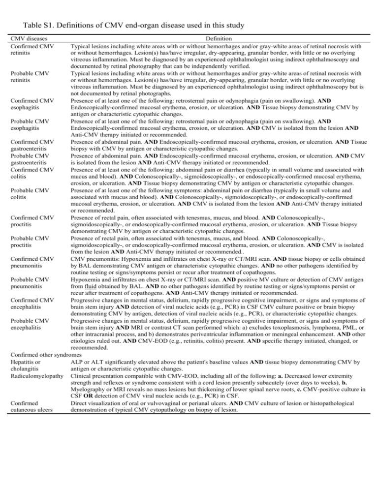
Table S1. Definitions of CMV end-organ disease used in this study CMV diseases Confirmed CMV retinitis Definition Typical lesions including white areas with or without hemorrhages and/or gray-white areas of retinal necrosis with or without hemorrhages. Lesion(s) has/have irregular, dry-appearing, granular border, with little or no overlying vitreous inflammation. Must be diagnosed by an experienced ophthalmologist using indirect ophthalmoscopy and documented by retinal photography that can be independently verified. Probable CMV Typical lesions including white areas with or without hemorrhages and/or gray-white areas of retinal necrosis with retinitis or without hemorrhages. Lesion(s) has/have irregular, dry-appearing, granular border, with little or no overlying vitreous inflammation. Must be diagnosed by an experienced ophthalmologist using indirect ophthalmoscopy but is not documented by retinal photographs. Confirmed CMV Presence of at least one of the following: retrosternal pain or odynophagia (pain on swallowing). AND esophagitis Endoscopically-confirmed mucosal erythema, erosion, or ulceration. AND Tissue biopsy demonstrating CMV by antigen or characteristic cytopathic changes. Probable CMV Presence of at least one of the following: retrosternal pain or odynophagia (pain on swallowing). AND esophagitis Endoscopically-confirmed mucosal erythema, erosion, or ulceration. AND CMV is isolated from the lesion AND Anti-CMV therapy initiated or recommended. Confirmed CMV Presence of abdominal pain. AND Endoscopically-confirmed mucosal erythema, erosion, or ulceration. AND Tissue gastroenteritis biopsy with CMV by antigen or characteristic cytopathic changes. Probable CMV Presence of abdominal pain. AND Endoscopically-confirmed mucosal erythema, erosion, or ulceration. AND CMV gastroenteritis is isolated from the lesion AND Anti-CMV therapy initiated or recommended. Confirmed CMV Presence of at least one of the following: abdominal pain or diarrhea (typically in small volume and associated with colitis mucus and blood). AND Colonoscopically-, sigmoidoscopically-, or endoscopically-confirmed mucosal erythema, erosion, or ulceration. AND Tissue biopsy demonstrating CMV by antigen or characteristic cytopathic changes. Probable CMV Presence of at least one of the following symptoms: abdominal pain or diarrhea (typically in small volume and colitis associated with mucus and blood). AND Colonoscopically-, sigmoidoscopically-, or endoscopically-confirmed mucosal erythema, erosion, or ulceration. AND CMV is isolated from the lesion AND Anti-CMV therapy initiated or recommended. Confirmed CMV Presence of rectal pain, often associated with tenesmus, mucus, and blood. AND Colonoscopically-, proctitis sigmoidoscopically-, or endoscopically-confirmed mucosal erythema, erosion, or ulceration. AND Tissue biopsy demonstrating CMV by antigen or characteristic cytopathic changes. Probable CMV Presence of rectal pain, often associated with tenesmus, mucus, and blood. AND Colonoscopically-, proctitis sigmoidoscopically-, or endoscopically-confirmed mucosal erythema, erosion, or ulceration. AND CMV is isolated from the lesion AND Anti-CMV therapy initiated or recommended.. Confirmed CMV CMV pneumonitis: Hypoxemia and infiltrates on chest X-ray or CT/MRI scan. AND tissue biopsy or cells obtained pneumonitis by BAL demonstrating CMV antigen or characteristic cytopathic changes. AND no other pathogens identified by routine testing or signs/symptoms persist or recur after treatment of copathogens. Probable CMV Hypoxemia and infiltrates on chest X-ray or CT/MRI scan. AND positive MV culture or detection of CMV antigen pneumonitis from fluid obtained by BAL. AND no other pathogens identified by routine testing or signs/symptoms persist or recur after treatment of copathogens. AND Anti-CMV therapy initiated or recommended. Confirmed CMV Progressive changes in mental status, delirium, rapidly progressive cognitive impairment, or signs and symptoms of encephalitis brain stem injury AND detection of viral nucleic acids (e.g., PCR) in CSF CMV culture positive or brain biopsy demonstrating CMV by antigen, detection of viral nucleic acids (e.g., PCR), or characteristic cytopathic changes. Probable CMV Progressive changes in mental status, delirium, rapidly progressive cognitive impairment, or signs and symptoms of encephalitis brain stem injury AND MRI or contrast CT scan performed which: a) excludes toxoplasmosis, lymphoma, PML, or other intracranial process, and b) demonstrates periventricular inflammation or meningeal enhancement. AND other etiologies ruled out. AND CMV-EOD (e.g., retinitis, colitis) present. AND specific therapy initiated, changed, or recommended. Confirmed other syndromes Hepatitis or ALP or ALT significantly elevated above the patient's baseline values AND tissue biopsy demonstrating CMV by cholangitis antigen or characteristic cytopathic changes. Radiculomyelopathy Clinical presentation compatible with CMV-EOD, including all of the following: a. Decreased lower extremity strength and reflexes or syndrome consistent with a cord lesion presently subacutely (over days to weeks), b. Myelography or MRI reveals no mass lesions but thickening of lower spinal nerve roots, c. CMV-positive culture in CSF OR detection of CMV viral nucleic acids (e.g., PCR) in CSF. Confirmed Direct visualization of oral or vulvovaginal or perianal ulcers. AND CMV culture of lesion or histopathological cutaneous ulcers demonstration of typical CMV cytopathology on biopsy of lesion.
