Red Blood Cell Metabolism
advertisement
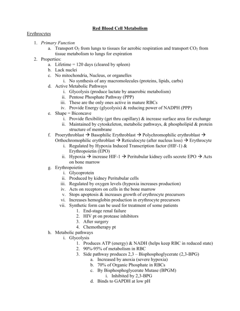
Red Blood Cell Metabolism Erythrocytes 1. Primary Function a. Transport O2 from lungs to tissues for aerobic respiration and transport CO2 from tissue metabolism to lungs for expiration 2. Properties: a. Lifetime = 120 days (cleared by spleen) b. Lack nuclei c. No mitochondria, Nucleus, or organelles i. No synthesis of any macromolecules (proteins, lipids, carbs) d. Active Metabolic Pathways i. Glycolysis (produce lactate by anaerobic metabolism) ii. Pentose Phosphate Pathway (PPP) iii. These are the only ones active in mature RBCs iv. Provide Energy (glycolysis) & reducing power of NADPH (PPP) e. Shape = Biconcave i. Provide flexibility (get thru capillary) & increase surface area for exchange ii. Maintained by cytoskeleton, metabolic pathways, & phospholipid & protein structure of membrane f. Proerythroblast Basophilic Erythroblast Polychromophilic erythroblast Orthochromophilic erythroblast Reticulocyte (after nucleus loss) Erythrocyte i. Regulated by Hypoxia Induced Transcription factor (HIF-1) & Erythropoietin (EPO) ii. Hypoxia increase HIF-1 Peritubular kidney cells secrete EPO Acts on bone marrow g. Erythropoietin i. Glycoprotein ii. Produced by kidney Peritubular cells iii. Regulated by oxygen levels (hypoxia increases production) iv. Acts on receptors on cells in the bone marrow v. Stops apoptosis & increases growth of erythrocyte precursors vi. Increases hemoglobin production in erythrocyte precursors vii. Synthetic form can be used for treatment of some patients 1. End-stage renal failure 2. HIV pt on protease inhibitors 3. After surgery 4. Chemotherapy pt h. Metabolic pathways i. Glycolysis 1. Produces ATP (energy) & NADH (helps keep RBC in reduced state) 2. 90%-95% of metabolism in RBC 3. Side pathway produces 2,3 – Bisphosphoglycerate (2,3-BPG) a. Increased by anoxia (severe hypoxia) b. 70% of Organic Phosphate in RBCs c. By Bisphosphoglycerate Mutase (BPGM) i. Inhibited by 2,3-BPG d. Binds to GAPDH at low pH i. Low pH in tissues due to metabolism ii. Causes 2,3-BPG production to increase O2 release e. Phosphoglycerate kinase binds to GAPDH at high pH i. High pH in lungs ii. Less 2,3-BPG made = O2 binding ii. PPP 1. Produces CO2 (oxidative phase), Fructose – P & Glyceraldehyde – P (for glycolysis from nonoxidative phase), & NADPH (major player in keeping RBC in the reduced state oxidative phase) 2. 5%-10% of metabolism in RBC 3. NADP+ increases Glucose – P DH activity to produce more NADPH i. NADPH i. Inhibits Glucose-P dehydrogenase (PPP) ii. Reduces Oxidized Glutathione 1. Reverses/prevents oxidative damage in RBCs iii. Reduces MetHb (Fe3+) to Hb (Fe2+) j. Glutathione i. Synthesis: ATP + Glutamate + Cysteine Glutamyl Cysteine Synthetase Glutamyl Cysteine + Glycine + ATP Glutathione Synthetase Glutathione ii. Oxidized form reduced by Glutathione Reductase 1. Flavin protein (FAD) 2. Uses NADPH iii. Functions: 1. Reduce H2O2 H2O & O2 (Glutathione Peroxidase) 2. Reduce MetHb (Fe3+ Ferric) to Hb (Fe2+ Ferrous) 3. Reduce oxidized protein thiols Reduction of MetHb 1. MetHb Reductase II a. About 5%-10% reduction (minor) b. Uses NADPH 2. Nonenzymatic reduction by Glutathione a. About 12% reduction (minor) b. Reduced glutathione regenerated by NADPH 3. MetHb Reductase I (Cytochrome b5 Reductase) a. About 67% of the reduction b. Electrons transferred to cytochrome b5 by NADH*** Cyt b5 reduces MetHb Clinical Conditions 1. Oxidative Stress a. Pt has Cyanosis = blue color to tissues (prominent in lips, nails, & gums) i. Defect = more MetHb 1. 2% MetHb = normal 2. 10%-12% = Cyanosis 3. Does not bind O2 ii. Causes for MetHb generation: 1. Superoxide anion (O2-) 2. Drugs (nitrites, anti-malarial, topical anesthetics) 3. Toxic oxidants (Tobacco smoke chemicals) 4. Inherited disorder 2. Methemoglobinemia/ Methemoglobincytemia a. Defects: i. MetHb Reductase II Mild prone to oxidative stress ii. Cytochrome b5 Reductase severity varies iii. Cytochrome b5 MetHb Reductase I Mild iv. Hemoglobin M α & β chain defects Heme Fe prone to oxidation v. Glutathione Peroxidase Mild less H2O2 broken down b. Treatments: i. Methylene Blue ii. Vitamin C & E iii. All reduce MetHb to Hb c. How cells normally deal with Oxidative Stress i. O2- produced (redox enzymes such as in ETC) reduced by Superoxide Dismutase to H2O2 converted to H2O & O2 by Glutathione Peroxidase ii. Vitamin C iii. Vitamin E converted to peroxyvitamin E after reducing something 1. Converted back to Vitamin E by Vitamin C 3. Glucose-P Dehydrogenase Deficiency a. X-Linked, Malarial Region Origin (Tropical Africa, Asia, Mediterranean) b. Conveys Malarial Resistance when heterozygous c. More sensitive to Oxidative stress i. Caused by common drugs and flava beans d. Tested for a birth e. Presentations: i. Neonatal Jaundice Urea cycle disfunction ii. Acute Hemolytic Anemia fatal in children iii. Hemolytic Attack caused by a bunch of drugs (Anti-malarial to cancer) O2 Transport by Hb 1. 2,3-BPG a. Produced by glycolytic side pathway (BPGM) b. Stabilizes T state of Hb to release more O2 c. Causes 66% release of O2 at tissues when present d. Increased by i. High Altitude, Exercise, Suffocation, Heart failure, Anemia, Obstructive Pulmonary Disease, Cystic Fibrosis, Congenital Heart Disease, Hyperthyroidism ii. Inherited Deficiencies: 1. Pyruvate Kinase Build up of PEP = more 2,3-BPG a. Hemolytic anemiaATP production=Na+/K+ ATPase defect 2. Hexokinase less glycolysis = less 2,3-BPG a. This also causes neurologic problems b. Hemolytic anemiaATP production=Na+/K+ ATPase defect 2. Different Hemoglobins a. Embryonic = ε2ζ2 b. Fetal = α2γ2 c. Adult = α2β2 d. Adult Hb transfers oxygen to Fetal Hb i. Fetal Hb = less affinity for 2,3-BPG & higher oxygen affinity 3. CO2 a. 2 Fates for transport i. Converted to HCO3- in RBCs (Carbonic Anhydrase) 1. 85% of transport CO2 2. Produces H+ ii. Forms covalent bond with N-terminus of Hb (Carbamylated Hb) 1. Stabilizes T state of Hb = more Hb released in tissues 2. 15% of transported CO2 iii. Free CO2 = <1% of CO2 transported b. Both H+ and Carbamylated Hb decrease the affinity of Hb for O2 (Stabilize T-State) Membrane 1. Component: a. Inner & outer leaflets have different compositions b. Lipids: i. Phospholipids 1. Phosphatidylethanolamine = inner-leaflet 2. Phosphatidylcholine = outer-leaflet 3. Phosphatidylserine = outer-leaflet = cellular damage (RBC removed by Macrophages in spleen) ii. Cholesterol provides some rigidity (within lipid bilayer) c. Lateral diffusion = fast & passive; Vertical diffusion = slow & uses ATP (Flipases/flopases) d. Proteins asymmetric in membrane i. Integral/Transmembrane 1. Traverse bilayer (hydrophobic residues face lipids) 2. Frequently form channels 3. Cytoplasmic & EC domain (C-& N-terminus can be on same side) 4. Typically glycosylated on exterior side (Example = glycophorins) 5. Cannot be removed with milt salt ii. Peripheral 1. On one side of the membrane 2. Does not contact lipid bilayer significantly a. Some have an anchoring region such as a dilochol or other isoprene units) 3. Removed by mild salt 2. SDS Page a. SDS = Sodium Dodecyl sulfate strong detergent b. SDS PAGE i. Mix membrane or protein mixture with hot SDS & a strong reducing agent (β-mercaptoethanol) 1. Denatures and reduces sulfide bonds in proteins 2. Solubolizes lipids 3. 1 SDS for every 2 peptide bonds all proteins have same charge ii. Run in a cross-linked polyacrylamide gel in an electric field 1. Can change cross-linking ratio to create a mass gradient iii. Separates proteins based on molecular masses iv. Commassie (Proteins) Blue or Periodic acid Schiff (Glycoprotiens) to stain 3. RBC Membrane proteins a. Utilize Ghost RBCs have cellular membranes with attached cytoskeletal elements i. Put in hypotonic solution to let burst and then put back on isotonic solution b. Proteins i. α-spectrin Peripheral ii. β-spectrin Peripheral iii. Actin Peripheral iv. Band 4.1 Peripheral v. Tropomyosin Peripheral vi. GAPDH Peripheral vii. Ankyrin Peripheral viii. Band 3 (Cl--HCO3- Exchanger) Integral ix. Glycophorins (Blood group proteins) Integral c. Interactions i. Spectrin – Ankyrin – Band 3 links to band 3 (Cl--HCO3- Exchanger) ii. Spectrin – Band 4.1 – Actin links spectrin to glycophorin C d. Spectrin & Defects i. RBCs distorted traveling thru capillaries ii. Return to normal shape in veins 1. Shape maintained by RBC cytoskeleton iii. Spectrin 1. α2β2 anti-parallel helix 2. Key component of cytoskeleton of RBC 3. Linked to Band 3 by Ankyrin 4. Linked to Glycophorin C by Band 4.2 5. Maintains RBC shape iv. Hereditary Spherocytosis 1. Defect = α (rare) or β spectrin/Band 3/ Ankyrin 2. Loss of vertical interaction 3. Spherical RBCs 4. Cleared by spleen quicker = Splenomegaly 5. Treatment = Splenectomy v. Ovalocytosis/Elliptocytosis 1. Defect = α or β spectrin/ Band 3/ Band 4.2 2. Loss of horizontal interaction = ellipse or oval RBCs 3. Southeast Asian Ovalocytosis a. Affects Malay archipelago and Philippines b. Lysine Glutamate in Band 3 c. Heterozygotes = little affect; Homozygotes = embryonic lethal d. Malarial resistance e. Band 3 i. Cl--HCO3- Antiporter ii. 12 transmembrane regions iii. Heavily glycosylated on extracellular side (glycoprotein) iv. N-terminus interacts with Ankyrin to attach to spectrin v. C-terminus involved in transport vi. GAPDH, Phosphoglycerate Mutase, & Aldolase attached at N-terminus vii. Isoforms found in Brain & Muscle, and Heart & Liver viii. Dimmer f. Other transmembrane proteins i. Carbohydrates give negative charge to surface of RBC ii. Glycophorins Glycoproteins 1. Basis for blood groups with certain sugars added iii. Other transporters 1. Types a. Uniporter transport one thing with its gradient no ATP b. Symporter Transport 2 things with gradients No ATP c. Antiporter Transport 2 things in opposite directions 2. Cl -HCO3- Antiporter a. Exchanges Cl- into RBCs for HCO3- out of RBCs at tissues b. Exchanges HCO3- into RBCs and Cl- out of RBCs at lungs c. Cytoplasmic domains i. N-terminus attached to GAPDH, Aldolase, PGK ii. C-terminus attached to Carbonic Anhydrase IV 1. CO2 + H2O H2CO3 H+ + HCO33. Glucose Transporter (Glut1) a. Passive uniporter (Not transport glucose against its gradient) b. Not sensitive to insulin c. 12 transmembrane regions d. Highly specific for glucose 4. Na+/K+ ATPase a. Maintains erythrocyte integrity b. Pumps 3 Na+ out & 2 K+ in c. Helps maintain concentration gradients d. Uses ATP e. Phosphorylation state determines confirmation i. Na+/K+ ATPase-Phosphorylated = Na+ let out & K+ binds ii. Na+/K+ ATPase-Dephosphorylated = K+ released & Na+ Binds 5. Aquaporins a. Allows RBCs to regulate amount of H2O within RBC b. Water moves in an out in a single file based on concentration c. Facilitates faster diffusion of H2O d. Passive facilitated diffusion 6. Glycophorins a. Glycoproteins that span the membrane b. Glycophorin A, B, C, D, & E c. A = majority present d. Help provide negative charge to RBC surface i. Prevents sticking to other RBC & Blood Vessels ii. If A or B absent Band 3 sugars compensate e. Have sugar groups attached by glycosyl transferases f. Usually about 60% carbohydrates by mass g. Receptors for Malaria pathogen (P. falciparum) g. Blood groups i. Sugars added to vast range of molecules to determine blood group ii. Fucosyl transferase fucose sugar (H group/gene) iii. N-acetlygalactosyl transferase N-acetlygalactose sugar (A group) iv. Galactosyl transferase Galactose sugar (A Group) 1. iii & iv sugars added to fucose sugar v. 5 blood groups 1. Bombay Blood Group a. No sugars present (Fucosyl transferase = defective) b. Can only receive from Bombay blood donors 2. O a. Fucose only b. Can donate to any Blood group c. Can only receive O blood group 3. AB a. Fucose + Galactose & Fucose + N-Acetlygalactose b. Can donate to AB blood groups only c. Can receive all blood donors 4. A a. Has Fucose + N-Acetlygalactose b. Can donate to AB and A blood groups c. Can receive A or O blood donors 5. B a. Fucose + Galactose b. Can donate to AB and B blood groups h. RBC Aging (Senescence) i. Reactive Oxygen Species (ROS) damage RBC ii. Hb gets damaged by ROS and other oxidants 1. Begins partially unfolded = hemichrome 2. Hemichrome associates with N-terminus of Band 3 3. Hemichrome-Band 3 complexes aggregate 4. Detected by Macrophages of spleen and cleared a. Autologous IgG binds to membrane surface of aggregated Band 3 complexes b. Macrophages detect IgGs

