Answer - Molecular and Cell Biology
advertisement
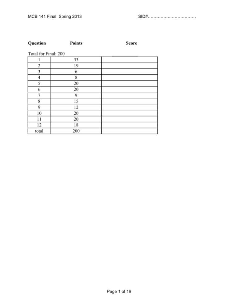
SID#…………….……………… MCB 141 Final Spring 2013 Question Total for Final: 200 1 2 3 4 5 6 7 8 9 10 11 12 total Points Score ___________ 33 19 6 8 20 20 9 15 12 20 20 18 200 Page 1 of 19 SID#…………….……………… MCB 141 Final Spring 2013 Question 1 (33 points) 1a) (10 points) Name that phenotype! Assuming tissue-specific knockouts are possible when needed, what is the consequence of knocking out the following genes in Drosophila? BAM, in the GSCs BAM promotes differentiation into a gamete when the GSC is removed from the source of Dpp (cap cells) – knocking it out will prevent oogenesis. GSCs will continuously proliferate out of control “Bag of Marbles” phenotype bicoid, in the nurse cells Bicoid is a maternally contributed mRNA (produced by the nurse cells) required for anterior patterning. The embryo will not have anterior features or structures (including anterior expression of giant, hunchback, even striped). Antp Antp is usually expression in the T2 segment to drive T2 leg and wing construction. Without Antp to exert posterior prevalence, Scr will expand to be expressed in both T1 and T2. T2 will adopt a T1 phenotype (T1 legs, no wings). Scr and abd-A No posterior prevalence by these missing genes Dfd gets expressed in H3 and T1 and generates H3 identity in both; Ubx gets expressed in T3 and A1-4 and conferes T3 identity in all of these. PolyComb repressor binding sites that regulate Labial Polycomb can no longer maintain an anterior border for this Hox gene… but, as the most anterior Hox gene anyway, it can’t expand further. No phenotype. 1b) (8 points) You suspect Dpp, the Drosophila homolog of BMP, is acting as a morphogen in the wing imaginal disc, turning on spalt at high concentrations, fictional (fic) at intermediate concentrations, and optomotor-blind (omb) at intermediate and lower concentrations. i) Propose a complete model for how different concentrations of Dpp can induce different expression patterns of these target genes. Careful! Affinity Threshold Model: Drosophila homologue of Smad (we didn’t tell them this) binds sites of varying affinity to generate different expression patterns. spalt: low affinity “Smad” sites fictional: intermediate affinity “Smad” sites AND repressed by Spalt omb: high affinity “Smad” sites AND repressed by Spalt (but not Fictional) More detailed: Dpp can signal through its receptors to activate the Drosophila homologue of Smad (they may say Smad1/5/8 – we didn’t talk about fly BMP signaling enough for them to know better), which Page 2 of 19 SID#…………….……………… MCB 141 Final Spring 2013 will bind its recognized motifs in the enhancers of these genes. At high concentrations, near the source, it can bind enhancers elements of any “perfectness” (it can tolerate sites that aren’t the ideal sequence). Since spalt is only expressed here, it likely contains poor binding sites. fictional is expressed a little further away, so probably contains sites of intermediate perfectness/affinity OR a combination of high- and low-affinity sites, but must be repressed somehow in the spalt+ domain – this could be accomplished if Spalt is a transcriptional repressor of fictional. Finally, omb must have high affinity/very ideal sites, since it is expressed furthest away, where the activate “Smad” concentration is low. It must be repressed in the spalt+ domain – again, Spalt may act as a transcriptional repressor on the omb enhancer to accomplish this. ii) Design one key experiment (you don’t need to explain technical details, since we didn’t talk much about fly genetic tools) to test whether Dpp is acting as a morphogen or through a relay mechanism, and predict the results for each case. Based on what we discussed in the course, the following is a complete answer: Generate a clone that is not responsive to Dpp. This could be a clone of cells that contain a nonfunctional or dominant negative “BMP receptor.” Then, use reporter genes, in situ hybridization, or antibody staining to see if Dpp target genes are turned on past the clone (the other side of the clone from the Dpp source). If Dpp is not a morphogen, it will not be able to bypass the clone (normally, it would have to signal from cell to cell within the clone), so Dpp target genes will not be activated on the side of the clone opposite the source. If Dpp is a morphogen, it can bypass the clone and act directly on the further-away cells to turn on target genes. If it can activate these 3 targets under these circumstances, it is a morphogen. 1c) (12 points) You swap the Bicoid binding sites that regulate transcription of giant and hunchback (i.e., you’ve modified the endogenous genomic loci), without changing the coding sequences of these genes. Answer the following questions about the consequence for this mutant Drosophila embryo. Use the axes here as the questions below indicate. Page 3 of 19 SID#…………….……………… MCB 141 Final Spring 2013 i) What will the expression patterns of Giant and Hunchback be? Draw and label curves representing protein concentration from Anterior to Posterior on the axes above. Explain why you drew the curves where you did here. The relevant regulatory regions of giant and hunchback have been switched here (although the protein coding sequences have not been altered), so their expression patterns will also be switched. Giant will be expressed in the normal Hunchback pattern due to its new, high-affinity Bicoid binding sites. Conversely, Hunchback is expressed where Giant would normally be, since it now has low-affinity sites and can only be activated by high concentrations of Bicoid. ii) What will the expression patterns of Kruppel and Knirps be? Draw and label curves representing protein concentration from Anterior to Posterior on the axes above. Explain why you drew the curves where you did here. Giant and Hunchback proteins are normal, so they have their normal effects. As a result, the Kruppel and Knirps expression patterns also get switched! iii) What will the expression pattern be of a reporter gene (lacZ, in this case) driven by the even skipped stripe 2 enhancer (eve 2) be? As usual, this enhancer is activated by Bicoid and Hunchback, and repressed by Giant and Kruppel. Shade in the bar above the axes labeled “eve2::lacZ” wherever lacZ will be expressed. If lacZ will not be expressed, write “not expressed” below. Either way, explain your answer here. Not expressed. Bicoid and Hunchback could activate this reporter gene by binding this enhancer if Giant were ever absent. However, in our mutant, there is no region where Bicoid and Hunchback are present that is free of Giant! Giant engages is short-range repression to prevent activation by Bicoid and Hunchback, so this construct never gets expressed. iv) What will the expression pattern of a reporter gene (GFP, in this case) driven by the eve 3 enhancer be? As usual, this enhancer is activated by DSTAT and repressed by Hunchback and Knirps. Shade in the bar above the graph labeled “eve3::GFP” wherever GFP will be expressed. If GFP will not be expressed, write “not expressed” below. Either way, explain your answer here. We know DSTAT as a ubiquitous activator. This construct will thus be activated everywhere Hunchback and Knirps are both absent. So, GFP will get turned on in the indicated regions. (As usual, we are ignoring the ends of the embryo – no points off for bands drawn (or not drawn) there.) 1d) (3 points) Name three key transcription factors required for the induction of pluripotent stem cells (iPS cells). Propose a mechanism for how they might function synergistically to activate gene expression. Sox2, Oct4, and Nanog are what we learned as the Yamanaka cocktail. These may bind their respective motifs and recruit… any of the following: CBP (a histone aceyltransferase), Trx/SET (a DNA methylase), or Swi/Snf (a histone-displacing ATPase), all of which make DNA more accessible to RNA polymerase, or recruit MEDIATOR, which directly binds and recruits RNA pol. Page 4 of 19 SID#…………….……………… MCB 141 Final Spring 2013 Question 2 (19 points total) 2a. (6 points) Below is shown a schematic drawing of a 128-cell mouse blastocyst shortly before it hatches and implants in the wall of the uterus. Into each box, put the appropriate letter or letters from the list to best identify each region. A letter may be used in one box, more than one box, or not used. A. blastocyst cavity (also called “blastocoel”) B. cells that will later develop into extraembryonic endoderm C. cells of the mural trophoblast D. cells of the polar trophoblast E. cells of the epiblast F. zona pellucida (must be destroyed before implantation occurs) G. cells that later develop into the mouse H. cells that first invade the uterine wall I. cells of the hypoblast (or visceral endoderm) J. cells that proliferate as embryonic stem cells when cultured in a Petri dish K. cells derived from the outermost cells of the 32-64 cell stage embryo L. cells derived from inner cells of the 3264 cell stage embryo Page 5 of 19 SID#…………….……………… MCB 141 Final Spring 2013 2b. (5 points) By the 32-64 cell stages, the mouse embryo contains irreversibly different populations of trophoblast and inner cell mass cells expressing different genes, such as cdx2 only in the trophoblast cells. Briefly describe how compaction (which begins at the late 8-cell stage, see figure) and ongoing cleavage contribute to the formation of these two populations. At the late 8 cell stage, compaction occurs—cells undergo apical-basal polarization and form and epithelium of cells adjoined by tight junctions (and adhesion junctions) with neighbors. Part of the surface of each cell is exposed to the outside medium (apical end), and part is exposed internally (basal end). At cleavage (8 cell to 16; 16 cells to 32), some cells divide vertically (to the surface) and both daughter cells remain in the surface, are polarized, and form more tight junctions. Other cells divide horizontally, and one daughter cell remains in the surface layer polarized and adjoined by tight junctions, and one is entirely inside with no apical-basal polarization and no tight junctions (nor does it form any). At the next divisions to 32 and 64 cells, surface cells again divide vertically or horizontally, the latter divisions releasing more inner cells, unpolarized and without tight junctions. Surface cells with few cell–cell contacts do not activate Hippo signaling; these fail to inhibit YAP, so Cdx2 gets activated (and inhibits Oct4), so the cells become trophoblast. Inner cells with many cell-cell contacts activate Hippo signaling; these inhibit YAP, so Cdx2 is not activated, and Oct4 is expressed. So, these cells form the “inner cell mass” of non-epithelial cells, and later become the epiblast and hypoblast. 2c. (2 points) It has been recently found that cdx2 gene expression is activated in trophoblast cells by the Hippo signaling pathway, which is itself activated by tight junction proteins and adhesion junction proteins. Is this consistent with your explanation above? Indicate the consistency or inconsistency. Two possible answers here… Consistent Their answer describes Hippo signaling pathway activity as a prevention of Yap phosphorylation. The kinases are off and Yap is active in the trophoblast, and activates Cdx2, which represses Oct4 (this should match their answer to part b, if they discuss Hippo and Yap there). Inconsistent Their answer describes Hippo signaling pathway activity as a promotion of Yap phosphorylation. This means the kinases are still off and Yap is active in the trophoblast, and turns on Cdx2, which represses Oct4 (this should also match their answer to part b, if they discuss Hippo and Yap there). Page 6 of 19 SID#…………….……………… MCB 141 Final Spring 2013 2d. (6 points) By the 128 cell stage, the mouse blastocyst contains irreversibly different epiblast cells and hypoblast cells (“visceral endoderm”) expressing different genes, such as gata6 in the hypoblast cells. Briefly describe the cell activities leading to blastocyst cavity formation, and tell how it and cell sorting contribute to the formation of the epiblast and hypoblast cell populations: Blastocyst cavity formation: polarized cells of the outer layer of the 16, 32, 64, and 128 cell stages pump Na+ through the basal cell surface into the internal intercellular space, and Clfollows (OK to say inner cells also contribute some ions), creating a higher salt concentration in the intercellular fluid relative to the outside fluid. The tight junctions in the trophoblast prevent the escape or entry of ions. Water moves in by osmosis. As the cavity expands, the trophoblast layer expands, but the inner cell mass remains a single compact clump of cells, keeping contact with only one part of the trophoblast inner surface. Most of the inner volume is osmosed fluid, the blastocyst cavity. Thus, the inner cell mass has one side against the inner surface of the trophoblast layer, and the opposite side facing the blastocyst cavity fluid. Sorting out: Inner cell mass cells become heterogeneous at the 64 and 128 cell stages, some cells expressing a gata6 gene. These sort out, moving toward the cavity and away from the trophoblast contact region, to form a cell layer (the hypoblast) between the remaining inner cell mass cells (the epiblast) and the cavity fluid. One model for this sorting event is that it occurs by differential cell adhesion. Question 3: (6 points) As you know, Chordin is a neural inducer released by the organizer of Xenopus, and other chordates, at gastrula and neurula stages. Predict outcomes for the following experimental interventions: 3a) An animal cap is cut from an early gastrula stage embryo developing from an egg injected in the animal hemisphere with chordin mRNA, and this cap is allowed to develop in isolation. It will form (circle one letter): a) epidermis b) posterior enural tissue c) anterior neural tissue 3b) Chordin mRNA is injected in the ventral equatorial region of a 4-cell Xenopus embryo, which is then allowed to develop. The ventral ectoderm of this embryo develops as (circle one letter): a) epidermis b) posterior neural tissue c) anterior neural tissue Explain your answer briefly: Chordin protein, when produced on the ventral side, neuralizes the nearby ectoderm by antagonizing BMP (i.e., accomplishes neural induction) but this ectoderm receives high levels of Wnt signals from somite mesoderm, and so the neural tissue is posteriorized. Page 7 of 19 SID#…………….……………… MCB 141 Final Spring 2013 3c) An anti-sense morpholino to chordin mRNA is injected in the dorsal equatorial region of a 4cell Xenopus embryo, which is then allowed to develop. Even though the production of Chordin protein is effectively knocked down by the morpholino, the embryo develops a nearly normal nervous system. Suggest why, and suggest what you might do to interfere more completely with neural induction. The Xenopus organizer produces not only Chordin, but also Noggin and Follistatin. All three are Bmp antagonists and individually capable of neural induction of ectoderm. When Chordin is depleted by knockdown of the chordin mRNA by antisense morpholinos, the nervous system still develops well, almost normally, due to induction by Noggin and Follistatin. For full interference with neural induction, these must also be knocked down by use of specific morpholinos against them, in addition to Chordin knockdown. Question 4. (8 points) The notochord is a distinguishing trait of members of the chordate phylum. Summarize your knowledge of notochord development by writing T (true) or F (false) next to each of the following statements: The notochord will develop in a Xenopus embryo depleted for maternal beta-catenin F mRNA. The notochord will develop in a Xenopus embryo depleted for maternal VegT mRNA. F The notochord will not develop in a Xenopus embryo injected with anti-sense T morpholinos to Nodals (anti-xnr1,2,4,5,6, and derriere). A second notochord will develop in a Xenopus embryo injected on the ventral side with T Wnt mRNA at the 4 cell stage. The trunk-tail organizer of the amphibian embryo is composed of notochord precursor T cells. The head organizer of the amphibian embryo is composed of notochord precursor cells. F Hensen’s node of the chick embryo, and of the mouse embryo, contains notochord T precursor cells. Notochord precursor cells release Bmp antagonists such as Chordin, Noggin, and T Follistatin in the Xenopus embryo. Notochord precursor cells release Wnt antagonists such as Frzb, Dkk, and Crescent in F the Xenopus embryo. Notochord precursor cells engage in convergent extension morphogenesis. T F T T F T T Notochord precursor cells engage in spreading migration morphogenesis. The differentiated notochord is located beneath the neural tube and between the left and right rows of somites. The notochord of the chick embryo is laid down behind the regressing Hensen’s node. Roofplate formation in the Xenopus neural tube depends on signals released by the notochord. Floorplate formation in the Xenopus neural tube depends on signals released by the notochord. Sclerotome formation from the somite depends on signals released by the notochord. Question 5 (20 points) Page 8 of 19 SID#…………….……………… MCB 141 Final Spring 2013 Below is a diagram of the somites in three vertebrates, along with the position of the large spinal nerves that innervate the forelimb (black bars - the brachial plexus) and the expression of hoxc6 (shading). 5a) (2 Points)In the age of dinosaurs, some plesiosaurs had 76 cervical (neck) vertebrae, compared the standard mammalian number of 7. At what somite would you expect hoxc6 expression to begin in the plesiosaur? Somite 80 (+ or -) 5b) (4 points)The forelimb (brachial) plexus contains far more cell bodies than the spinal nerves and ganglia in the neighboring somites. However, at the early stage of development these structures contain similar numbers of cell bodies. What mechanisms might account for the difference in cell number between the brachial plexus and the neighboring nerves? What might be the principal regulator? After the early stage (same number) there must be increased growth of neural stem cells in the brachial plexus, which then differentiate, or increased cell death outside the brachial plexus. Either-The principal regulator might be that there is a target (limb) derived protection from cell death/neurotrophic factor to maintain survival. Or-Alternatively (if growth) there may be a localized program of increased production of a trophic/growth factor in the spinal cord. (Its unlikely that the limb could provide a growth factor, since the axons would have to get there to sense it, and if they don’t grow first, then they could not get there) Page 9 of 19 SID#…………….……………… MCB 141 Final Spring 2013 5c) (6 points) The brachial nerves emerge periodically from the spinal cord. What determines the periodicity? Name two ligand-receptor pairs that are involved in this process, and where they may be expressed. The periodicity derives from the initial migration of neural crest cells over the anterior part of each somite, which leads to local condensation of the Dorsal Root Ganglion, which prefigures the spinal nerve root. The migration is local because NC cells are repelled from the posterior somite by ephrin /semaphorin expression in the posterior somite. Ephrin ligand expressed by posterior somite, Eph receptor on the neural crest cells Semaphorin ligand on the posterior somite, neuropilin receptor on the neural crest cells 5d) (8 points) Compared to a chicken, ducks have an A/P axis of similar length, but have more somites, and each of these is smaller than chicken somites. What single change to the duck somite segmentation “clock and wavefront” might have caused the changes in somite number and size? Describe this change conceptually, and propose a potential underlying molecular mechanism. If the clock increases its rate and the wavefront stays the same, then there will be more Hairy expression cycles, and more somites before the differentiation wavefront passes Underlying mechanism: leave FGF expression and growth the same Hairy cycle must increase its rate: the activator can stay the same, but the turnover of Hairy repressor must increase to arrest the cycle quickly and allow the activator to restart the cycle again. This could be achieved by greater instability of the mRNA (built into a destabilizing sequence in the 3’UTR of hairy) Or increased protein turnover Or a picture would be nice. Clock faster Hairy destabilized Negative feedback loop accelerated Wavefront same Wavefront recession slower Tailbud proliferation/growth slowed Molecular basis? Clock same Page 10 of 19 SID#…………….……………… MCB 141 Final Spring 2013 Question 6 (20 points) You are interested in determining what signals induce placode formation in the epidermis during lens development. You have sequenced the transcriptomes of the retina and lens, and notice several interesting features: 1) Wnt3, BMP, and Shh are expressed in the retina, though they are also expressed in other regions of the embryo. 2) Tbx7 and FoxH1 are expressed exclusively in the retina at this stage, and nowhere else. 3) Nodal receptor type II subunit, Frizzled8, Cdx9, and Ret are expressed exclusively in the region of the epidermis that is competent to form the lens placode at this stage. 6a) 4 points Which of these genes might you expect to be a signal that induces lens placode formation, and why? Wnt3, BMP and Shh are all secreted so might be the signal But Fz8 is a receptor for Wnt3 (nodal type II also a receptor for BMP7 class of BMPs) So favor Wnt/Fz3 pair (BMP7/nodalRII pair) 6b) (8 points) How would you test this gene’s activity? Address the following points: Expression of this gene, by the retina, is required for lens placode induction. This gene acts directly on the presumptive lens placode. (You don’t have to show it’s a morphogen). To test requirement, use knockdown (of Wnt3) by a morpholino oligonucleotide, or if the mouse, could knock out the gene. Ask whether lens placode no longer forms despite presence of the optic cup/retina, and the epidermis overlying it. Best to do this by transplanting in a lineage traced optic cup ectopically (within the placode field) with lineage tracer, to ensure no carryover of presumptive placode! Transplanted cup with control MO still induces lens, transplanted cup with MO knowckdown of Wnt3 no lens. (need to state either that one must check that the retina is still there, or do a location specific ko by targeted injection of MO –with fluorescent MO lineage tracer), or conditional KO to ensure that the retina still forms. In the mouse wnt3 is required for gastrulation!). Is it direct? If the receptor is required in the placode, then probably direct. Knock out the fz8 receptor in the placode by targeted injection of a MO with lineage tracer. Control: make a recombinant of wt versus MO injected optic cup into wt or MO depleted host- predict placode still forms except in the case of MO depleted host. Implies receptor in present in the presumptive placode, and the signal is therefore direct. Could also use recombinant Wnt3 on a bead implanted into the presumptive placode area, with control bead as well. Or, tissue specific KO of Fz8: drive Cre using one of the lens placode-specific genes above to recombine out floxed Fz8. Page 11 of 19 SID#…………….……………… MCB 141 Final Spring 2013 6c) (4 points) What two tools or assays could you use to determine whether lens placode formation occurs in the experiment you proposed in part B? You only have to explain what tools you will use (you don’t have to describe how they work, or mention particular gene names, but use whole sentences!). Only 2 tools required: Histological sections of the fixed animals can be used to recognize the lens tissue (morphology or histological stains) In situ hybridization can monitor expression of lens genes (crystalline or a gene listed above) Immunohistochemistry can monitor lens protein production (eg crystalline or a gene listed above) 6d) (4 points) As part of your experiments, you determine that an early step in lens placode induction is activation of the protein Shroom3. What morphogenetic activity would you expect to see in the lens placode cells, and why would you expect this? Lens placode cells are epithelial, with an Apical Basal polarity, so we expect Shroom to induce apical constriction, and invagination of the placode cells on the way to becoming a lens vesicle. Might predict this because Shroom has a similar activity in the hingepoint cells of the neural epithelium during primary neurulation. (a drawing would be nice) Page 12 of 19 SID#…………….……………… MCB 141 Final Spring 2013 Question 7 (9 points) Describe 3 instances of Delta/Notch signaling we encountered this semester. What happens in each system if Notch is knocked out or knocked down in that tissue? In fly neurogenic ectoderm: Notch is a “neurogenic gene,” so when knocked out there is excess neurogenesis - instead of making both neuroblasts and epidermis in the ventrolateral domain, all of this makes neuroblasts, and not epidermis. In tracheole formation, Notch signaling is required for lateral inhibition by the growing tips cells of the neighbors, so in the absence of Notch signaling, expect an excess growth at the tip to make ?a bulb? Too many sprouts?, something wacky In frog/fish embryos primary neurogenesis, Notch is used in lateral inhibition to moderate the number of cells that undergo neurogenesis. In the absence of Notch too many cells in the presumptive neurogenic areas differentiate. In somite formation Notch is used to coordinate the behavior of cells to be in the same phase of the Hairy cycle. In the absence cells lose synchrony and this messes up the sharp boundaries of segments/somites. Page 13 of 19 SID#…………….……………… MCB 141 Final Spring 2013 Question 8 (15 points) Describe the steps you would take to reprogram cells isolated from a muscular dystrophy patient to generate a pool of muscle cells. Include gene names. Take fibroblasts, e.g. from skin. Transfect with the Yamanaka cocktail (3 factors, according to Mike Levine), and select for stable expression of these (sox2, Oct4, Nanog) (myc, Klf4) Select cells that have induced Pluripotent stem cell morphology/express Nanog (if the latter need to do sib selection or use a live Nanog expression marker, or other reporter gene as long as it’s driven by a stem cell-specific enhancer/promoter ) Put these through a muscle differentiation regimenA sprinkle of Nodal to induce mesoderm/presomitic mesoderm A soupcon of Wnt and Shh in the absence of BMP. At each stage, need to show that the desired program is in effect, either using a sib selection of colonies with assay of gene expression, or using fluorescent reporters in the cells. Select cells that are expressing muscle precursor genes, like MyoD or Myf5 or even Pax3. OR once you have the iPS cells, transfect with a modest dose of myoD/myf5 expression construct to induce muscle fate (most likely answer) OR directly induce muscle cells from fibroblasts by expression of Pax3/MyoD/Foxc1 etc. Page 14 of 19 SID#…………….……………… MCB 141 Final Spring 2013 Question 9 (12 points) You are working in Professor Levine’s lab, and think you may have found a neural crest-like population of cells in a non-vertebrate chordate, the sea squirt Ciona intestinalis. Based on what you know about NC cells in vertebrates, describe 2 experiments you might do to determine exactly how similar these are to authentic NC cells. Neural crest cells migrate from the dorsal neural folds to diverse positions, where they differentiate into different derivatives, such as pigment cells, peripheral neurons. One- do these cells migrate from the dorsal neural tube? Use a lineage tracer injection, e.g. rhodamine dextran, DiI, into a cell at the edge of the neural plate. Follow its development- does it migrate to a new position? Does it show properties of a migrating cell ? (filopodia, lamellipodia) Is their migration influences by chemorepellents such as ephrin or semaphorin – stripe assay Two- do these cells form Neural crest like derivatives? Use the lineage traced cells, and ask whether they populate the pigment cells, or the peripheral nervous system of the tadpole. Three- label these cells using an appropriate Neural crest like gene promoter, sort and do a transcriptome analysis. Do a control from another region of the tadpole or neurula stage. Do these cells express a neural crest-like transcriptional program while other cells of the tadpole do not?? Page 15 of 19 SID#…………….……………… MCB 141 Final Spring 2013 Question 10 (20 points) Provide ten examples where BMP signaling is used in development. What is the target tissue and what is the result? Only 10 required 1. Drosophila: Dpp is used in patterning the dorsal part of the embryo. The epidermis becomes dorsal epidermis (and amnioserosa – did not discuss) 2. Dpp is used in Germ cell formation. Dpp signals to maintain the stem cell niche 3. Frog Ectoderm patterning. BMP signaling specifies epidermis instead of neural tissue 4. Mesoderm Patterning: BMP signaling specified ventral mesoderm instead of dorsal mesoderm 5. Neural tube: BMP signaling in the dorsal neural tube specifies dorsal interneurons/neural crest 6. Somites: BMP signaling specifies lateral somite/hypaxial muscle 7. Somites: BMP signaling inhibits sclerotome formation 8. Neural crest: BMP signaling from the aorta induces Sympathetic ganglion neurons 9. Muscle: Myogenin represses myoblast proliferation and muscle formation. 10. Limb: Skeleton: BMP signaling recruits mesenchyme into chondrocytes/skeletal precursors 11. BMP signaling inhibits joint formation 12. BMP inhibits AER survival 13. BMP induced interdigital mesenchyme apoptosis Page 16 of 19 SID#…………….……………… MCB 141 Final Spring 2013 Question 11 (20 points) The enhancer of the Nodal gene in mouse contains several enhancers. One of them is upstream of the gene, and is called the proximal epiblast enhancer. Another, containing FoxH1 binding sites, is within the first intron of the gene. The proximal epiblast enhancer is initially active radially around the proximal epiblast, but its activity becomes amplified and restricted to the prospective primitive streak, on one side of the embryo. a) (4 points) At the time of primitive streak formation, what region/tissue prevents amplification in the prospective head region, and how? The anterior visceral endoderm expresses Lefty and Cerberus, both extracellular Nodal antagonists, which prevents nodal activity pre-emptively and therefore blocks the amplification b) (4 points) What two positive feedback events are important for amplifying the expression of Nodal in the primitive streak region? 1. Positive feedback by production of the Nodal processing protease PACE (or furin, kexlike – did not discuss) in the Extraembryonic ectoderm, which cleaves Nodal precursor produced in the Proximal epiblast 2. Positive feedback by activation of Smad2/3 signaling through the nodal receptor. PSmad2/4 complex displaces co-repressors from the FoxH1 bound to the nodal enhancer in the first intron, and recruits activators, so activating more expression of Nodal c) (3 points) what transgene would you construct to monitor the activity of the FoxH1 enhancer from the Nodal gene Take the FoxH1 enhancer and place upstream of a reporter with a minimal promoter, such as the heat shock promoter, driving lacZ/GFP, with a polyadenylation site. Inject this into the mouse fertilized egg/incorporate into ES cells and use to make mouse chimeras. The GFP/lacA activity can be measured to monitor the activity of the gene Page 17 of 19 SID#…………….……………… MCB 141 Final Spring 2013 d) (2 points) How would you expect the activity of this enhancer to compare to the FoxH1 enhancer from lefty2? Probably not at all different, since they both contain FoxH1 binding sites that activate the respective gene in response to Nodal signaling. There might be a slight difference in the efficiency if the FoxH1 sites are differentially bound due to different affinity. e) (4 points) If you used the lefty2 FoxH1 enhancer to drive the expression of Nodal, what phenotype would you expect? If they think it’s an additional transgene: the FoxH1 enhancer would respond to Nodal signaling in the primitive streak to express more Nodal, so would result in an increase in mesoderm formation relative to the control embryo Or, if they think the endogenous Nodal enhancer was completely replaced with the lefty2 FoxH1 enhancerL Nodal will lack the epiblast enhancers, and not be turned up in the proximal epiblast. Primitive streak may form later, but will likely be reduced. f) (3 points) How would you test if the lefty2 gene is required cell-autonomously? Need a marked lefty2 mutant line of ES cells (mark with GFP or lacZ), and a control line (also marked but Lefty2 heterozygous or wildtype). Make chimeric mice by injecting this line into a wild type blastocyst. Assay at the end of gastrulation to compare the overall phenotype of the mice, and the specific phenotype of the marked lefty- cells. To see if the gene is needed cell autonomously, we need to see cases where the population of lefty2 mutant cells is sufficient to cause excess mesoderm formation in the mouse. In these mice we examine to see whether the lefty2 marked cells behave differently from wild type- do they contribute more effectively to mesoderm than the unmarked wild type cells? If some then this is evidence for a cell autonomous effect. If no evidence for a selective increase in mesoderm formation by the lefty2- cells relative to wild type, then there is no evidence for cell autonomy (this is what we expect – Lefty2 is secreted, is diffusible, and acts in the extracellular space). In the control chimeras, we can see if the cells contribute equally to mesoderm to provide a baseline of the variation in behavior of cells which should not be phenotypically different. Page 18 of 19 SID#…………….……………… MCB 141 Final Spring 2013 Question 12 (18 points) Shown below is a classic drawing of the skeleton of the Coelacanth, a lobe-finned fish, that is thought to be similar to the fish ancestors of the tetrapods. The fish has some paired lobe fins, and some in the midline (indicated by the asterisks). Assuming that you are easily able to obtain embryos at any stage for these fish, and can isolate any gene sequence that you want, what experiments (at least three) would you do to test whether the outgrowth of these midline fins is similar in developmental mechanism to the tetrapod limb? Restrict your answer to a discussion of FGF signaling. 1. descriptive: Is FGF (FGF8 and 4) expressed in the apical ectodermal ridge of the fin bud? 2. If we cut off the AER, the source of FGF does fin growth arrest, 3. Can excision of AER be rescued by application of an FGF soaked bead? 4. If we apply FGF beads to the midline ectopically, does it induce an ectopic fin? 5. If a loss of function experiment is proposed- how? Small molecule drug on a bead- OK 6. MOs - are they effective this late? Need control 7. genetic KO- need ES cells etc. Page 19 of 19
