Supplemental Material - Springer Static Content Server
advertisement
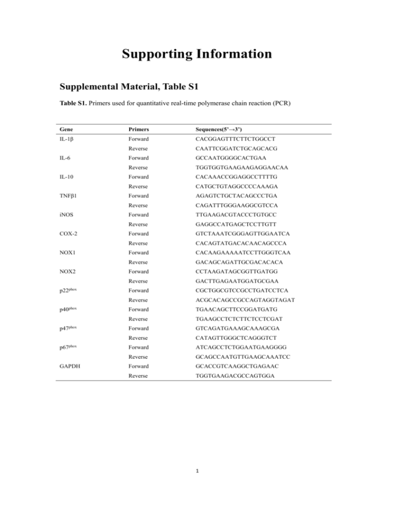
Supporting Information Supplemental Material, Table S1 Table S1. Primers used for quantitative real-time polymerase chain reaction (PCR) Gene Primers Sequences(5’→3’) IL-1β Forward CACGGAGTTTCTTCTGGCCT Reverse CAATTCGGATCTGCAGCACG Forward GCCAATGGGGCACTGAA Reverse TGGTGGTGAAGAAGAGGAACAA Forward CACAAACCGGAGGCCTTTTG Reverse CATGCTGTAGGCCCCAAAGA Forward AGAGTCTGCTACAGCCCTGA Reverse CAGATTTGGGAAGGCGTCCA Forward TTGAAGACGTACCCTGTGCC Reverse GAGGCCATGAGCTCCTTGTT Forward GTCTAAATCGGGAGTTGGAATCA Reverse CACAGTATGACACAACAGCCCA Forward CACAAGAAAAATCCTTGGGTCAA Reverse GACAGCAGATTGCGACACACA Forward CCTAAGATAGCGGTTGATGG Reverse GACTTGAGAATGGATGCGAA Forward CGCTGGCGTCCGCCTGATCCTCA Reverse ACGCACAGCCGCCAGTAGGTAGAT Forward TGAACAGCTTCCGGATGATG Reverse TGAAGCCTCTCTTCTCCTCGAT Forward GTCAGATGAAAGCAAAGCGA Reverse CATAGTTGGGCTCAGGGTCT Forward ATCAGCCTCTGGAATGAAGGGG Reverse GCAGCCAATGTTGAAGCAAATCC Forward GCACCGTCAAGGCTGAGAAC Reverse TGGTGAAGACGCCAGTGGA IL-6 IL-10 TNFβ1 iNOS COX-2 NOX1 NOX2 p22phox p40phox p47phox p67phox GAPDH 1 Figure S1. The effect of PM2.5 on cell viability of K562 cells. (a) MTT assay was used to determine the cell viability of K562 cells with PM2.5 exposure. (b) Morphological photo of cells were shown. Cells were examined with light microscope (10×). Values were representative of at least three biologically independent experiments with similar results. *p < 0.05, **p < 0.01, compared to controls. 2 Figure S2. The effect of PM2.5 on the localization of NF-κB in nucleus. The localization of NF-κB p65 through indirect immunefluorescence using FITC conjugated secondary antibody. The nucleus was stained with DAPI. Cells viewed at 60×magnification (Delta Vasion, USA). 3 Figure S3. PM2.5 activates the NADPH oxidase. RT-PCR assay was carried out to measure the expressions of NAPDH oxidase subunits. (a) The effects of PM2.5 on NOX1, NOX2 and p22phox expressions in HL-60 cells. (b) The effects of PM2.5 on p40phox, p47phox and p67phox expressions in HL-60 cells. (c) The mRNA levels of NOX1, NOX2 and p22phox expressions in K562 cells upon PM2.5 exposure. (d) The mRNA levels of p40phox, p47phox and p67phox expressions in K562 cells upon PM2.5 exposure. Data represented were mean ± SD of three identical experiments made in four replicates. *Statistically significant differences as compared to controls (*p < 0.05, **p < 0.01). 4 Figure S4. The effect of NAC on PM2.5-enhanced localization of NF-κB in nucleus. After co-treatment with NAC, the localization of NF-κB p65 through indirect immunofluorescence using FITC conjugated secondary antibody. The nucleus was stained with DAPI. Images were captured by a fluorescence microscope and viewed at 60×magnification (Delta Vasion, USA). 5


