Correction for image degrading effects
advertisement
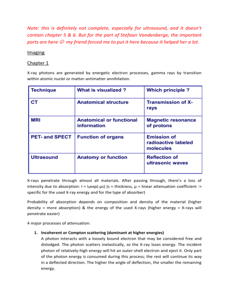
Note: this is definitely not complete, especially for ultrasound, and it doesn’t contain chapter 5 & 6. But for the part of Stefaan Vandenberge, the important parts are here my friend forced me to put it here because it helped her a lot. Imaging Chapter 1 X-ray photons are generated by energetic electron processes, gamma rays by transition within atomic nuclei or matter-antimatter annihilation. Technique What is visualized ? Which principle ? CT Anatomical structure Transmission of Xrays MRI Anatomical or functional information Magnetic resonance of protons PET- and SPECT Function of organs Emission of radioactive labeled molecules Ultrasound Anatomy or function Reflection of ultrasonic waves X-rays penetrate through almost all materials. After passing through, there’s a loss of intensity due to absorption: I = I0exp(-µs) (s = thickness, µ = linear attenuation coefficient -> specific for the used X-ray energy and for the type of absorber) Probability of absorption depends on composition and density of the material (higher density = more absorption) & the energy of the used X-rays (higher energy = X-rays will penetrate easier) 4 major processes of attenuation: 1. Incoherent or Compton scattering (dominant at higher energies) A photon interacts with a loosely bound electron that may be considered free and dislodged. The photon scatters inelastically, so the X-ray loses energy. The incident photon of relatively-high energy will hit an outer-shell electron and eject it. Only part of the photon energy is consumed during this process; the rest will continue its way in a deflected direction. The higher the angle of deflection, the smaller the remaining energy. ℎ (1 − 𝑐𝑜𝑠𝜃) 𝑚𝑒 𝑐 Compton scattering mostly depends on the density of the object. 1 𝑃(𝐸𝛾 , 𝜃) = 𝐸𝛾 1+ 2 (1 𝑚𝑒 𝑐 − 𝑐𝑜𝑠𝜃) 2. Coherent scattering = Rayleigh effect Photon interacts with a strongly bounded electron that may be set into vibration at the frequency of the radiation. While the atom returns to its undisturbed state, it will emit radiation of the same wavelength but in a random direction. The process is elastic so no energy is lost. 3. Photoelectric effect (dominant at low energies) Photon interacts with orbital electron (photon loses all its energy) and ejects several fluorescent photons and an orbital electron with a binding energy e lower than the energy of the incident photon. Due to the ejection of the electron, the atom is ionized and brought into an excitation state. The kinetic energy transferred to the atom will be negligible, while for the electron this energy is independent on the scattering angle θ and will be equal to hv – e. Probability increases if the photon energy is only slightly higher than the electron binding energy. 4. Pair production Photon interacts with a nucleus and in transformed into an electron and a positron, with any excess of photon energy transferred into kinetic energy in the produced couple of particles. 𝛿𝜆 = 𝜆2 − 𝜆1 = Scattered photons do not carry any useful information but can cause noise. X-ray production: In Rontgen tube, fast moving electrons ejected from a heated cathode filament are retarded by a metal object and cause the production of X-rays. More electrons will be released as the T of the cathode filament increases. Inside the vacuum tube, which contains the cathode and the anode of the X-ray source, an electrical potential forces the electrons to accelerate towards the anode. The electron current between the filament and the anode is expressed in mA. The vacuum is needed to prevent early burning of the cathode filament and to make the electrons reach the anode in an undisturbed way. To create X-rays, the electrons need a sufficiently high velocity when they hit the anode. This velocity depends on the voltage difference between the cathode and the anode. (higher voltage -> higher velocity). X-ray quality mainly depends on the voltage difference, quantity depends on both voltage and tube current. Electrons will either interact with orbital electrons or with nuclei of the target atoms. These interactions will result in a conversion of kinetic energy into thermal and electromagnetic energy in the form of X-rays. This production is caused by two different processes, creating two types of rays: brehmsstrahlung, created by the retardation of electrons when they interact with the anode, and a second type that is related to characteristic X-rays, emitted from heavy elements when their electrons make transitions between lower atomic energy levels. Two parameters when we use X-ray source: 1) number of photons, determined by the current of the cathode 2) energy of the emitted photons, determined by the voltage Beam hardening: Lower energy photons will be absorbed more than higher energy photons. The average energy of the output spectrum is higher than the input beam. The beam is more reduced at lower energies than at higher energies. In own words: higher average energy as you go through the material. Higher energy beams go through easier and “survive” while low energy beams die out. The patient absorbs all the low energy photons: they contribute to the dose but not to the image quality -> preferentially absorbed before hitting the patient: filter (aluminium) + collimator (limits the area of the patient that is irradiated). Collimating scatter grid: improves the measured date to make it more close to a projection. Absorbs scattered photons. Detectors: p. 17 Computed tomography (CT) Non-destructive visualization of slices of a 3D object, using X-rays. A CT image is a pixel-bypixel map of X-ray beam attenuation (essentially density). A bright value (white in grayscale) shows an area of high attenuation (dense material). Scaled in Hounsfield Units. Voxel value CT (x,y) = 1000 * (µ(x,y) - µwater)/ µwater Principle: measure line integrals at different angles; calculate pixel values in slice • • Parallel beam: rotation angle vs radial distance Fan beam: rotation angle vs angle in fan Multislice: wider in z direction beam & detector matrix; reduction scan time; more scatter Cone-beam: even wider; complex algorithms. Max. distance between subsequent slices: determined by Full Width at Half Maximum of the slice sensitivity profile (SSP) (= variation of the relative sensitivity in axial direction in the center of a slice, ideally rectangular). Effective slice thickness Dz -> max. distance: Dz/2. For helical mode: Nyquist gives us Table Feed (axial distance which table shifts during one rotation) = Dz/2, pitch = 0.5. Due to symmetry 180°, interpolation is used TF = Dz, pitch = 2. In practice pitch = 1-2: higher pitch results in shorter scan time, lower dose and less motion artifacts. Analytical reconstruction Analytical methods reconstruct the image from their projections directly and are based on the mathematical inverse of the tomography process. They are based on a continuous model. Iterative methods use a computational model for the forward and back projection. By an iterative process the image is improved at each iteration. They are based on a discrete model. Forward projection 𝑝θ (𝑡) = ∫ ∫ 𝑓(𝑥, 𝑦)𝛿(𝑥 𝑐𝑜𝑠 𝜃 + 𝑦 sin 𝜃 − 𝑡)𝑑𝑥 𝑑𝑦 Radon transform Transformation consisting of the integral of a function over straight lines. Define the straight line in image space by: (𝑥(𝑡), 𝑦(𝑡)) = 𝑡(sin 𝜃, − cos 𝜃) + 𝑠 (cos 𝜃, sin 𝜃) Then the tf is given by +∞ +∞ 𝑅[𝑓](𝜃, 𝑠) = ∫−∞ 𝑓(𝑥(𝑡), 𝑦(𝑡))𝑑𝑡 = ∫−∞ 𝑓( 𝑡(sin 𝜃, − cos 𝜃) + 𝑠 (cos 𝜃, sin 𝜃))𝑑𝑡 The goal of image reconstruction is to calculate the image from its projections at different angles. Sinogram = a visual representation of the raw data obtained in a CT scan. Back projection Distributes the activity in sinogram bin over the ray in image space: +∞ 𝑏𝜃 (𝑥, 𝑦) = ∫ 𝑝𝜃 (𝑡)𝛿(𝑥 cos 𝜃 + 𝑦 sin 𝜃 − 𝑡) 𝑑𝑡 −∞ By adding up the images at all the angles, we obtain a back projected image: 𝜋 𝜋 +∞ 𝑓𝑏 (𝑥, 𝑦) = ∫0 𝑏𝜃 (𝑥, 𝑦)𝑑𝜃 = ∫0 ∫−∞ 𝑝𝜃 (𝑡)𝛿(𝑥 cos 𝜃 + 𝑦 sin 𝜃 − 𝑡) 𝑑𝑡 𝑑𝜃 (1) Difference between original image and back projection: in the original image all activity was concentrated in one point. When we back project, we do not know where along the line this point was located (we do not know the object that was measured). Our best guess is to uniformly distribute the sinogram value along the ray. Therefore not everything will end up in the central point. By this process the maximum activity will end up in the original point (all sinogram rays cross there) but also quite some activity will end up outside the original point. It can be shown that the impulse response of the back-projection algorithm is equal to: ℎ𝑏 (𝑟) = 1 𝑟 The reconstructed image can be expressed as the convolution of the original image with this impulse response: 𝑓𝑏 (𝑥, 𝑦) = 𝑓(𝑥, 𝑦) ∗ 1 𝑟 Therefore back projection is not sufficient and we have to use the inverse of the Radon transform to reconstruct images from the measured transmission data: Option 1: 2D Fourier based recon Transform image to Fourier domain and remove blurring effect Not efficient Option 2: filtered back projection and central section theorem Fourier slice/central section theorem Fourier transform (1D) along the radial coordinate. 𝐹1 (𝑝𝜃 (𝑡)) = ∫ 𝑝𝜃 (𝑡) 𝑒 −𝑗2𝜋𝜌𝑡 𝑑𝑡 𝑝𝜃 (𝑡) = ∫ 𝐹1 (𝑝𝜃 (𝑡))𝑒 𝑗2𝜋𝜌𝑡 𝑑𝜌 (2) in (1): If we use the 2D inverse Fourier tf of the original image: Change the integration limits Then the back projected image can be rewritten (2) And we can replace F(ρ,θ) by F1(pθ(t)) in f(x,y) (CENTRAL SECTION THEOREM): Difference between backprojected and original: for the backprojected, the Fourier tf of each projection pθ(t) is weighted by the inverse of |ρ| along radial line. This causes the blurred effect of the backprojected algorithm and the blurring effect can be removed by weighting each transformed projection with |ρ| prior to the backprojection. This brings us to the final equation for the filtered backprojection algorithm: FBP is really fast, but assumes exact projection data and a large number of angles in the datasets. Iterative reconstruction methods are more flexible to include corrections and nonexact projections. Linear attenuation coefficients of X-rays: Difference between measured date and line integrals because: 1. Limited number of projections and spatial resolution in each projection: sampling 2. Measurement errors (noise) 3. Deviation between simplified mathematical model (line integrals) and real physical data acquisition system: a. Due to finite resolution of the detector, integrals are along “tubes” rather than lines b. X-ray and gamma scattering c. Beam polychromaticity in CT 4. Displacements of the patient during data acquisition NO FBP IN SPECT AND PET BECAUSE iterative construction methods have replaced analytical methods. Datasets are smaller for these type of scanners and the acquisition times are longer, so there is more time for image reconstruction (normally iterative is slower, but here it’s okay because there’s more time and smaller datasets). + larger deviation from the line integral model and a larger resulting gain in image quality when switching from analytical to iterative. So: iterative is slower, but better image quality which is better for pet & spect. Chapter 2: Spect & Monte Carlo Characteristic molecule is labeled by chemical coupling to a radioactive isotope, small dose, image (= scintigram) concentration of molecules at certain position, physical processes or metabolism of specific organs. Most sensitive but limited spatial and temporal resolution. Radioactive decay = Unstable atomic nucleus loses energy by emitting ionizing particles and radiation. This decay results in an atom of one type, the parent nuclide, transforming to an atom of a different type, the daughter nuclide. The decay rate on average is predictable. Particles: - alpha = strong ionization, very short penetration, do not escape body so not possible to use for imaging - Beta: destructive, not used for imaging Gamma: tissue penetrating property, can be used for detection and imaging Requirements isotope: Practical halflife (hours-days) Suitable chemical properties to attach it to a pharmaceutical High purity (minimal amount of other isotopes) High specific activity (high concentration of molecule after labeling) Molecule determines resolution, not isotope! Gamma camera: - Mechanical Collimator: photons which are capable of leaving the body of the patient and travel in the direction of the gamma camera are mechanically selected by the collimator. With a collimator, the direction of incidence of the photons to the detector is mechanically limited. A mechanical collimator consists of many narrow air holes separated by thin septa, which are made of a material with a high attenuation coefficient to ensure that photons hitting a septum will be absorbed. Therefore only photons reaching the collimator along the direction of the holes will pass the collimator and are able to reach the scintillation crystal. Septal penetration: max. 0.5%; more lead in collimator reduces septal penetration so the spatial resolution is improved, but at the cost of sensitivity. Increasing the hole size improves the sensitivity but at the expense of resolution. - Detectors: Scintillation crystals: convert gamma radiation into scintillation light (NaI(Tl) crystal = sodium iodide crystal doped with thallium). Every incident photons will deposit its energy in the crystal through the photo-electric effect. The resulting photonelectron travels through the crystal and distributes its kinetic energy by multiple collisions with other electrons. These electrons are brought to a higher energy level which brings the entire crystal to an excited state. When the crystal subsequently returns to its ground state, it releases its energy and approx. 10% of the energy loss gives rise to light flashes, also called scintillations. The amount of scintillations is proportional with the level of excitation of the crystal and thus with the energy of the incident photon. Scintillation time: time between excitation and emission of secondary photons, should be short so crystal can process next photon Photon Multiplier Tube: convert light into an electrical signal and amplify this signal. Light photons from the scintillation flash dislodge photoelectrons from the photocathode in the PMTs. These photoelectrons are accelerated to the nearest dynode, where they further dislodge electrons. This generates a measurable current at the output of the PMT. The electrical signal at the end of the PMT is sent towards an Anger logic readout for energy discrimination and localization of the incoming photon. The total charge S collected from the back of all PMTs i is a measure of the energy deposited by the incident gamma ray. How can we find back position of scintillation with sufficient accuracy? = Anger logic: relative amount of scintillation light on PMT. The most intense signal is obtained from the PMT nearest to the event and progressively weaker signals are found as the distance increases. The total signal of the PMTs at the same X distances ad Y distances is first summed. By weighting the position Xi of the PMTs at the same X-position with the relative strength of the signal SXi at these PMT I, the location of the interaction point in the X direction can be determined to within N a few mm. X SX i X i / E i 1 N Y SYi Yi / E i 1 N E SX i i 1 - Solid state detectors = semiconductor detectors: direct conversion from gamma photons to an electrical signal without the intermediate step of visible light. Each gamma ray creates a number of electron-hole pairs; by applying a voltage over the semiconductor, the holes and the electrons will drift towards their corresponding electrode, hereby inducing an electric charge on them. + large number of information carriers => superior energy resolutions. - expensive Image degradation: caused by unwanted photon interaction in the detection system and in the object. Mostly due to - - Imperfect detection system • Limited energy resolution • Limited spatial resolution Interaction of photons with matter (patient) • Photoelectric effect • Compton Scatter 1. Sensitivity: determined by solid angle subtended by detectors, geometric properties of the collimator and material properties of the photon detectors. Major influence is by the collimator as it determines the acceptance angle. (number of emitted photons that will hit the detector) 2. Energy resolution 3. Spatial resolution and partial volume effect The spatial resolution of a gamma camera is determined by the collimator parameters ad the intrinsic detector resolution. Intrinsic resolution Ri Total: S ~ Rc2 -> resolution cannot be optimized without compromising sensitivity and vice versa. Correction and simulation techniques Spectrum-based methods: inexpensive, but increase noise Model-based methods: highly accurate, but computationally very demanding -> integrated in iterative reconstruction Monte carlo simulations Phantom studies: Advantages – Predict quality of patient scans – Known distribution – Allows to compare different systems Disadvantages – Difficult to fill – Time consuming – Does not always represent patient well Monte carlo = numerical solution to a problem that models objects interacting with other objects or their environment based upon simple object-object or object-environment relationships. A solution to a macroscopic system through simulation of its microscopic interactions. – Statistical simulation techniques – Based on random numbers produced by generators – System described by probability density functions – Many repetitions realistic simulation Needed : Probability density function (pdf) – Describes physical interaction in a system – Probability of interaction as a function of variable – Discrete or continuous Random number Generator – Method to determine random numbers from interval between 0 and 1 Rule for sampling pdf Sampling method 1: cpdf – inverse cpdf F-1 needs to be known – Random number R1 in [0,1] x1 – Repeat this process (x1,x2, … xn) Sampling method 2: rejection method – Pdf limited to [a,b] – Random number R1 in [0,1] r =a +(b-a)R1 random number between a en b – calculated pdf(r) – Random number R2 in [0,1] accept if R2 < pdf(r) Chapter 3: PET & iterative reconstruction PET: position emission tomography. Positrons produced in cyclotrons, labeled, injected, … Position emission results in the atomic number of the nucleus reduced by one. Released positron annihilates with a nearby electron -> 2 photons in opposite directions -> detection. Electronic collimation instead of mechanical. Advantage over SPECT: No use of mechanical collimator, Results in higher sensitivity, Better spatial resolution. Disadvantage: But limited to positron emitters (short halflife and cyclotron) Production in cyclotron: target sample is bombarded by charged particles. These particles need sufficient energy to overcome the nuclear Coulomb Force. A cyclotron is composed of the D’s with a gap between them. In the center in between both D’s positive charged particles are generated in bursts by a positive ion source. The D’s are placed between the poles of an electromagnet and the magnetic field makes the ions follow a circular path. A cyclotron is based on two effect: a charged particle in an electrical field will experience a force in the direction of the field and a charged particle in a magnetic field will encounter a force perpendicular on movement and perpendicular on magnetic field. The positive particles are attracted towards the negatively charged D. as soon as the group of particles reaches the other D, the polarity is changed and the particles accelerate towards the other D. Every time the particles cross the gap, they gain energy and the radius increases. At the end of the spiral path (radius = radius D) the particles are directed onto a target. The accelerated positive particles will add positive charge to the target resulting in an element with another atomic number. This is in general an unstable element which decays by positron emission. Detection: * no rotation required (full ring) * no simultaneous imaging possible because energy is always 511 keV * pixellated detectors Coincidence detection: Two photons: assume path and the annihilation point are at the same line (colinearity) and that both photons arrive around the same time (simultaneity). When one of the photons is detected, a coincidence circuit is opened: if a second photon arrives within a very short time, the system has registered a coincidence. By registering multiple of these coincident lines, projections of the emission distribution can be formed and the distribution is calculated from these by image reconstruction techniques. Scintillators: Should stop the incoming gamma ray with a high probability and produce a light signal that allows to determine the energy, time and position. Now: pixellated scintillators. Geometry & collimation: Optimizing geometry, detection efficiency, axial FOV and collimation. Remove septa: 3D mode, combinations between different rings are possible. Better sensitivity, more image degrading effects Full ring scanners: remaining mechanical collimation are lead shields at the axial ends to reduce effects from outside field of view activity Increase axial extent: larger FOV, higher sensitivity, higher cost and more image degrading effects Measure more info about the coincidence itself: time difference between detections: determine position annihilation -> improves image quality, shorter acquisition time => time of flight pet! Image degrading effects Resolution and partial volume effect: limited by the physics of position emission: position range (= distance positron travels through object to lose enough kinetic energy before the annihilation takes place, depends on the energy of the emitted positions (different for each isotope) and on the material of the object) and photon acolinearity (= caused by the momentum of the positron and the electron at annihilation. Due to conservation of momentum the resultant photons will not be emitted in exactly opposed directions). Dominant factor in spatial resolution is the detector itself. Additionally: parallax effect degrades spatial resolution: depth of interaction is not measured. Partial volume effect: the activity of small hot structures will be underestimated and the activity in small cold structures will be overestimated. (difficult to follow up small tumors: is reduced activity cause of reduction in size or uptake?) Attenuation: photons have to go through the body before being detected: probability they will undergo Compton scatter (deflection leading to non-detection) or Photoelectric effect (absorption). Photon pair is attenuated when one of the photons (or both) is not detected due to an interaction in the body. 511 keV, probability of absorption is relatively high, for scatter somewhat lower. Due to the attenuation effect annihilation from the center of the body will have a much lower probability to escape the body than annihilations from the edge -> cupping effect. Attenuation map is derived from a low dose CT (acquired before the PET scan) Scatter and randoms: Scattered coincidences = wrong coincidences: as one (or both) photons deviate due to Compton effect, the registered photon pair deviates from the correct one. Compromise: only coincidences from a certain energy window around 511 keV are accepted. 3D PET systems have a much larger scatter fraction than 2D. scatter is removed by combining estimation techniques and correction in iterative reconstruction. Random coincidences: two photons are registered in coincidence if they are detected within a given coincidence time window τ. Larger time window than max. time difference between two photons to avoid loss of true coincidences -> gives us also incorrect coincidences as two separate annihilations within the coincidence time window can each contribute a single. The combination of these two singles is called a random coincidence. When the activity in FOV increases, the random coincidences increase. This amount can be estimated from a singles measurement. A more common practice is to use a delayed coincidence window: when a single hits a detector, only combinations with another single occurring in the time frame [τ, 2τ] are measured. Countrate: No mechanical collimator is used -> detectors have to process singles at much higher rates. The process of scintillation and determination of the energy and position requires a certain integration time = deadtime. Detectors should not process more singles during the deadtime because if they do, the single will be registered as a higher energy photon and therefore will fall outside the energy window and not lead to a coincidence. -> countrate curves operate in first half of the curve. Correct for loss by applying countrate loss factors based on measured singles rate. Pet in practice PET-CT: CT helps in localization. Combinations gives us shorter study time + patient is in exact same position -> more accurate. Pet and iterative reconstruction Discretize the image into pixels and treat each pixel value as an unknown. The forward projection is then represented by a matrix multiplication. This system allows correction for image degrading effects. Matrix from: P = HF, each element fj in F is a pixel value, each element pi in P is a projection measurement, and hij is a coefficient which represents the probability of detecting a photon, originating from pixel j in the projection bin i. We are interested in finding a vector F that is a solution of P = HF -> successive estimates: compare with measured data, corrected, backprojected, image updated. Different algorithms: Deterministic iterative methods: incorporate the system characteristics Statistical iterative methods: e.g. Maximum Likelihood Expectation Maximization (MLEM) Based on the fact that the number of measured photons follows a Poisson distribution. The goal of MLEM is to find a “general” solution as the best estimate for f(x,y): the mean number of radioactive disintegrations in the image that an produce the sinogram with the highest likelihood. The MLEM is thus an optimization method, meaning that the best estimate for the solution fitting a given criterion (= maximization of the likelihood of the reconstructed image) is found. This can be done used the Poisson law that allows one to predict the probability of a realized number of detected counts, given the mean number of disintegrations. Each iteration of this algorithm is divided into 2 steps: 1. Expectation step (E step): the formula expressing the likelihood of any reconstructed image given the measured date is formed. 2. Maximization step (M step): the image that has the greatest likelihood to give the measured date is found first image F(0) can be initialized to 1 or by FBP. Once the forward projection becomes equal to the measured projection data the ratio in projection space becomes equal to 1 -> the update image in image space also becomes 1 = This is called convergence. Properties: - - Multiplicative algorithm • Starting image should always be different from zero • Positivity constraint: once a initial positive pixel value is chosen it will stay positive Slow convergence of high frequencies Noise amplifies with iteration number, due to Poisson charac. Difficult to choose the right iteration number as convergence depends on • Size of object • Distribution • Noise Solution to slowness = Ordered-subsets expectation maximization (dividing problem into subsets) Correction for image degrading effects: Correct for attenuation: Two ways of correcting: precorrect (scale factor per detector pair), or include it in iterative reconstruction. Correction for randoms: correction for scatter: Chapter 4: Ultrasound Fundamental physics Propagation: acoustic waves with a frequency above audible range cause mechanical stresses and vibrations in matter. When a stress is applied normal to the material surface, a longitudinal pressure wave is excited, i.e. the oscillatory motion of the particles in the medium is parallel to the wave propagation direction. The ultrasound imaging applications used in clinical practice are based on longitudinal waves because the shear waves at diagnostic imaging frequencies vanish very quickly. Speed c with which the waves propagate: 1 𝐾 𝑐= √ = √ 𝜅𝜌 𝜌 With K = bulk modulus, κ = compressibility. c ≈ 1540 m/s. image degradation may occur if the sound in certain regions is too much differing from 1540 m/s, with improper focusing of the ultrasound beam as a result, also called phase aberration. Bone is one of the structures which should be avoided in this sense (3500 m/s) Scattering: energy of the ultrasonic vibrations scatters with changes in acoustic impedance of the medium, or more specifically with changes in density or compression. This process is crucial, since it ‘s the info (amplitude, phase, frequency) contained in the backscattered waves which is used during imaging. Different types of scattering: 1. Specular reflections: when the dimensions of the reflecting object are much larger than the ultrasonic wavelength. Z = ρc, 1 = incident, 2 = transmitted (refracted = deviation of a beam when it crosses a boundary between two media in which the sound speed is different) 2. Diffuse scattering (most common): when the wave is scattered by an inhomogeneity of dimensions smaller than the ultrasound wave. The roughness features on the scattering object then fail to construct any significant interference pattern. e.g. red blood cells. In particular Rayleigh scattering occurs, which mean that the scattered intensity has a fourth power dependence on frequency. 3. Diffractive scattering: between 1 & 2. Size of the scattering object is then comparable to the wavelength and ultrasound is scattered directionally. Attenuation: An ultrasonic wave propagating through a heterogeneous medium loses energy, or is attenuated as a function of depth z and frequency f: p(z) = p(z=0)exp(-αfz) p = pressure, α = attenuation coeff. compensation in the receiving amplifier of the ultrasound transducer using time gain compensation (TGC) Basic imaging principles Pulse-echo imaging: an ultrasonic probe (= transducer) excites ultrasonic pulses, which travel through the imaged medium and reflect/scatter when changes in acoustic impedance are met. The backscattered signals (= radio-frequent signals) are received by the transducer and further processed to image and characterize the imaged medium. Depth d = ct/2 2 because wave has to travel back and forth Piezo-electric effect: Transducer creates US waves via piezo-electric effect = Conversion of electrical to acoustic energy and vice versa. Piezo-electric materials produce an electrical signal when experiencing a vibration and on the other hand vibrate when a time-varying electrical field is applied (reverse effect). Piezo-electric transducer (PZT) has an Z = 20*Ztissue -> 80% of the energy is reflected back. This causes internal vibrations inside the piezo-electric slab, continuing long after the electrical signal has stopped = ringing. Solution: backing layer to absorb (limited amount of) ultrasound waves. Acoustic matching layers necessary at front of piezo-electric slab to increase transmitted acoustic power. Size of piezo-electric slab also influences the efficiency of energy transmission, since the transducer resonance occurs at the frequency for which the slab thickness is half a wavelength. Combined effect: transducer bandwidth (large ones are desirable)

