WHOLE MOUSE FIXATION VIA TRANSCARDIAL PERFUSION
advertisement
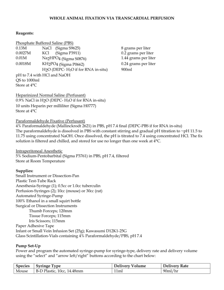
WHOLE ANIMAL FIXATION VIA TRANSCARDIAL PERFUSION Reagents: Phosphate Buffered Saline (PBS) 0.13M NaCl (Sigma S9625) 0.0027M KCl (Sigma P3911) 0.01M Na2HPO4 (Sigma S0876) 0.0018M KH2PO4 (Sigma P0662) H2O (DEPC- H2O if for RNA in-situ) pH to 7.4 with HCl and NaOH QS to 1000ml Store at 4°C 8 grams per liter 0.2 grams per liter 1.44 grams per liter 0.24 grams per liter 900ml Heparinized Normal Saline (Perfusant) 0.9% NaCl in H2O (DEPC- H2O if for RNA in-situ) 10 units Heparin per milliliter (Sigma H0777) Store at 4°C Paraformaldehyde Fixative (Perfusant) 4% Paraformaldehyde (Mallinckrodt 2621) in PBS, pH 7.4 final (DEPC-PBS if for RNA in-situ) The paraformaldehyde is dissolved in PBS with constant stirring and gradual pH titration to ~pH 11.5 to 11.75 using concentrated NaOH. Once dissolved, the pH is titrated to 7.4 using concentrated HCl. The fix solution is filtered and chilled, and stored for use no longer than one week at 4°C. Intraperitoneal Anesthetic 5% Sodium-Pentobarbital (Sigma P3761) in PBS, pH 7.4, filtered Store at Room Temperature Supplies: Small Instrument or Dissection-Pan Plastic Test-Tube Rack Anesthesia-Syringe (1); 0.5cc or 1.0cc tuberculin Perfusion-Syringes (2); 10cc (mouse) or 30cc (rat) Automated Syringe-Pump 100% Ethanol in a small squirt bottle Surgical or Dissection Instruments Thumb Forceps; 120mm Tissue Forceps; 115mm Iris Scissors; 115mm Paper Adhesive Tape Infant or Small Vein Infusion Set (25g); Kawasumi D12K1-25G Glass Scintillation-Vials containing 4% Paraformaldehyde/PBS, pH 7.4 Pump Set-Up Power and program the automated syringe-pump for syringe-type, delivery rate and delivery volume using the “select” and “arrow left/right” buttons according to the chart below: Species Mouse Syringe Type B-D Plastic, 10cc, 14.48mm Delivery Volume 11ml Delivery Rate 90ml/hr Rat B-D Plastic, 30cc, 21.59mm 33ml 90ml/hr Procedure: 1. Set up the automated syring-pump according to the Pump Set-Up section on the previous page. 2. Label and fill the perfusion syringes to the hilt with each perfusant. 3. Attach the infant infusion set to the heparinized-saline-syringe, prime it to expel air through the needle tip and lock the primed syringe into the syringe pump cradle. 4. Fill the anesthesia-syringe with sufficient pentobarbital for all animals. 5. Place the plastic test-tube rack upside-down in the instrument or dissection-pan. 6. Administer anesthetic to one animal via intraperitoneal injection; 0.1cc for mouse, 0.4cc – 1.6cc for rat. 7. Assess and assure surgical-plane anesthesia by toe pinch. 8. Without delay upon reaching surgical plane, place the anesthetized animal on top of the inverted testtube rack. 9. Wet the fur of the ventral skin surface with 100% ethanol and wipe with gauze. 10. Superficially cut the ventral skin with scissors, just below the zyphoid-process, being careful not to perforate the peritoneal membrane. 11. Expand the ventral skin-opening by manually pulling the opposing skin segments rostrally and caudally. 12. With the ventral skin pulled to expose the thoracic and peritoneal membrane surfaces, superficially cut the peritoneal membrane with scissors, just below the zyphoid-process and expose the diaphragm and visceral organs, being careful not to lacerate any significant vasculature. 13. Performing a combination of blunt dissection and scissor-assisted dissection techniques, open the thoracic cavity by cutting the diaphragm from one lateral aspect to the other lateral aspect while avoiding cutting any visceral organs. 14. Continuing with a combination of blunt dissection and scissor-assisted dissection, carefully cut both lateral aspects of the rib cage, in a caudal to rostral direction up to 2nd rib, while avoiding lung, heart and mammary arteries. 15. Fold-back or invert the ventral rib-cage surface to meet the ventral skin surface of the animal’s neck area and gently tape it into place to expose thoracic organs. 16. Orient the dissection-pan so that the apex of heart resides at 6 o’clock and the aortic outflow resides at 12 o’clock. 17. Using thumb forceps, gently grasp and hold the right ventricular free-wall and visualize the delineation of the darker, blood-filled-coloration of right ventricle and the redder, muscularcoloration of left ventricle. 18. Lacerate the right atrial-chamber with scissors and carefully (but quickly) insert the 25g needle of the infant infusion set attached to the heparinized-saline-syringe into the left ventricular chamber at the 5:30 position. The needle should be angled towards a 2 o’clock position. 19. Release the forceps grasp of the right ventricular free-wall. 20. Begin perfusion by depressing the “run-stop” button on the automated syringe-pump. Blood and heparinized saline effluent should flow from the right atria laceration and drip into the instrument or dissection pan. The pump should read out amount of perfusant dispensed. 21. Visualize the heart and liver as perfusion commences. The right ventricular chamber should remain somewhat darkened in color when compared to the left ventricular chamber. The liver should begin to blanch as blood is replaced with heparinized-saline. 22. At the completion of the saline perfusion, carefully change-out the syringes without dislodging the 25g infant infusion needle from the left ventricle. 23. Reset the syringe-pump and reinitiate perfusion with 4% paraformaldehyde/PBS syringe, by depressing the “run-stop” button on the automated syringe-pump. 24. Visualize the animal’s extremities for evidence of tremors resulting from the aldehyde-crosslinking of nerves and muscle. 25. At the completion of the paraformaldehyde perfusion, remove the 25g infant infusion needle from the left ventricle and begin dissection of needed tissues. If further perfusions are to be performed, the infant infusion catheter and needle must be reprimed (flushed) with heparinized saline. 26. All collected tissues are then grossly trimmed and immersed in 4% paraformaldehyde/PBS filled glass scintillation vials. Fixation is continued by immersion with agitation, and is carried out at 4°C for 1016 hours. 27. At the conclusion of fixation, fixative from the vials of tissues for immunohistochemistry or in-situ hybridization are decanted and replaced with PBS (DEPC-PBS for RNA in-situ) and paraffin processed as soon as possible.
