Calcium Oxalate Worksheet Answer Key
advertisement
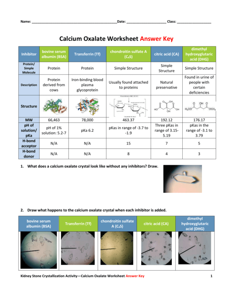
Name: _____________________________________________Date: _____________________ Class: __________________ Calcium Oxalate Worksheet Answer Key Inhibitor bovine serum albumin (BSA) Transferrin (Tf) chondroitin sulfate A (C4S) citric acid (CA) dimethyl hydroxyglutaric acid (DHG) Protein/ Simple Molecule Protein Protein Simple Structure Simple Structure Simple Structure Description Protein derived from cows Iron-binding blood plasma glycoprotein Usually found attached to proteins Natural preservative Found in urine of people with certain deficiencies 66,463 78,000 463.37 pH of 1% solution: 5.2-7 pKa 6.2 pKas in range of -3.7 to -1.9 192.12 Three pKas in range of 3.155.19 176.17 pKas in the range of -3.1 to 3.79 N/A N/A 15 7 5 N/A N/A 8 4 3 Structure MW pH of solution/ pKa H-bond acceptor H-bond donor 1. What does a calcium oxalate crystal look like without any inhibitors? Draw. 2. Draw what happens to the calcium oxalate crystal when each inhibitor is added. bovine serum albumin (BSA) Transferrin (Tf) chondroitin sulfate A (C4S) citric acid (CA) Kidney Stone Crystallization Activity—Calcium Oxalate Worksheet Answer Key dimethyl hydroxyglutaric acid (DHG) 1 Name: _____________________________________________Date: _____________________ Class: __________________ Inhibitor bovine serum albumin (BSA) Transferrin (Tf) chondroitin sulfate A (C4S) citric acid (CA) (binds to the top and bottom faces) dimethyl hydroxyglutaric acid (DHG) Mark on the crystal where each molecule binds (Answers to this question are in the PowerPoint presentation file, if students pay attention) 3. Compare groups of inhibitors. (two groups: proteins and simple structures) Which were the most effective at blocking growth? Speculate why one would be more effective. Simple structures were more effective at blocking growth because they were able to bind to the surface of the crystal face better. The proteins were also much larger and folded causing them to be less effective. 4. Compare inhibitors within their own group. Which ones were the most effective? Explain why this might be. C4S was the best inhibitor of the simple structures group because it had the largest molecular weight and structure meaning it could block more of a crystal face with one molecule than the other two inhibitors. It also has more H bond acceptors and donors in comparison. BSA was the best inhibitor of the proteins because it binds to four faces of the crystal. 5. Do the crystals have different shapes when inhibitors are added? What might cause these different shapes? (Think about how steps grow on a crystal surface in all directions but at different rates. How does an inhibitor affect those rates?) The crystals have the same general shape, but the length of the crystal varies depending on the additive. If the crystals are shorter in length than the original crystal, the growth of the crystal in one direction is blocked, but not the others. The same can be said if the crystals are thinner. Therefore, the crystal can have different shapes, but the general shape remains the same for these inhibitors. 6. Of all the inhibitors used today, which do you think would be the best choice as a possible drug? Why? C4S was the best inhibitor overall because the crystal was almost completely blocked from growing. The molecule has the most hydrogen bond acceptors and donors in comparison to the other molecules that are not proteins and the proteins could be configured in a way that does not allow them to bind as effectively as the smaller molecules. Kidney Stone Crystallization Activity—Calcium Oxalate Worksheet Answer Key 2


