4. bragg scattering with microwaves
advertisement
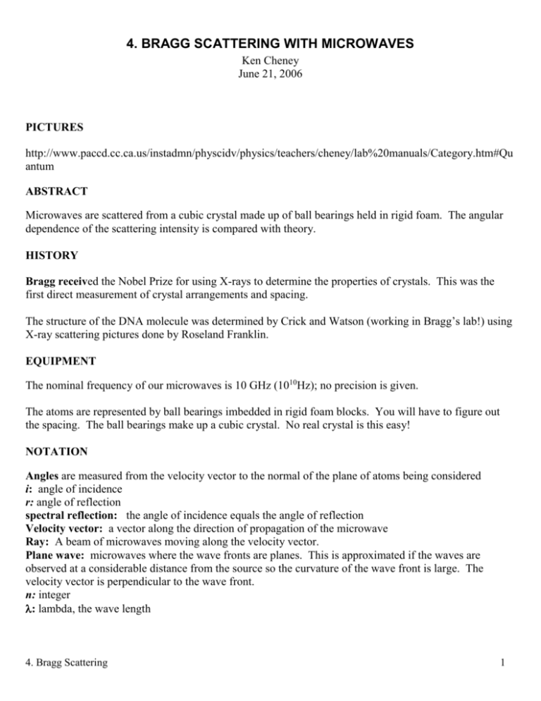
4. BRAGG SCATTERING WITH MICROWAVES Ken Cheney June 21, 2006 PICTURES http://www.paccd.cc.ca.us/instadmn/physcidv/physics/teachers/cheney/lab%20manuals/Category.htm#Qu antum ABSTRACT Microwaves are scattered from a cubic crystal made up of ball bearings held in rigid foam. The angular dependence of the scattering intensity is compared with theory. HISTORY Bragg received the Nobel Prize for using X-rays to determine the properties of crystals. This was the first direct measurement of crystal arrangements and spacing. The structure of the DNA molecule was determined by Crick and Watson (working in Bragg’s lab!) using X-ray scattering pictures done by Roseland Franklin. EQUIPMENT The nominal frequency of our microwaves is 10 GHz (1010Hz); no precision is given. The atoms are represented by ball bearings imbedded in rigid foam blocks. You will have to figure out the spacing. The ball bearings make up a cubic crystal. No real crystal is this easy! NOTATION Angles are measured from the velocity vector to the normal of the plane of atoms being considered i: angle of incidence r: angle of reflection spectral reflection: the angle of incidence equals the angle of reflection Velocity vector: a vector along the direction of propagation of the microwave Ray: A beam of microwaves moving along the velocity vector. Plane wave: microwaves where the wave fronts are planes. This is approximated if the waves are observed at a considerable distance from the source so the curvature of the wave front is large. The velocity vector is perpendicular to the wave front. n: integer : lambda, the wave length 4. Bragg Scattering 1 a: the atom-to-atom spacing along the sides of a cubic crystal SPECTRAL REFLECTION Normals to the top plane of atoms Incoming rays Plane wave front Outgoing rays D C r i a B E Atoms THEORY, SPECTRAL REFLECTION FROM A PLANE OF “ATOMS” Angles are measured from the normal to the plane under consideration. It will be shown below that these special cases are really generally applicable! Consider a plane through the “top” side of the cubic crystal; let this be the surface of our crystal. This is the plane reflecting the microwaves in this step. Assume the microwaves (or X-rays) are approaching with a velocity vector parallel to another side of the crystal cube. Take a view of a plane through the third side of the crystal cube. We see the atoms making up a lattice of squares, say with the microwaves approaching from the top left of the top surface. Presumably the microwaves will scatter in many directions from each “atom” but there will only be intense scattering in directions that produce “in phase” contributions from scattering from many atoms. To be in phase the path differences between the rays must be n, where n is an integer and is the wavelength of the microwaves. The simplest, and most effective case, is when the angle of incidence (from the direction of the incoming microwaves to the normal to the surface) is equal to the angle of reflection (normal to outgoing 4. Bragg Scattering 2 microwaves). A little sketching will show that for equal angle, spectral, reflection all parts of an incoming plane wave travel equal distances to make up an outgoing plane wave. In the figure “SPECTRAL REFLECTION” the incoming wave front BC is reflected into the outgoing wave front DE. For all parts of the wave front to be in phase the incoming distance CE must equal the outgoing distance BD, which implies that i equals r. From now on we will assume spectral reflection. Notice that the arguments so far don’t restrict the incoming angle, all angles will reflect equally well. But the outgoing angle will always equal the incoming angle. THEORY, REFLECTION FROM PARALLEL PLANES OF ATOMS For the cubic crystal considered above consider reflection from the surface plane of atoms and also from the next parallel plane down below the surface. See the figure "SCATTERING FROM PARALLEL PLANES OF ATOMS" Consider an atom from each plane, one directly above the other. We want to find out what incidence/reflection angle will result in path differences of n between reflections from the top atom and the bottom atom. We know from the considerations above that the reflection must be specular to be in phase with other atoms in the same plane. Light striking the bottom atom travels farther than light striking the top atom both before and after the reflection. The symmetry of the reflection means that the total path difference is just twice the incoming path difference. We draw a right triangle: one side is between the atoms, the other sides are determined by the incoming velocity vector of the wave striking the bottom atom and the wave front of this velocity vector when the wave front passes the top atom. Call the intersection of the velocity vector and the wave front W, the top atom T and the bottom atom B. WB is the incoming path difference, half the total path difference, or n/2. The angle between WB and TB is the incoming angle i. Since the velocity vector is perpendicular to the wave front, the angle between WB and WT is a right angle. Finally (!): WB=TB*cos (i) Remembering that TB is a, WB = n/2 we get for the total path difference Path Difference = 2WB=n=2a*cos (i) cos (i) =n/2a 4. Bragg Scattering 3 SCATTERING FROM PARALLEL PLANES OF ATOMS Outgoing Wave Front T W Atoms i Outgoing Ray – Velocity Vector r B OTHER PLANES The next most interesting planes are those you obtain by rotating the “crystal” by 45 degrees and taking planes separated by the diagonals of the “atomic” cubes. In experimental X-ray diffraction we know, measure i for many angles depending on the crystal structure and deduce the interatomic spacing and geometry from the pattern of reflections. MIXED CRYSTALS 4. Bragg Scattering 4 Generally real crystals are not one perfect “single crystal” like our ball bearings in foam but are random mixes of perfect crystals oriented every which way. You might well wonder then what application our analysis and experiment have in the real world?? Imagine a beam of X-rays striking a real, mixed crystal and scattering in all directions. In most directions the waves will not be in phase and there will be only a small intensity. Some crystals however will be oriented just right so the incoming waves make the proper angle with the crystal planes and constructive interference can occur between the rays. This can happen for any plane whose normal makes the proper angle with the incoming ray. The result will be a cone of in phase rays centered on the incoming ray. If the detector is a film normal to the incoming ray then there will be circles exposed on the film. Naturally since real crystals are not simple cubic the resulting pattern will be more complex!! We will do a matter wave experiment (electrons providing the waves and carbon providing the random crystals) which will indeed let us see such circles on a CRT type tube! EXPERIMENTAL PROCEDURE The plan is to map out the intensity of the reflected microwaves by moving the microwave source and detector around so they are always making equal angles to the normal to the plane of atoms of interest. Both source and detector need to be moved to keep i and r equal. If you draw lines from the center of the “crystal” plane to the receiver you can make a polar plot of angle verses intensity as you take your data. To keep the distance from the crystal constant draw an arc a half meter to a meter from the crystal and move the source and detector along this arc. There is a trade off between sensitivity and angular resolution (the farther away you are the better the angular resolution). Experiment and decide what works best for you. Report your findings in your report to help others. You are only interested in the location of the maximum so don’t waste your time carefully measuring away from the maximum. Equipment You will probably receive an amp/power supply with the microwave source plugged into it. The only switch that affects the microwaves is the on/off switch. The detector has a small blue diode you can see at the back of the “antenna”. Presumably the diode will work best if oriented along the electric fields of the microwaves. This diode changes the 1010Hz AC microwaves into 2 1010 Hz pulsed DC. Be sure the voltmeter is set on DC! EXPERIMENTAL FINE POINTS! 4. Bragg Scattering 5 The microwaves are polarized. Check that the detector is rotated to get the largest signal when pointed directly at the microwave source. Remember that the velocity of electromagnetic waves is perpendicular to the electric field. For significant reflection the electric field must be suitable both for the incoming and reflected wave, i.e. parallel to the reflecting surface. Try rotating the source and detector (together) to find what orientation reflects the best! The microwave source and detector are quite directional. If the source or detector get twisted (around an axis through the source or detector) accidentally while they are being moved from one angle of incidence to another the intensity will change (perhaps lots) just due to this twisting. You can carefully line up the source and detector with a string from the center of the crystal. And/or you can twist the source and detector to obtain the maximum reading for each angle of incidence. The frequency is not well known. It might be worthwhile to measure by facing the microwave source toward the detector and carefully moving the detector away while you record the location of maximum. There should be a maximum every half wavelength. ANALYSIS Graphical: Plot the locations of the theoretical maximum on top of your experimental plot. Percent: Find the percent difference between the locations of the maximum. More? Do you see evidence of maximum from other planes? Sketch the planes, calculate the theoretical angles, and show on your plot if they look interesting. Discussion: Do your results seem reasonable? Why (%)? Do you have any explanation of missing or extra maxima? THANKS: My thanks to Bill Schramm for many discussions as to the how to clarify the english and physics. Version: Wednesday, June 21, 2006, 5:06 PM 4. Bragg Scattering 6
