Density Functional Theory Calculations of the Structures, Binding

Brian A. Harkins
05/06/99
Density Functional Theory Calculations of the Structures, Binding
Energies, and Infrared Spectra of Methanol Clusters
Research performed in this paper dealt with the structures, binding energies, and infrared spectra of methanol clusters. The methanol clusters ranged form 2- 5 monomers.
There were a variety of conformers studied. Trends among families of conformers and groups of different conformers were discussed in the literature along with comparisons to experimental data. Calculations were all performed on the Gaussian 92 and Gaussian 94 suite of programs. B3LYP was the method used and the basis set consisted mainly of the
6-31+G(d) set with references to the 6-311++G(3df,2p). My purpose is to explain the calculations performed in this paper by reproducing their results for the monomer and dimer.
Perhaps the first and most important aspect of this study concerns the selection of which Ab Initio Theory to use and along with that what basis set. To understand the decision making that goes on when choosing these items we must first gain an understanding of what Ab Initio Theory is and how does this correspond to the data and calculations obtained. Ab Initio molecular orbital theory is connected with predicting the properties of atomic and molecular systems. It is based upon the fundamental laws of
Quantum mechanics and uses a variety of mathematical transformations and approximations.
1
In quantum mechanics the
Ψ is a description of the amplitude of the matter wave and is called the wave function of the particle.
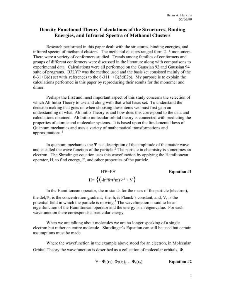
2 The particle in chemistry is sometimes an electron. The Shrodinger equation uses this wavefunction by applying the Hamiltonean operator, H, to find energy, E, and other properties of the particle.
H
Ψ
=E
Ψ
Equation #1
H=
{(
-h
2
/8
π
2 m)
▽
2
+ V
}
In the Hamiltonean operator, the m stands for the mass of the particle (electron), the del,
▽ , is the concentration gradient, the, h, is Planck’s constant, and, V, is the potential field in which the particle is moving.
3
The wavefunction is said to be an eigenfunction of the Hamiltonean operator and the energy is an eigenvalue. For each wavefunction there corresponds a particular energy.
When we are talking about molecules we are no longer speaking of a single electron but rather an entire molecule. Shrodinger’s Equation can still be used but certain assumptions must be made.
Where the wavefunction in the example above stood for an electron, in Molecular
Orbital Theory the wavefunction is described as a collection of molecular orbitals,
Ф
.
Ψ
=
Ф
1
(r
1
),
Ф
2
(r
2
),… Ф n
(r n
) Equation #2
1
Brian A. Harkins
05/06/99
The (r n
) designation stands for the position vectors of the electrons. The position vector for the nuclei of the molecules would also have to be included if not for the assumption that we can make because of the Born-Oppenheimer Approximation.
According to this approximation the velocities of the electrons move at such a greater speed than the nuclei that the of the nuclei appear to be fixed. For this reason we are allowed to use only the positions vectors of the electrons.
Two electrons that switch positions in a system should result in a sign change.
This is not the case in the Equation #2 . To compensate for this discrepancy spin states are applied to the molecular orbitals. There are two spin states for an electron, spin up,
α
(i)
, and spin down,
β
(i), where ,i, stand for the which electron. A pair of spin orbitals is prepared by applying both spin states to a molecular orbital,
Ф
1
(r
1
)
α
(1) and
Ф
1
(r
1
)
β
(1).
A matrix with n columns and n/2 rows, where n is the number of electrons, is created.
The determinant mixes all the possible orbitals of all of the electrons in the molecular system to form the wavefunction.
4
Next we say that the molecular orbital is defined as:
Ф
I
=
∑
C ui
X u
Equation #3 where the coefficients C ui are the molecular orbital expansion coefficients and the X u are the basis functions.
5
The u stands for which basis function the coefficient belongs to and the i stands for which molecular orbital it belongs to. This is what brings us to the Ab
Initio theory part of out discussion.
Different theories or methods take this information and process it to solve the wavefunction. One method squares the different coefficients and then takes their sum.
The sum of the squares is known as the density matrix. This density matrix is a very important part of the energy that is obtained from the operator. The values of the coefficients are not known going in but Gaussian allows for a guess of the values. Now the program tries to minimize the energy of the system that is obtained from the
Hamiltonean while at the same time minimizing the molecular coefficients. When this point is reached the system is optimized and properties such as frequencies, binding energies, bond enthalpies, and structures can be obtained. This method of Ab Initio theory is called the Hartree- Fock Theory (HF).
The Hartree- Fock theory is a very poor model. The reason is the way it uses the density matrix. In Hartree- Fock it takes into account the interaction of an electron and all its neighboring electrons. This is an acceptable approximation but is not a very accurate picture of what the surface is actually like. Other models have been developed that take the Hartree- Fock theory one step further and actually use functions of the electron density. It looks at the electron correlation of the molecule. In this process a grid is set up around the molecule and the program traces all around the molecule observing the electron density at each particular point. This method of calculations is called Density
Functional Theory (DFT) and is a much more accurate method than HF. The BeckeLYP model is an example of a DFT method.. The DFT is so effective that it actually
2
Brian A. Harkins
05/06/99 overshoots the experimental value. For this reasons methods have been created to compensate for this dilemma.
Since the development of DFT a number of hybrid models have been developed.
Examples are the B1LYP and the B3LYP. The B3LYP is the model used in the research paper. It incorporates both HF and the BLYP model. The hybrid can be thought of as a linear combination of both the HF and BLYP including a cross product of the two:
HYBRID = C
1
(HF) + C
2
(BLYP) + C
3
(HF)(BLYP)
A percentage, C, is assigned to each model to determine the weight from each that is used. The percentages came about based on data that was obtained using the combination of these two methods and than varying the percentages until they got good agreement with the experimental data. The number in the models, 1 and 3, correspond to how many of the percentages are fixed. In the B1LYP only the HF is fixed. In the
B3LYP all three are fixed. These models are quite accurate but there are other models just as accurate or more so.
Moller-Plesset perturbation theory is a model that is actually applied to the HF calculation and acquires great accuracy. Examples are the MP2 and MP4 models. This theory takes electrons that are in the ground state and puts them into higher excited orbitals. The number designates the number of excited states to use in the calculation.
This theory is a one time correction to the HF energy and are generally less-time consuming than the approaches that iteratively solve for the actual weights of the higher configurations in the total wave function.
6
This being true it is still rather time consuming compared to the other two models mentioned above. Graph #1 7 actually shows the comparison of time it takes for each model to perform a calculation of the molecule C
5
H
12
. Graph #1
Relative Times for the Calculations of C5H12
70
60
50
40
Relative Times (factors of
6.4 sec.)
30
20
10
0
3-21G
6-31G(d)
Basis Sets
6-31+G(d)
6-311++G(2d,p)
HF
MP2
B3LYP
Theories
HF
B3LYP
MP2
3
Brian A. Harkins
05/06/99
The values on the left-hand side are relative time factors. The job actually took 6.4 seconds and these are all factors of that time. Methods are listed on the right hand side.
Notice that the times increase greatly going from the HF method to the MP2 method.
Time has to be taken into account when deciding on what model to use for an experiment. Only cases where extreme accuracy is needed would a time consuming method be used. The labels along the bottom are the basis sets and they are described in the following section.
The basis sets are chosen along with the method to solve the wavefunction. They can be thought of as the molecular orbitals (s,p,d,…) that were discussed in
Equation #3 .
Larger basis sets mean longer calculations. Basis sets such as the ones listed in
Graph #1 can be broken down to determine the number of basis functions involved in the calculation. Take for example the basis sets that were discussed in the paper: 6-
31+G(d) and 6-311++G(3df,2p). The table below breaks down each of these basis sets and explains what each number, letter, and symbol refers to in the set. Total Basis
Functions can be found in the bottom row.
Table #1
Basis
Set
ORBITAL Basis
Set
ORBITAL
6 1s 6 1s
3 2s, 2p 3 2s, 2p
1
+
3s, 3p
4s, 4p
1
1
3s, 3p
4s, 4p d 3d +
+
5s, 5p
(non-hydrogen)
2s (hydrogen)
3df
2p
3d, 4d, 5d, 4f
(non-hydrogen)
3s, 3p, 4s, 4p
(hydrogen)
Total
Basis
Functions
19 Total
Basis
Functions
54
The ,+, in the basis set is like the normal s and p orbitals except it allows the orbitals to occupy more space. This is called a diffuse function and is good for systems with lone pairs as there are in methanol. The 6-31+G(d) has a total basis function value of
4
Brian A. Harkins
05/06/99
19. This means that there would be 19 basis functions used for each atom, not to mention the 19 coefficients that go along with those functions. That would mean a wavefunction consisting of 1,805 (19X19X5) molecular orbtials for methanol. The size and complexity of these calculations would be far to great to solve without the advent of modern computers with speeds and memory size capable of performing such calculations. For as
P.A.M. Dirac realized in 1929:
The underlying physical laws necessary for the mathematical theory of a large part of physics and the whole of chemistry are thus completely known, and the difficulty is only that the exact application of these laws leads to equations much too complicated to be soluble.
8
Referring back to Graph #1 it is clear to see that the basis set is a major contributor to the time it takes to perform a calculation of a molecule. Notice also that the larger basis set, 6-311++G(3df,2p), that the researchers compared their findings to is not even listed on the graph. It would be an even larger set and take an even greater amount of time. It is for these reasons that the researchers might have chosen to use the
B3LYP model along with the 6-31+G(d) basis set. It should be noted that these might have not been the original model and basis sets planned for use in the experiment, but rather the ones that were in most agreement with experimental findings and the larger basis set, 6-311++G(3df,2p).
Understanding the background involved in Ab Initio Molecular Theory brings us to the next step. That step is the explanation of the work done in the paper and how they came about their findings. I have taken two of the simplest molecules used in the paper to explain how Gaussian is used and how the data is collected in this paper. The two molecules are the methanol monomer and the methanol dimer cluster. I used the Gaussian
94 program with the B3LYP model and 6-31+G(d) basis set, as done in the paper.
The first calculation that I reproduced too much success was the binding energy of the dimer. The binding energy that they are referring to in the paper involves the hydrogen bonds between the hydrogen of the -OH group on one methanol with the oxygen of an –OH group on an adjacent methanol.
Figure #1
5
Brian A. Harkins
05/06/99
The dotted lines in Figure #1 shows the hydrogen bond. An important part of solving for these energies is feeding the coordinates of each atom into the Gaussian program. There are a number of ways to do this step. I have chosen to build the molecule in a program called Hyperchem. The steps are as follows:
I.
Build the molecule by double clicking on the uppermost left button that looks like a target. This brings up a menu with all the elements of the periodic table that are used to build the molecule. Begin by picking the element and then clicking on the screen. For my methanol monomer I chose the hydrogen atom and then clicked on the screen in four different places, one for each hydrogen. I then chose the carbon and clicked once on the screen and did the same for oxygen. By clicking on one atom on the screen and then dragging it to another you can form the bonds. Figure #2 is a snapshot of my monomer:
Figure #2
II.
Select the second button that resembles a bull’s eye and then by clicking on two or three atoms at a time, bond lengths or bond angles can be highlighted for the molecule. After the atoms have been highlighted, go to the menu bar at the top of the screen and click on Select. Scroll down to Set Bond Length or Set Bond
Angle and input the measurements. The OH bond length and C-O-H bond angle were 0.969 A and 109.2 respectively. These were the only two values I was given from the literature. These values are all I need because Gaussian will optimize the rest of the coordinates. Figure #3 shows the bond angle input:
Figure #3
6
Brian A. Harkins
05/06/99
III.
After all the bond lengths and bond angles are set, it is time to save the molecule.
In order for Gaussian to be able to read the file, the image must be saved as a
*.pdb file. Now start up Gaussian and under the Utilities selection choose
NewZMat. This will convert the *.pdb file into a *.gjf file that the Gaussian program can read. Once this is done go under File and click Open. Choose the gjf file and open it. Now all the coordinates are loaded. In the route section choose the appropriate model and basis set. In the experiment they used B3LYP and 6-31+G(d). The route section must also contain the operations you want it to perform. The researchers did calculations for energies, frequencies, and normal modes. For that reason I chose the command that would have Gaussian run a frequency and optimization job. Figure #4 is the input commands:
Figure #4
The Guess=Read command in the route section is there because I ran the monomer first with the 6-31G(d) basis set and then used those optimized conditions to run the 6-31+G(d) basis set. Without the diffuse function my binding energy deviated by 81% from the experimental value. With the diffuse function it deviated only by 14%. This demonstrates the importance of diffuse functions in molecules with lone pairs.
IV.
When Gaussian is finished running it creates an output file that contains all the information that is needed to calculate the properties discussed in the paper.
Only the frequencies and the normal modes of vibration can be viewed at this time. The binding energy cannot be calculated until the Gaussian runs the dimer.
In order to see the frequencies and the normal modes the output file is edited so that the program Gaussview can read it. Once the information is loaded the vibrations and the modes can be viewed, Figure #5 . I went through each of the
7
Brian A. Harkins
05/06/99 modes of vibrations until I found the one that corresponded to the OH stretch discussed in the literature.
Figure #5
The OH stretch frequency is shown in the lower right hand corner. The frequency is 3753.5 cm
–1
compared to the literature value that they found to be 3762.9 cm
–1
. The shift of the dimer frequency from the monomer frequency is what they compared to the experimental findings and the larger basis set. In order to do this I had to build and run the dimer as I had done with the monomer. Again I started in Hyperchem and built the dimer cluster. The procedure was as follows: i.
The molecules for the methanol dimer was built in the same way as shown in
Figure #2 , except that once I had built the monomer I made a copy of it. Going under Select in the top menu bar and then clicking on the Molecule option did this.
I then clicked on the second button on the left hand side that resembles a bull’s eye.
Clicking on the methanol monomer highlighted the entire molecule. I then went under Edit and clicked on Copy this produced another monomer exactly as the first.
Figure #6 shows the window that contains the two monomers:
8
Brian A. Harkins
05/06/99
Figure #6 ii.
Now that I had the two monomers it was time to set up the cluster to agree with the geometry that was given in the literature. The first part was to arrange them as the molecules appear in Figure #1. Clicking on one of the molecules highlights it. Then by clicking on the selection on the left hand side that resembles a bent paper clip you can rotate it. To move it left , right , up or down, choose the selection that looks like four arrows pointing outward. When the two monomers are in the correct arrangement, the hydrogen bond must be formed. Go under Build and click on Allow Ions. Then go back under Select and choose the Atom setting. Now simply click on the hydrogen of one monomer and the oxygen of the other and connect the two. The bond will be highlighted. Figure #7 shows an example:
Figure #7
9
Brian A. Harkins
05/06/99 iii.
The bond angle and lengths are entered the same as they were in Figure #3 .
The bond angle and lengths are given in the Table #2 .
Table #2 Methanol Dimer
MeOH
Unit
0-H
Bond
Length
C-O-H
Bond
Angle
O-O separation
O-H-O
Bond
Angle
1 0.969 A 109.4 deg
## ##
2 0.977 A 109.3 deg
2.862 174.2
The Values for the table come from Zwier*, Timothy S., et al, Journal of Physical Chemistry,
102, 82-84.
The O-O separation is the distance from one oxygen on a methanol to the
oxygen on the adjacent methanol. To do this the hydrogen bond
must be broke and in its place a oxygen- oxygen formed. Figure #8
shows the bond angle of the O-H-O bonds.
Figure #8 iv.
Now that all the coordinates have been set, it is time to feed it into Gaussian as we did in step III for the monomer. Figure #9 shows the Gaussian input command. Notice that beside the FOPT FREQ command we also show
10
Brian A. Harkins
05/06/99
Figure #9
{CalcFC} and Guess=Read. When I originally tried to run the dimer using just the
FOPT FREQ command the linked died. The reason is that when Gaussian is trying to minimize the geometric coordinates and the energy of the system, it is unable to locate the minimum because the interactions of the hydrogen bonds are so much softer than those of actual bonds. The result is a parabola that is extremely wide and Gaussian cannot predict the minimum, Graph #2 . For normal bonds the parabola is narrow and Gaussian simple walks down the graph till the minimum is found, Graph #3 .
Graph #2
Hydrogen Bond
Hydrogen Bond
Geometric Coordinates
11
Brian A. Harkins
05/06/99
Graph #3
Normal Bond
Normal Bond
Geometric Coordinates
The values in the charts are arbitrary. The graph is just to show what Gaussian is trying to work with and how in Graph #2 it can’t locate the minimum and ends up bouncing back and forth on the peak until the link dies. The {CalcFc} command compensates by telling it to calculate the Force Constants instead of guessing at them.
This would normally take a long amount of time but since I had already optimized my geometry it took only a few hours. v.
Gaussian again creates an output file for the dimer and it can be converted to a gjf file as was done in IV. Now it is ready to be ran in Gaussview. Figure #10 shows the frequency spectrum and structure for the dimer. The frequencies that I calculated were in good agreement with those from the literature and with the experimental. Going through each normal mode of vibration and finding the one that corresponded to the OH stretch identified the frequency. The vibrations were numbers 29 and 30 on Figure #10.
Figure #10
12
Brian A. Harkins
05/06/99
The frequencies that correspond to the vibrational modes are 3616.7 cm
-1
and 3765.5 cm
-1
, respectively. The literature does not compare the frequencies
directly with those of the experimental and larger basis set but rather the shifts of
the dimer frequencies from the monomer. In the case of my calculations, the OH
stretch of my monomer was 3753.5 cm
-1
. This means that the shifts from the
monomer's frequencies are –136.8 cm -1 and 12 cm -1 , respectively. These are
in good agreement with the experimental. In fact it is almost more accurate
than the findings in the literature. I have created a table at the end of the
discussion on my findings, Table #3
. The table lists my results, the researcher’s
results, as well as the experimental data.
Before continuing onto the binding energies, I should mention that the normal modes of vibration in the above section are the same as those that would be obtained by doing a complete symmetry analysis of the molecule(s). Gaussian performs these operations and the vibrations that I view are the different stretches and bending of the molecule(s). The table that is given in the book assigns a value to the size of the stretch of the one OH bond on a methanol unit in comparison to the OH bond on the adjacent methanol. The stretch value of one OH bond is seven times larger than that of the adjacent methanol in both vibrational modes.
9
Consequently, the same held true in my findings.
When Gaussian performed the calculations for the monomer and dimer it created an output file. In the previous section the file was edited and fed into Gaussview to get the frequencies. Binding energies can be found directly from the output file. The paper makes several comparisons of different energies and their significance against larger basis sets. As an example I chose only to use the “ Sum of electronic and zero-point Energies” from the output file and compare it to the ZPE corrected energies in the paper. In order to do this the energy from monomer must be subtracted from the dimer. Since the dimer is made up of two methanol units, the monomer must be multiplied by a factor of two. The energy from the monomer was –115.673892 hartrees and the energy from the dimer was –231.355378 hartrees. The difference must then be multiplied by a factor of 627.5095.
10
This is how many kcal/mol are in one hartree. The
ZPE corrected energy that I found was 4.76 kcal/mol. Applying the scaling factor 11 of
0.7863071 to the binding energy gave me 3.74 kcal/mol.
Table #3 Comparison of Frequencies, Frequency Shifts, and Binding Energies
My Calculations
B3LYP 6-31+G(d)
B3LYP
6-31+G(d)
12
B3LYP
6-311++G(3df,2p)
13
Experimental
Freq. Freq. Bind Freq. Freq. Bind Freq. Freq. Bind Freq Freq
14
Monomer
Unit 1
Shift
3753 0.0
Ener
#
Shift Ener
3762 0.0 # #
Shift Ener
0.0 # #
Shift
0.0
3616 -137 # 3612 -150 # # -148 # # -170 # Dimer
Unit 1
Dimer
Unit 2
3765 12 -4.76
-3.74
3758 -4.3 -4.82
-3.79
# -3 -3.55
-3.08
# +3
Bind
15
Ener
#
3.2
13
Brian A. Harkins
05/06/99
Table #3 shows the data I obtained from my calculations, the literature’s data, those of the larger basis set, and the experimental. All frequencies are in reciprocating centimeters and all binding energies are in kcal/mol. All my calculations were very close to those obtained in the literature. In fact my value for the binding energy was one percent closer to the experimental than the literatures. The importance of this comparison is that although the larger basis set produces values closer to the experimental, the smaller basis set is close enough that it is an acceptable model as long as great accuracy is not required.
The calculations that I performed above could be applied to any one of the different methanol clusters in the paper. The only difference would be the amount of time the program would take to complete the task and the amount of memory needed to store all the results. What is the importance of being able to do these calculations and obtain this data? The researchers in this paper use the findings from the different clusters to show trends and relationships between their sets of data. The data, if correct, should show agreement between structural data, binding energies, and infrared spectra.
One example is when the researchers took the binding energies that they had calculated for cyclic methanol clusters and compared them to the structural data. It appeared that as the ring increased (trimer- pentamer) the binding energies did so as well.
Structural data indicated that steric interference due to the hydrogens caused the O-H-O bond angles to approach 180 degrees and the oxygen-oxygen distance to decrease greatly from the trimer to the pentamer.
16 The two trends are in excellent agreement. As the distance of a bond decreases (to an extent) and the rigidity of the molecule increases, the strength of that bond increases. Take for an example a single bond compared to a double bond (ethane vs. ethene). The sigma bond is longer than the double bond and is free to rotate. The double bond is stuck in place due to the pi bonds. The amount of energy needed to break that double bond is extremely higher than the energy needed to break the single bond.
Another example from the paper is that, even though the average H bond energy in the chain conformer is higher than that of the cyclic with the same amount of methanols, the cyclic geometry is favored. The increased cooperativity of the extra H bond stabilizes the cyclic form so much that it is more stable than the chain. The result is a higher total binding energy for the cyclic form than the chain. The total binding energies from the paper show a higher value for the cyclic conformer than the chain.
Frequency comparisons can be made directly from experimental spectra. The researchers found that the results collected for the different calculations were in agreement with each other and with experimental data.
17
In this paper I explained Ab Initio Theory, how the calculations could have been obtained, and briefly how the results are used to validate the whole of the data. My purpose, along with creating a clearer understanding of the processes involved in this type of work, was to bestow upon the reader a greater appreciation of the time, patience, and complexity of this type of work.
14
Brian A. Harkins
05/06/99
R e e f f f e e r r r e e n c c e e s s s
1) Foresman, James B.; Frisch A.E.; Exploring Chemistry with Electronic Structure
Methods ; Gaussian Inc.: Pittsburgh, 1993, 253.
2) McQuarrie, Donald A.; Quantum Chemistry ; University Science Books.: 1983, 79.
3) Foresman and Frisch, 253.
4) Foresman and Frisch, 260.
5) Foresman and Frisch, 261.
6) Foresman, James B.; Ab Initio Techniques in Chemistry: Interpretation and
Visualization ; American Chemical Society.: 1997, 249.
7) Foresman and Frisch, 123.
8) Foresman and Frisch, 258
9) Hagemeister, Frederick C.; Gruenloh, Christopher J.; Zwier, Timothy S . J. Phys.
Chem. 1998, 102 , 89.
10) Foresman and Frisch.
11) Boys, S. F.; Bernardi, F. Mol. Phys . 1970 , 19 , 553.
12) Hagemeister, Gruenloh, and Zwier, 90-91.
13) Mo, O.; Yanez, M.; Elguero, J. J. Chem. Phys . 1997 , 107 , 3592- 3601.
14) Huisken, F.; Kulcke, A.; Laush, C.; Lisy, J. M. J. Chem. Phys . 1991 , 95 , 3924-29.
15) Bizzari, A.; Stolte, S.; Reuss, J.; Rijdt, J. G. C. M. V. D.-V.D.; Duijneveldt, F.B.V.
Chem. Phys . 1990 , 143, 423-35.
16) Hagemeister, Gruenloh, and Zwier, 86.
17) Hagemeister, Gruenloh, and Zwier, 92.
15


