IP大會病理切片討論個案簡短病史 湯子殷
advertisement

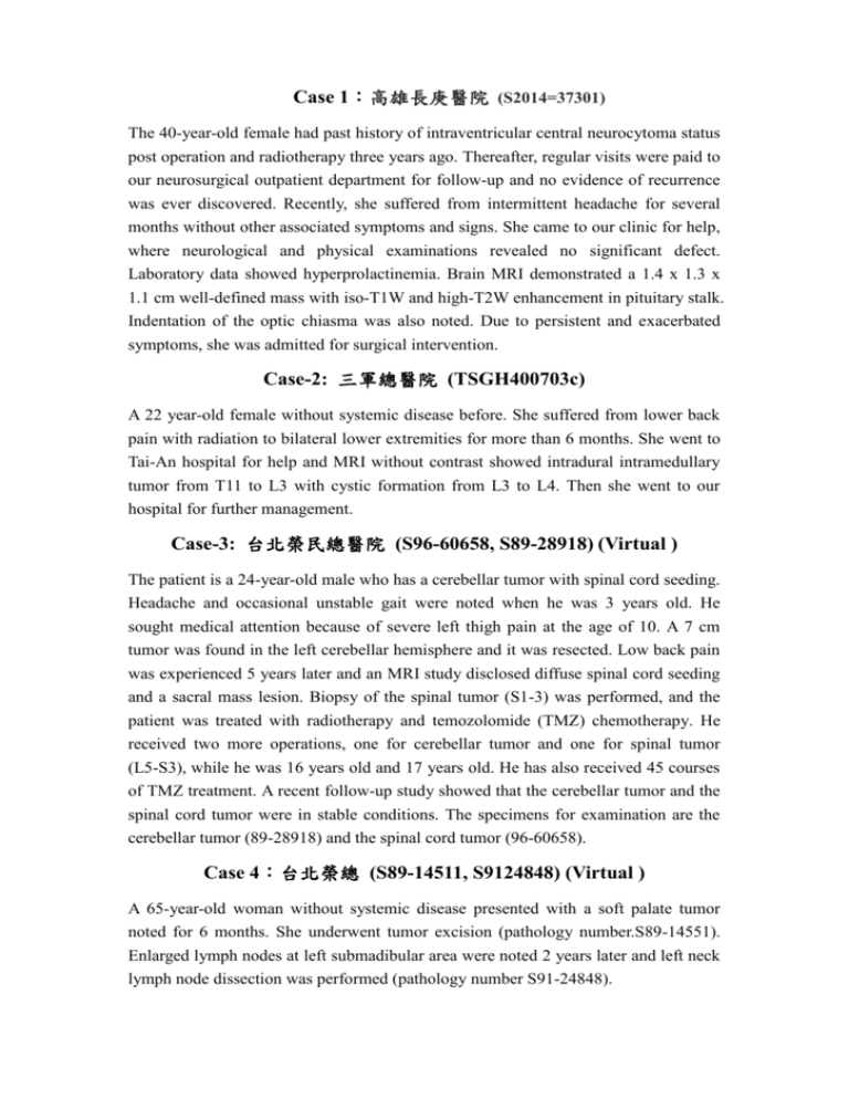
Case 1:高雄長庚醫院 (S2014=37301) The 40-year-old female had past history of intraventricular central neurocytoma status post operation and radiotherapy three years ago. Thereafter, regular visits were paid to our neurosurgical outpatient department for follow-up and no evidence of recurrence was ever discovered. Recently, she suffered from intermittent headache for several months without other associated symptoms and signs. She came to our clinic for help, where neurological and physical examinations revealed no significant defect. Laboratory data showed hyperprolactinemia. Brain MRI demonstrated a 1.4 x 1.3 x 1.1 cm well-defined mass with iso-T1W and high-T2W enhancement in pituitary stalk. Indentation of the optic chiasma was also noted. Due to persistent and exacerbated symptoms, she was admitted for surgical intervention. Case-2: 三軍總醫院 (TSGH400703c) A 22 year-old female without systemic disease before. She suffered from lower back pain with radiation to bilateral lower extremities for more than 6 months. She went to Tai-An hospital for help and MRI without contrast showed intradural intramedullary tumor from T11 to L3 with cystic formation from L3 to L4. Then she went to our hospital for further management. Case-3: 台北榮民總醫院 (S96-60658, S89-28918) (Virtual ) The patient is a 24-year-old male who has a cerebellar tumor with spinal cord seeding. Headache and occasional unstable gait were noted when he was 3 years old. He sought medical attention because of severe left thigh pain at the age of 10. A 7 cm tumor was found in the left cerebellar hemisphere and it was resected. Low back pain was experienced 5 years later and an MRI study disclosed diffuse spinal cord seeding and a sacral mass lesion. Biopsy of the spinal tumor (S1-3) was performed, and the patient was treated with radiotherapy and temozolomide (TMZ) chemotherapy. He received two more operations, one for cerebellar tumor and one for spinal tumor (L5-S3), while he was 16 years old and 17 years old. He has also received 45 courses of TMZ treatment. A recent follow-up study showed that the cerebellar tumor and the spinal cord tumor were in stable conditions. The specimens for examination are the cerebellar tumor (89-28918) and the spinal cord tumor (96-60658). Case 4:台北榮總 (S89-14511, S9124848) (Virtual ) A 65-year-old woman without systemic disease presented with a soft palate tumor noted for 6 months. She underwent tumor excision (pathology number.S89-14551). Enlarged lymph nodes at left submadibular area were noted 2 years later and left neck lymph node dissection was performed (pathology number S91-24848). Case 5: 彰化基督教醫院 (圖片) This 46-year-old woman is a housewife without previously known illness. She presented with recurrent epistaxis 5-6 times for weeks in 2011. Then she visited our ENT OPD and nasopharyngoscopic exam revealed reddish mass from right osteomeatal complex with blood clot. Initial biopsy revealed spindle cell lesion. After FESS removal of the tumor, she has regular follow up and tumor recurrent is noted 3 years later. This time, the symptoms are nasal obstruction with epistaxis off and on. Sinoscopy reveals a mass on right septum. MRI revealed right-sided nasal septum lesion. Biopsy revealed tumor recurrent and further excision is performed. Case 6: 台中榮總 (S0200495E) A 44-year-old male patient complained of left thigh swelling mass for 2 months. He went to our clinics and MRI shows a 17x10 cm heterogeneous with high signal for both T1 and T2 view mass at left thigh medial aspect. Under the impression of sarcoma, he was admitted for tumor excision. Case 7: 台中榮總 (S0321747) This 42-year-old has a soft tissue mass on right popliteal fossa for several years. MRI shows a well-enhanced subcutaneous soft tissue mass at popliteal fossa, 13x24x25 mm in size. The patient undergoes tumor wide excision. Grossly, the tumor is yellow-white and well demarcated. Case 8:高雄長庚醫院 (103-170-3) This is a 19-year-old man who denied any systemic disease and past medical history. He found a painless mass over right buttock 5 years ago. The mass was movable with mildly soft consistency, and the size progressively enlarged. No other obvious symptoms were noted. He went to orthopaedic OPD. The MRI image study revealed a well-defined lobulated tumor mass measuring 6.1 x 6.0 x 3.6 cm with heterogeneous enhancement, in the right buttock, between the subcutis and gluteus maximus. No destructive bony lesion of pelvic bones is found. He was then admitted, and received tumor excision surgery. Case-9: 高雄醫學大學附設醫院 (KMU-14-11272) A 29-year-old female patient was well-being before and denied any systemic disease. She complained of epigastralgia for two weeks. She had no symptoms of vomiting, diarrhea, fever or body weight loss. No jaundice or peripheral lymphadenopathy was noted on physical examination. Computed tomography of abdomen demonstrated a 10cm subhepatic peripancreatic lesion with hypovascularity, hemorrhage and cystic change. CT-guided biopsy was performed and then she underwent tumor excision after diagnostic biopsy. Case 10:高雄榮民總醫院 (VGHKS PATH 01-34256) (Virtual) This male neonate was born via caesarean section at 36+5 weeks gestation due to suspected congenital intestinal obstruction on antenatal ultrasound examination. At six days after birth, frequent watery diarrhea up to 10-15 times per day was documented. The diarrhea persisted even after fasting. Severe dehydration and metabolic acidosis were noted thereafter. Barium study of UGI and small intestine suggested the possibility of congenital intestinal lymphangiectasia. Endoscopic biopsy was then obtained at duodenum for evaluation. Case 11: 三軍總醫院 A 35-year-old women presents with right flank pain and gross hematuria. The abdominal CT revealed one well-defined cystic mass measuring 5 x 4.5 cm in the middle and lower pole of right kidney. Under the impression of rupture of arterio-venous malformation, she underwent embolization two times (103/07/16 and 103/08/17). Due the persistent symptoms and acute urine retention, the radical nephrectomy was performed. Case 12:和信醫院 (S12-44150) A 33 year-old man experienced painless gross hematuria since 2012.11. A left renal tumor measuring 6 cm was detected by abdominal CT. No metastasis was found clinically. He received retroperitoneal hand-assisted laparoscopic radical nephrectomy at NTUH on 2012.11.21. Tumor cells were positive for PAX8, CK(AE1/AE3) and negative for CK7 and CD10. Other stains [p504S, HMB45, CD117, CK(34BE12)] were equivocal due to strong background staining. An unclassified renal cell carcinoma was diagnosed. Case 13:臺大醫院 (S1420675C) This 43-year-old white man had fever, arthralgia, dry cough, generalized malaise and weight loss (3 kilograms in two months) for three months. He visited our hospital in April, 2014. Laboratory data showed leukocytosis with left shift, eosinophilia, elevated ESR and CRP, hyponatremia, hypoalbuminemia and microcytic anemia. Gallium scan with SPECT showed generalized lymphadenopathy. Under the impression of fever, weight loss, and generalized lymphadenopathy, lymph node biopsy was performed. Case 14: 成大醫院 (14-20437) A 62-year-old male presented with left anterior auricular mass since February 2014, followed by progressive enlargement of bilateral anterior and posterior auricular masses for two weeks. Other manifestations included pain, local heat and erythema of the overlying skin, and body weight loss of 2Kg in recent one month. He went to our emergency room and was treated as lymphadenitis. A nasopharyngeal biopsy followed by lymph node excision was performed. Case 15:亞東醫院 (S2014-07787) The patient is a 46 year old woman with a past history of asthma. She complained tarry stool for one month. Associated symptom includes general weakness, nausea, vomiting, body weight loss (from 50 kilograms to 46 kilograms) and pitting edema of bilateral legs. She visited our emergency department where ileus and anemia (hemoglobin 6.4g/dl) were noted. The chest plain film revealed a mass shadow in the right lower lobe. The abdominal and chest computed tomography showed a 12.4 x 9.5 x 8.3 cm polypoid tumor in the jejunum with adhesion to sigmoid colon, a 7.5 x 7.5 x 6.4 cm tumor at right lower lobe of lung, and a 3 cm nodule inside the muscular layer of right upper abdominal wall. Under the impression of jejunal cancer with metastasis, she underwent segmental resection of jejunum. During operation, a 12 cm tumor located at upper jejunum, 40 cm distal to Treitz ligament, adhered to sigmoid colon, and a 3 cm tumor at right upper abdominal wall were noted. Case 16: 奇美醫院 (2006-10-6543A) A 27-year-old male presented with two tumors of the skin, one in the right lower leg and the other in the left thigh. Both were biopsied. Tracing back his history, he was diagnosed with cutaneous anaplastic large cell lymphoma over the skin of his left calf 6 months ago at other hospital and was treated there with CHOP and local radiation. After the diagnosis of relapsed lymphoma in our hospital, he was treated with various chemotherapy regimens but the disease progressed with bone marrow involvement and leukemic change in 3 months. Case 17:馬偕紀念醫院 (S14-8538) A 37 year-old male with DM and HTN had a protruding mass over right upper lip for a month. It measured 2 cm in greatest dimension. He denied symptoms such as fever, body weight loss, and night sweating. He sought help at local clinic, where biopsy was done. And due to persistent oozing of the wound, he was referred plastic surgeon. Cytology Case A: 和信治癌中心醫院 The 45 years old male patient found a palpable lump over subareolar area of right breast for half a year. The lump was relatively well-demarcated and painless but persistent with size slightly increased. The sonography showed an ovoid iso- to hypo-echoic nodule about 1.3 cm at right nipple region. Then, fine needle aspiration was done. Cytology Case B: 台北榮民總醫院 (C103-27506) The 49-year-old female presented with a palpable left breast mass. Physical and mammogram exams showed a 25 mm, freely movable, lobulated mass over left outer upper quadrant breast and fibroadenoma was suspected. Sonography showed a heterogeneous echogenic mass lesion measured 3.4 x 2.4 cm at left breast 11/5. Under the impression of malignancy, fine needle aspiration was performed. Cytology Case C: 台北榮民總醫院 The 63-year-old male has been diagnosed with rectal adenocarcinoma and finished the course of neoadjuvant concurrent chemoradiotherapy. A pre-operative CT revealed a 2.3 x 2.1 cm lesion (asterisk) in the sacral region. Under the impression of adenocarcinoma with bone metastasis, aspiration was performed.


