common-surgeries
advertisement

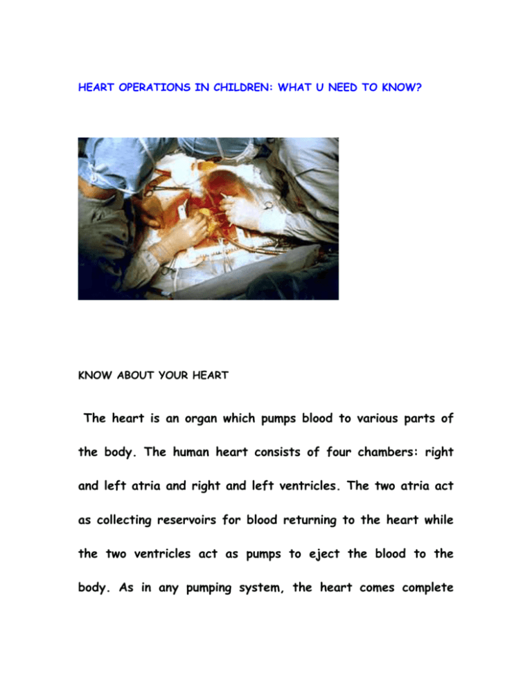
HEART OPERATIONS IN CHILDREN: WHAT U NEED TO KNOW? KNOW ABOUT YOUR HEART The heart is an organ which pumps blood to various parts of the body. The human heart consists of four chambers: right and left atria and right and left ventricles. The two atria act as collecting reservoirs for blood returning to the heart while the two ventricles act as pumps to eject the blood to the body. As in any pumping system, the heart comes complete with valves to prevent the back flow of blood. Deoxygenated blood returns to the heart via the major veins (superior and inferior vena cava), enters the right atrium, passes into the right ventricle, and from there is ejected to the pulmonary artery on the way to the lungs. Oxygenated blood returning from the lungs enters the left atrium via the pulmonary veins, passes into the left ventricle, and is then ejected to the aorta. . WHAT IS CARDIAC SURGERY? Cardiac surgery is a surgery on the heart and/or great vessels performed by a cardiac surgeon. Heart surgery is done for a variety of reasons and ranges from minimally invasive procedures to actually removing the heart and replacing it with a donor heart. Frequently, it is done to treat complications of heart. Types of Heart Surgeries The common procedures which are performed on the heart can be grouped into several categories : -Closures -Shunts -Atrial Baffles -Relief of Stenosis -PA Banding -Conduits -Relief of Cyanosis Type Procedure Defects Treated Reduced pulmonary blood Description Classic: Anastomosis of BlalockShunts Taussig flow (TOF, TGA, pulmonary subclavian artery to PA (with atresia, tricuspid atresia) or without modification) TGA (early palliation), Atrial Surgical atrial mixing septostomy Also called Blalock-Hanlon mitral atresia, complex procedure congenital heart disease Percutaneous atrial Balloon atrial TGA, tricuspid atresia septostomy, also called septostomy Rashkind balloon procedure Closures Atrial septal ASD with significant shunt Primary or patch closure defect (ASD) Percutaneous closure closure Ventricular Isolated VSD or with other septal defect (VSD) closure Primary or patch closure anomalies (TOF) Patent ductus arteriosus Ligation ± division of PDA Patent ductus arteriosus Transcatheter technique (PDA) ligation Closure of ASD and VSD, Endocardial Endocardial cushion defect repair of atrioventricular valve cushion defect (also called atrioventricular abnormalities (e.g., cleft repair canal defect) mitral leaflet) Surgical PA reduction in Large left-to-right shunt PA band to decrease PA flow banding flow area of lesions and pressure TGA (replaced by arterial Dacron or pericardial baffle PA Atrial Mustard baffles switch procedures at many directs systemic venous return centers) to PA via anatomic LV, pulmonary venous return to aorta via anatomic RV TGA (replaced by arterial RA free wall and interatrial switch procedures at many septal tissue used for Senning centers). May be used as interatrial baffle similar to a part of “double switch” Mustard repair. procedure for L-TGA Various procedures including Aortic end-to-end anastomosis, patch Relief of coarctation Aortic coarctation enlargement, Gore-Tex graft; stenosis repair balloon dilation for recoarctation Pulmonic TOF, pulmonic stenosis Brock trans-RV approach valvotomy Direct surgical repair Balloon dilation Aortic Direct surgical valvotomy or Congenital aortic stenosis valvotomy percutaneous balloon dilation Surgical commissurotomy— Mitral repair Congenital mitral stenosis initially a “closed” procedure without cardiopulmonary Open heart surgery Open heart surgery is any surgery where the chest is opened and surgery is performed on the heart muscle, valves, arteries, or other heart structures (such as the aorta). The term "open" means that the chest is "cut" open . Most heart surgery is performed under general anesthesia, requiring that the patient be intubated and put on a ventilator(artificial breathing machine). Some less invasive procedures, such as placing stents or a pacemaker, may be performed with monitored anesthesia care, known as twilight sleep. CARDIOPULMONARY BYPASS A heart-lung machine (also called cardiopulmonary bypass) is usually used during open heart surgery. While the surgeon works on the heart, the machine helps provide oxygen-rich blood to the brain and other vital organs. HOW THE CHEST IS OPENED DURING CARDIAC SURGERY? The heart surgeon will make a 2-inch to 5-inch-long surgical cut in the chest wall. Muscles in the area will be divided so the surgeon can reach the heart. The surgeon can repair or replace a valve or perform bypass surgery. During endoscopic surgery, the surgeon makes one to four small holes in the chest. Then he uses special instruments and a camera to perform the surgery. During robot-assisted valve surgery, the surgeon makes two to four tiny cuts (about 1/2 inch to 3/4 inch) in the chest. The surgeon uses a special computer to control robotic arms during the surgery. The surgeon sees a three-dimensional view of the surgery on the computer. This method is very precise. Closed Heart Surgery Closed heart surgery implies that the "heart lung machine" or "bypass" machine is not used and the heart is visualized but not cut open. Listed below are details of three types of closed heart surgery: Closed Heart Surgery Patent Ductus Arteriosus Coarctation of the Aorta Blalock-Taussig Shunt Patent Ductus Arteriosus (PDA) PDA refers to an open vessel that allows blood to flow between the aorta and the pulmonary artery. The ductus arteriosus is open during fetal life to divert blood away from the unused lungs. Normally the ductus closes within the first day of life, but for unknown reasons it sometimes remains open. This occurrence is more common in premature infants. If the PDA is small, there may be no symptoms at all. Symptoms of a large PDA are rapid breathing, fatigue, and slow weight gain. After surgical correction, these symptoms will disappear. The surgery involves a left thoracotomy incision. The vessel is "ligated" and divided in half or clipped so that there will be no flow. This is a curative operation; no other surgery is required. WHEN IS PDA LIGATED? Usually PDA is closed without surgery by device closure. There are only 3 scenarios where PDA is operated: 1.Premature babies with large PDA and developing heart failure 2. PDA with infective endocarditis 3. Large PDA in babies <5kg causing recurrent chest infections or poor weight gain Coarctation of the Aorta Coarctation of the Aorta is a obstruction/narrowing of the aorta. It may present itself as early as birth or in late childhood. The signs are usually high blood pressure, or a higher blood pressure in the arms than in the legs. Older children sometimes complain of leg cramps. Surgery to correct this will equalize the blood pressure in the upper and lower extremities. The surgery involves opening the chest through a left thoracotomy incision, removing the narrowed portion of the aorta, and reattaching the two ends of the aorta together. This is also a curative operation. Blalock-Taussig Shunt A Blalock-Taussig Shunt or "BT Shunt" is used to help increase blood flow to the lungs in babies born with defects that obstruct blood flow to the lungs. The surgery entails opening the chest either through a left or a right thoracotomy approach and placing a Gore-Tex tube form the innominate artery to the pulmonary artery. This is a palliative procedure, meaning that in most cases the final repair will be done at a later date. Can a child lead normal life after cardiac surgery? In general, problems that can be corrected without the use of heart-lung bypass support may involve a shorter hospitalization and recovery time. Clearly, the length of recovery will depend partly on potential complications that may arise and partly on the health of the patient before surgery. A 6-8 week recovery period is not uncommon. Nutrition is a critical component of this recovery Postoperative period child as well. in ICU After surgery, most infants can be fed enterally (in the gut) after a day or two. But even when the child is not being fed formula or milk, nutrition is being delivered in an intravenous form. In more limited situations, simple "IV" fluids containing sugar-water will suffice. At other times, the IV nutrition can replace all the sugars, proteins and fats that the patient needs. That complex form of IV nutrition is called TPN (Total Parenteral Nutrition). Some babies can take a while to recover after surgery until they can be fed by mouth. This partly depends on how the child was feeding before surgery and whether there are any medical reasons affecting the ability of the gut to work. It is not unusual for even some kids who have been feeding normally before surgery to have a setback. They might require some sort of extra nutritional support. Nutrition is critical in the healing process. At times we place tubes, called feeding tubes, into the stomach (through the mouth or the nose) to make sure your child receives adequate calories to heal properly. Often children are discharged home on some medications. Typically these include diuretics (water-pills) and sometimes other heart medications. The dosage of these medications will be adjusted when you follow up with your surgeon and cardiologist. Most patients are seen within a week to two weeks after discharge. We will provide you with a set of instructions before your discharge to guide you on your child's medicines and postoperative care. We will teach you how to assess the wounds and what problems to look for. Sick people should not visit for the first few days. Good and frequent hand washing is critical, especially before examining the wound. The wound should be kept clean and dry for the first couple of weeks. Generally avoid immunization within the first 6-8 weeks after surgery. Finally, as with any surgical incision, a rest period helps ensure good wound healing. There will be a period of time when activity will be somewhat restricted to help with healing.


