Regulation of Respiration
advertisement
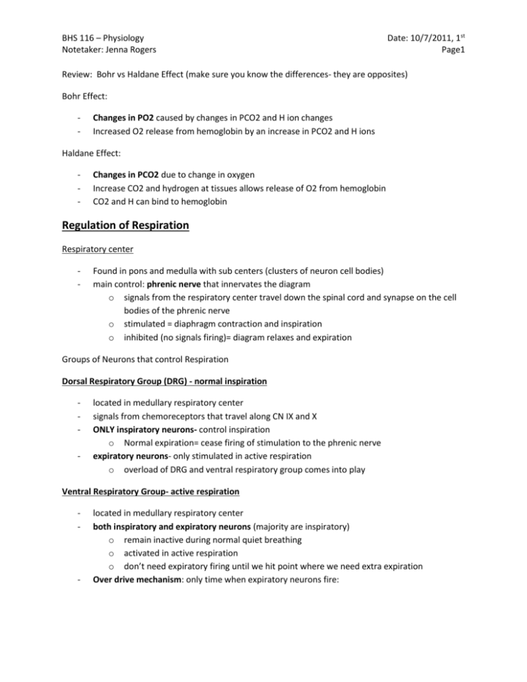
BHS 116 – Physiology Notetaker: Jenna Rogers Date: 10/7/2011, 1st Page1 Review: Bohr vs Haldane Effect (make sure you know the differences- they are opposites) Bohr Effect: - Changes in PO2 caused by changes in PCO2 and H ion changes Increased O2 release from hemoglobin by an increase in PCO2 and H ions Haldane Effect: - Changes in PCO2 due to change in oxygen Increase CO2 and hydrogen at tissues allows release of O2 from hemoglobin CO2 and H can bind to hemoglobin Regulation of Respiration Respiratory center - Found in pons and medulla with sub centers (clusters of neuron cell bodies) main control: phrenic nerve that innervates the diagram o signals from the respiratory center travel down the spinal cord and synapse on the cell bodies of the phrenic nerve o stimulated = diaphragm contraction and inspiration o inhibited (no signals firing)= diagram relaxes and expiration Groups of Neurons that control Respiration Dorsal Respiratory Group (DRG) - normal inspiration - located in medullary respiratory center signals from chemoreceptors that travel along CN IX and X ONLY inspiratory neurons- control inspiration o Normal expiration= cease firing of stimulation to the phrenic nerve expiratory neurons- only stimulated in active respiration o overload of DRG and ventral respiratory group comes into play Ventral Respiratory Group- active respiration - - located in medullary respiratory center both inspiratory and expiratory neurons (majority are inspiratory) o remain inactive during normal quiet breathing o activated in active respiration o don’t need expiratory firing until we hit point where we need extra expiration Over drive mechanism: only time when expiratory neurons fire: BHS 116 – Physiology Notetaker: Jenna Rogers o Date: 10/7/2011, 1st Page2 when there is increased pulmonary ventilation and we are breathing faster and harder, there is signal spillover from DRG and this stimulates Ventral group neurons causing inspiration and to a lesser extent expiration 2 Centers in the Respiratory Center - - Pneumotaxic Center- brake on DRG firing to allow relaxation to occur o In the pons respiratory center o Modulator of DRG o Sends signals to DRG that help switch off their firing o Limit the duration of inspiration- a check and balance system Prevents constant breathing in with no relaxation and expiration o Strong pneumotaxic signals= inspiration doesn’t last very long b/c the DRG neurons are constantly being inhibited by the pneumotaxic center Short inspiratory signals o Weak pneumotaxic signals= inspiration lasts longer b/c there is no brake o Role: increase the rate of inspiration by limiting the length of inspiration Apneustic Center o In pons o Inhibits the switching off of the DRG o Acts against Pneumotaxic o Encourages prolonged inspirations o Normal: pneumotaxic is more dominant, inspiration is limited o Nonfunctional pneumotaxic= prolonged inspirations with short bursts of expiration (apneusis) There are other receptors to trigger expiration Pre- Botzinger Complex- the SA note of respiration o o Sets the pace for respiratory rhythm Interacts with DRG Hering- Breuer Reflex - - Stretch receptors in the muscles of the walls of the bronchi and bronchioles send sensory nerve signals via the vagus nerve to the DRG Pulmonary stretch receptors sense and increased volume in the bronchioles o When apneustic center takes over and there is prolonged inspiration, the smooth muscles in the bronchi and bronchioles sense a stretch and sends a message to the DRG to switch off the inspiratory neurons Internal control in lungs and respiratory tract for expiration to occur when apneustic takes over BHS 116 – Physiology Notetaker: Jenna Rogers Date: 10/7/2011, 1st Page3 Peripheral Chemoreceptor Systems- control inspiration based on PO2 and PCO2 - - - - Found in same spots as baroreceptors Detect changes in the arterial blood PO2 o Not active until P02 drops below 60 mm Hg Simulate increase in ventilation o Nerve impulses really increase when PO2 drops below 60 mm Hg o Increased impulses sent back to respiratory center resulting in increased respiration Sense H ion concentration- Pathological high acid in the blood Weakly respond to increased PCO2 Only in emergency situations- in an area when there is a lot less oxygen or if we are climbing a mountain (atmospheric PO2 drops, alveolar PO2 drops, arteriole PO2 drops) Effects on alveolar ventilation o Only a dramatic increase of respiratory rate when we get below a PO2 of 60 o Need more O2 in b/c there is less in the alveoli to exchange with the blood. Need to increase respiratory rate to bring more O2 in Hemoglobin dissociation o PO2 below 60= hemoglobin is giving up O2 at a much faster pace o Need to increase ventilation to fill up the empty hemoglobin spots with O2 Summary: o Greatest effect at PO2 below 60 o No peripheral regulation- low PO2 would be inhibitory to the medullary respiratory center, decreasing ventilation o With peripheral Regulation- when it does get low, they send signals to increase ventilation to bring arteriole PO2 back up to normal levels o Receptors only respond to amount of O2 physically dissolved in blood All of O2 bound to hemoglobin is not sensed by the chemoreceptors Where peripheral chemoreceptors fail: Carbon monoxide- dissociates O2 from hemoglobin but chemoreceptors don’t sense the loss of O2 Anemia- decreased red cells are not sensed so there is still not much O2 being delivered Central Chemoreceptors - located in the medulla (very close to the DRG) sensitive to CO2 induced H ion changes in the brain don’t respond directly to CO2- only respond to H ions CO2= Trojan horse that brings the H ions across the blood brain barrier (H can’t cross barrier) o CO2 will bind with water to form carbonic acid which dissociates into bicarbonate and H o H binds to the central chemoreceptors o Signal sent to DRG BHS 116 – Physiology Notetaker: Jenna Rogers - - Date: 10/7/2011, 1st Page4 Elevated PCO2= increase in ventilation to blow off CO2 Decrease in PCO2= decrease in ventilation to decrease the amount of CO2 blown off to increase the amount that stays in the blood PCO2 is the primary driver of respiration rate until PO2 drops below 60 Why we can’t hold our breath forever o Central chemoreceptors sense that there is a PCO2 increase in the tissues because we are not blowing off the CO2 and it sends a signal to force expiration Desensitize o Patented short term regulators o Extended elevated PCO2- they don’t respond as easily anymore o Ex. Chronic lung disease Early disease- central chemo receptors work Later- desensitized and cannot respond as efficiently Pathological high acid in the blood- increase respiration - Hydrogen Ions from lactic acid or ketoacidosis (diabetes) Drive H concentration up and increases acidity of blood o stimulates the peripheral chemoreceptors to trigger medullary respiratory center to increase respiration to blow off more CO2 o Indirectly reducing the acidity in the blood by getting rid of CO2 Amount of H added to the blood from CO2 is adjusted Acclimatization - - When we climb a mountain and become acclimated to the altered O2 concentration at these altitudes Ascend too quickly= mountain sickness o Low PO2 – atmospheric P is low o low oxygenated hemoglobin- releasing oxygen more easily at PO2 below 60 o Decrease PCO2 levels Peripheral chemoreceptors sense low PO2 and increase ventilation which blows off more CO2 Slow ascension- allow acclimation- needs TIME (2-3 days) o No mountain sickness b/c: o Central chemoreceptors drive respiration normally but then peripheral chemoreceptors take over at high altitudes. We are giving peripheral chemoreceptors time to kick in o Erythropoietin production increases- increase # of RBC and increase binding capacity of blood. Takes a few days o RBC produce more BPG which stimulates O2 release from the hemoglobin easing the delivery of O2 to the tissue BHS 116 – Physiology Notetaker: Jenna Rogers Date: 10/7/2011, 1st Page5 Exercise - - O2 consumption o Moderate exercise increases O2 consumption and increase ventilation 4-8 fold o Severe exercise increases ventilation dramatically Body needs to adjust ventilation rate to bring in enough oxygen for muscles Regulation of respiration o No change in arterial PCO2 pH or PO2- not driving ventilation in exercise o The major drivers of ventilation during exercise Brain transmits impulses to muscles and respiratory center- we know we are exercising Body movements excite joint and muscle proprioceptors which send excitatory impulses to the respiratory center Increased body temperature Epinephrine release from adrenal glands Voluntary control of respiration - - - Hyperventilate- over breath or deep breaths to bring in more air o Take in more O2 and blow off more CO2 o Max amount brought in is 6000 ml/min Hypoventilation- take smaller or no breaths o PCO2 increases- stimulates central chemoreceptors and override voluntary control to force expiration o PO2 decreases Alter PO2 and PCO2 levels and changes pH Not mediated through respiratory center o Contraction and relaxation of diagram signals from the cortex- conscious centers Other involuntary controls - - Airway irritant receptors- triggers cough o Sensory nerve endings in epithelium of airway o Stimulated by irritants and goes directly to DRG to stimulate coughing and sneezing Lung J Receptors o J= in the lung in the alveolar wall in close juxtaposition to the pulmonary capillaries o Stimulated by Increase in BP in lungs engorgement of capillaries with blood causes capillaries to touch up against receptors to trigger them o Stimulated by edema Extra fluid places pressure on the receptors BHS 116 – Physiology Notetaker: Jenna Rogers o - - Gives the feeling of not being able to catch your breath Feeling of breathlessness= dyspnea Brain Edema o Increase fluid p in brain directly inactivates respiratory center o Fluid buildup causes pressure in brain and can alter the signals from the DRG to the diaphragm Anesthesia- depresses respiratory center o Why you have to be ventilated Clicker Question Peripheral Chemoreceptors most strongly respond to 1. 2. 3. 4. Date: 10/7/2011, 1st Page6 Decreased arterial PO2 Decreased arterial PCO2 Increased arterial PO2 Increased arterial PCO2 Ans: Decreased arterial PO2
