Laboratory Exercise 15: Anatomy of the Blood Vessels
advertisement
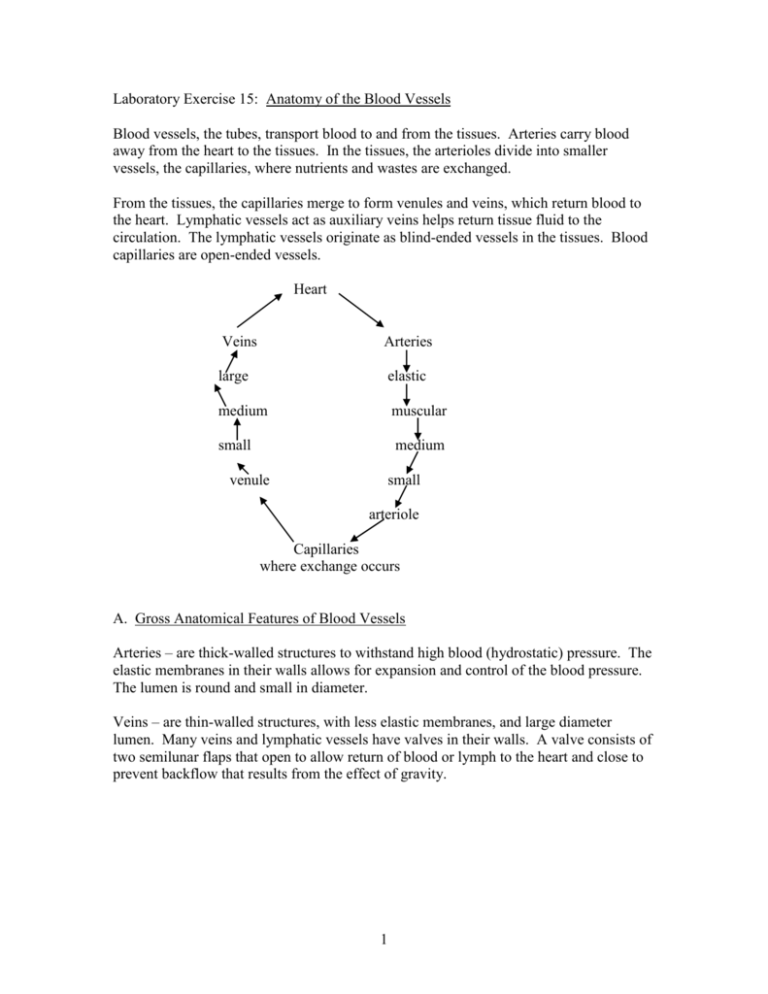
Laboratory Exercise 15: Anatomy of the Blood Vessels Blood vessels, the tubes, transport blood to and from the tissues. Arteries carry blood away from the heart to the tissues. In the tissues, the arterioles divide into smaller vessels, the capillaries, where nutrients and wastes are exchanged. From the tissues, the capillaries merge to form venules and veins, which return blood to the heart. Lymphatic vessels act as auxiliary veins helps return tissue fluid to the circulation. The lymphatic vessels originate as blind-ended vessels in the tissues. Blood capillaries are open-ended vessels. Heart Veins Arteries large elastic medium muscular small medium venule small arteriole Capillaries where exchange occurs A. Gross Anatomical Features of Blood Vessels Arteries – are thick-walled structures to withstand high blood (hydrostatic) pressure. The elastic membranes in their walls allows for expansion and control of the blood pressure. The lumen is round and small in diameter. Veins – are thin-walled structures, with less elastic membranes, and large diameter lumen. Many veins and lymphatic vessels have valves in their walls. A valve consists of two semilunar flaps that open to allow return of blood or lymph to the heart and close to prevent backflow that results from the effect of gravity. 1 B. Microscopic Anatomy of Artery and Vein The wall of a muscular artery is composed of three layers: 1. Tunica intima – inner layer of simple squamous epithelium (endothelium), a small amount of loose connective tissue, and the internal elastic membrane circling the lumen as the outer most part of the tunica intima. 2. Tunica media – thick middle layer composed of circular smooth muscle and broken elastic membranes (fenestrated membranes). The external elastic membrane is the outer most part of the tunica media. 3. Tunica externa (adventitia) – outer layer composed of loose connective tissue, containing small nutrient vessels, vasa vasorum and small nerves of the autonomic nervous system, nervi vasorum. The smooth muscle of the tunica media regulates the lumen diameter of the muscular arteries via the autonomic nervous system. Relaxation of the muscle increases lumen diameter – vasodilation – and blood volume to a particular area. Contraction of the muscle decreases lumen diameter – vasoconstriction – and blood volume to a particular area. The walls of the veins are composed of the same three layers, but are thinner as they are not subjected to as much pressure as the artery’s wall and do not distribute blood to the tissues but collects blood from the tissues. The vein walls are less muscular with a reduced tunica media but a relatively thick tunica externa. C. Major Blood Vessels Outlined below are the main vascular circuits by which blood travels to and from the heart. For arteries, begin with the heart, work distally. For veins, start peripherally and trace the venous return to the heart. Names of blood vessels often reflect their location. 2 Outline of Major Systemic Arteries A. Aorta and its Branches Left ventricle Aortic semilunar valve Ascending aorta left coronary artery right coronary artery Aortic Arch Brachiocephalic Right Common Carotid* right side of head and neck Right Subclavian → right upper extremity (see section B.) Left Common Carotid* left side of head and neck Left Subclavian left upper extremity (see section B.) Thoracic Aorta Intercostals intercostals and chest muscles, pleurae Superior Phrenics posterior and superior surfaces of diaphragm Bronchials bronchi of lungs Esophageals esophagus Abdominal Aorta Inferior Phrenics Celiac inferior surface of diaphragm Common Hepatic liver Left Gastric stomach and esophagus Splenic spleen, pancreas, stomach Superior Mesenteric small intestine, cecum, ascending and transverse colon Suprarenals → adrenal (suprarenal) glands Renals → kidneys Gonadals Testiculars Ovarians Inferior Mesenteric testes ovaries Transverse, descending, sigmoid colon, rectum Common Iliacs (see section C.) 3 B. Arteries of the Upper Limb Subclavian Artery Axilliary Artery Brachial Artery arm Radial Artery* (lateral) forearm Ulnar Artery (medial) Palmer Arches forearm hand C. Arteries of the Lower Limb Common Iliac Artery Internal Iliac Artery External Iliac Artery Femoral Artery* pelvis and perineum lower limb, lower abdominal wall thigh region Popliteal (behind the knee) Artery* Anterior Tibial Artery Tibioperoneal trunk knee joint and popliteal region Dorsalis Pedis Artery* posterior leg Posterior Tibial Artery Peroneal Artery (lateral) (fibular) plantar region of foot lateral leg region *Blood vessels where pulse can be taken. 4 dorsum of foot Outline of Major Systemic Veins A. Veins of the upper limb Venous Networks of Hand Medial Basilic Vein Cephalic Vein (Lateral) (superficial arm and forearm) Axilliary Vein Internal Jugular Veins (large) drains blood from the veins of brain, head and neck, these veins are medial. External Jugular Veins (small) drains blood from veins of superficial regions of head and neck, these veins are lateral Subclavian Vein Brachiocephalic Vein Superior Vena Cava Right Atrium Tricuspid valve Right Ventricle 5 B. Veins of the Lower Limb Dorsal / Plantar Venous Arches of Foot Small Saphenous Vein (posterior leg) Deep Veins (peroneal, anterior and posterior tibialis draining the leg) Great Saphenous Vein (medial leg, anterior thigh) Femoral Vein (thigh) External Iliac Vein Popliteal Vein (knee joint and popliteal region) Common Iliac Vein Inferior Vena Cave Gonadal (testes or ovary) Renal (kidney) Suprarenal (adrenal) Hepatic (liver) Phrenic (diaphragm) Right Atrium 6 Arteries of the Brain - Circle of Willis The major cerebral arteries form the circle of Willis. The circle of Willis surrounds the infundibulum of the pituitary. The anterior cerebral arteries are branches of the internal carotid arteries, the posterior cerebral arteries are branches of the vertebral arteries. The vertebral arteries run through the transverse foramen of the cervical vertebrae. From internal carotid arteries there are the; Anterior cerebral arteries, connecting the 2 anterior cerebral arteries is the anterior communicating artery Middle cerebral arteries Posterior communicating arteries From vertebral arteries the basilar artery branches off. The basilar artery then gives off the cerebellar arteries along the base of the cerebellum. From the basilar artery the posterior cerebral arteries branch off. Connecting the posterior cerebral arteries to the anterior cerebral arteries are the posterior communicating arteries. These 4 arteries form the circle of Willis. D. Portal Circulation For the most part venous blood follows a direct route to the right atrium. However, blood leaving the stomach and intestines contain nutrients absorbed from the gastrointestinal tract. The hepatic portal vein shunts this blood to the liver, to enable the liver to remove nutrients for later use by the body’s cells and to detoxify harmful substances. A portal system is one that begins and ends in capillaries. The hepatic portal system begins in the capillaries of the gastrointestinal tract and ends in capillaries of the liver. The hepatic portal vein forms from a merger of the superior mesenteric and splenic veins. The splenic vein receives branches of the inferior mesenteric, pancreatic and short gastric vein. The right and left gastric veins drain directly into the portal vein near the stomach’s pyloric region. Hepatic veins drain the blood from the liver into the inferior vena cava, which returns the blood to the heart. E. Lymphatic System Lymphatic vessels collect excess tissue fluid and return it to the venous system. The lymphatic system begins as blinded-ended capillaries within the intestinal villi. These lymphatic capillaries are called lacteals. From the lacteals the lymph fluid flows into larger and larger lymphatic veins. Along the path of the lymphatic veins are lymph nodes. The 7 lymph nodes house lymphocytes and macrophages that defend the body against foreign substances. The nodes are in a strategic position to engulf and destroy antigenic materials and so “sterilize” the lymph before it is returned to the blood circulatory system. The large lymphatic veins then lead into two collecting ducts. The right lymphatic duct drains the upper right side of the body and empties into the right subclavian vein. The major collecting duct, the left lymphatic or thoracic duct, drains the rest of the body and empties into the left subclavian vein. 8


