BOLD Response to Median-Nerve Stimulation: A
advertisement
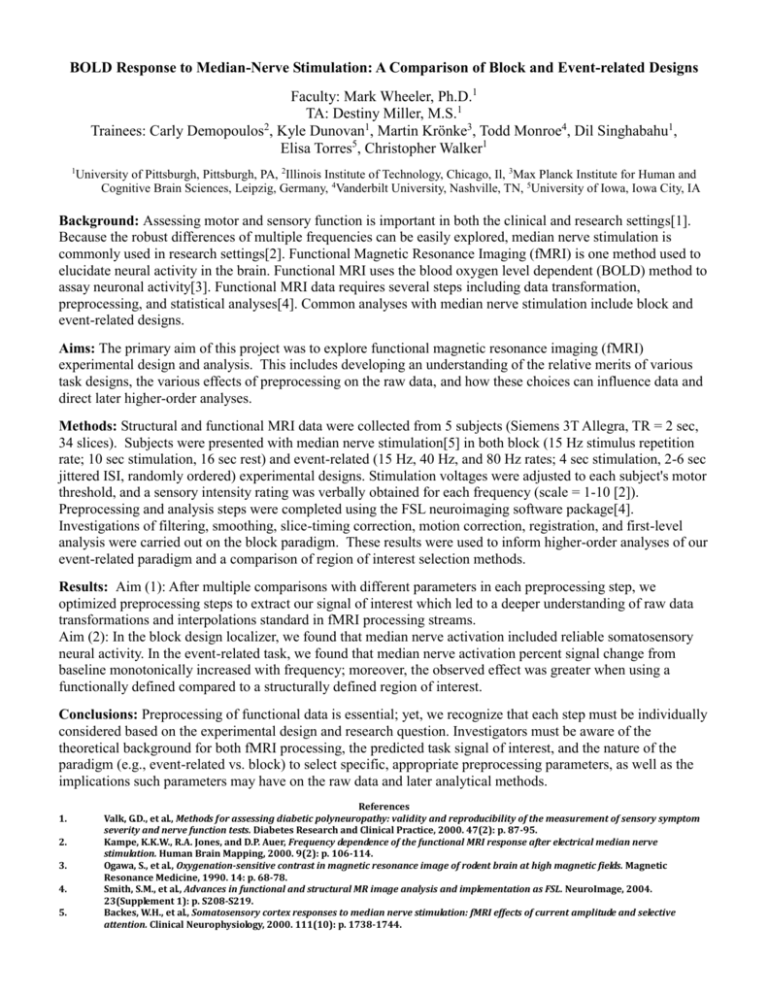
BOLD Response to Median-Nerve Stimulation: A Comparison of Block and Event-related Designs Faculty: Mark Wheeler, Ph.D.1 TA: Destiny Miller, M.S.1 2 Trainees: Carly Demopoulos , Kyle Dunovan1, Martin Krönke3, Todd Monroe4, Dil Singhabahu1, Elisa Torres5, Christopher Walker1 1 University of Pittsburgh, Pittsburgh, PA, 2Illinois Institute of Technology, Chicago, Il, 3Max Planck Institute for Human and Cognitive Brain Sciences, Leipzig, Germany, 4Vanderbilt University, Nashville, TN, 5University of Iowa, Iowa City, IA Background: Assessing motor and sensory function is important in both the clinical and research settings[1]. Because the robust differences of multiple frequencies can be easily explored, median nerve stimulation is commonly used in research settings[2]. Functional Magnetic Resonance Imaging (fMRI) is one method used to elucidate neural activity in the brain. Functional MRI uses the blood oxygen level dependent (BOLD) method to assay neuronal activity[3]. Functional MRI data requires several steps including data transformation, preprocessing, and statistical analyses[4]. Common analyses with median nerve stimulation include block and event-related designs. Aims: The primary aim of this project was to explore functional magnetic resonance imaging (fMRI) experimental design and analysis. This includes developing an understanding of the relative merits of various task designs, the various effects of preprocessing on the raw data, and how these choices can influence data and direct later higher-order analyses. Methods: Structural and functional MRI data were collected from 5 subjects (Siemens 3T Allegra, TR = 2 sec, 34 slices). Subjects were presented with median nerve stimulation[5] in both block (15 Hz stimulus repetition rate; 10 sec stimulation, 16 sec rest) and event-related (15 Hz, 40 Hz, and 80 Hz rates; 4 sec stimulation, 2-6 sec jittered ISI, randomly ordered) experimental designs. Stimulation voltages were adjusted to each subject's motor threshold, and a sensory intensity rating was verbally obtained for each frequency (scale = 1-10 [2]). Preprocessing and analysis steps were completed using the FSL neuroimaging software package[4]. Investigations of filtering, smoothing, slice-timing correction, motion correction, registration, and first-level analysis were carried out on the block paradigm. These results were used to inform higher-order analyses of our event-related paradigm and a comparison of region of interest selection methods. Results: Aim (1): After multiple comparisons with different parameters in each preprocessing step, we optimized preprocessing steps to extract our signal of interest which led to a deeper understanding of raw data transformations and interpolations standard in fMRI processing streams. Aim (2): In the block design localizer, we found that median nerve activation included reliable somatosensory neural activity. In the event-related task, we found that median nerve activation percent signal change from baseline monotonically increased with frequency; moreover, the observed effect was greater when using a functionally defined compared to a structurally defined region of interest. Conclusions: Preprocessing of functional data is essential; yet, we recognize that each step must be individually considered based on the experimental design and research question. Investigators must be aware of the theoretical background for both fMRI processing, the predicted task signal of interest, and the nature of the paradigm (e.g., event-related vs. block) to select specific, appropriate preprocessing parameters, as well as the implications such parameters may have on the raw data and later analytical methods. 1. 2. 3. 4. 5. References Valk, G.D., et al., Methods for assessing diabetic polyneuropathy: validity and reproducibility of the measurement of sensory symptom severity and nerve function tests. Diabetes Research and Clinical Practice, 2000. 47(2): p. 87-95. Kampe, K.K.W., R.A. Jones, and D.P. Auer, Frequency dependence of the functional MRI response after electrical median nerve stimulation. Human Brain Mapping, 2000. 9(2): p. 106-114. Ogawa, S., et al., Oxygenation-sensitive contrast in magnetic resonance image of rodent brain at high magnetic fields. Magnetic Resonance Medicine, 1990. 14: p. 68-78. Smith, S.M., et al., Advances in functional and structural MR image analysis and implementation as FSL. NeuroImage, 2004. 23(Supplement 1): p. S208-S219. Backes, W.H., et al., Somatosensory cortex responses to median nerve stimulation: fMRI effects of current amplitude and selective attention. Clinical Neurophysiology, 2000. 111(10): p. 1738-1744.

