MRI OF THE RIGHT KNEE
advertisement
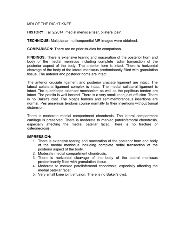
MRI OF THE RIGHT KNEE HISTORY: Fall 2/2014, medial meniscal tear, bilateral pain. TECHNIQUE: Multiplanar multisequential MR images were obtained. COMPARISON: There are no prior studies for comparison. FINDINGS: There is extensive tearing and maceration of the posterior horn and body of the medial meniscus including complete radial transection of the posterior aspect of the body. The anterior horn is intact. There is horizontal cleavage of the body of the lateral meniscus predominantly filled with granulation tissue. The anterior and posterior horns are intact. The anterior cruciate ligament and posterior cruciate ligament are intact. The lateral collateral ligament complex is intact. The medial collateral ligament is intact. The quadriceps extensor mechanism as well as the popliteus tendon are intact. The patella is well located. There is a very small knee joint effusion. There is no Baker's cyst. The biceps femoris and semimembranosus insertions are normal. Pes anserinus tendons course normally to their insertions without bursal distension. There is moderate medial compartment chondrosis. The lateral compartment cartilage is preserved. There is moderate to marked patellofemoral chondrosis, especially affecting the medial patellar facet. There is no fracture or osteonecrosis. IMPRESSION: 1. There is extensive tearing and maceration of the posterior horn and body of the medial meniscus including complete radial transection of the posterior aspect of the body. 2. Moderate medial compartment chondrosis. 3. There is horizontal cleavage of the body of the lateral meniscus predominantly filled with granulation tissue. 4. Moderate to marked patellofemoral chondrosis, especially affecting the medial patellar facet. 5. Very small knee joint effusion. There is no Baker's cyst.


