CD38 References - Janis V. Giorgi Flow Cytometry Labratory
advertisement
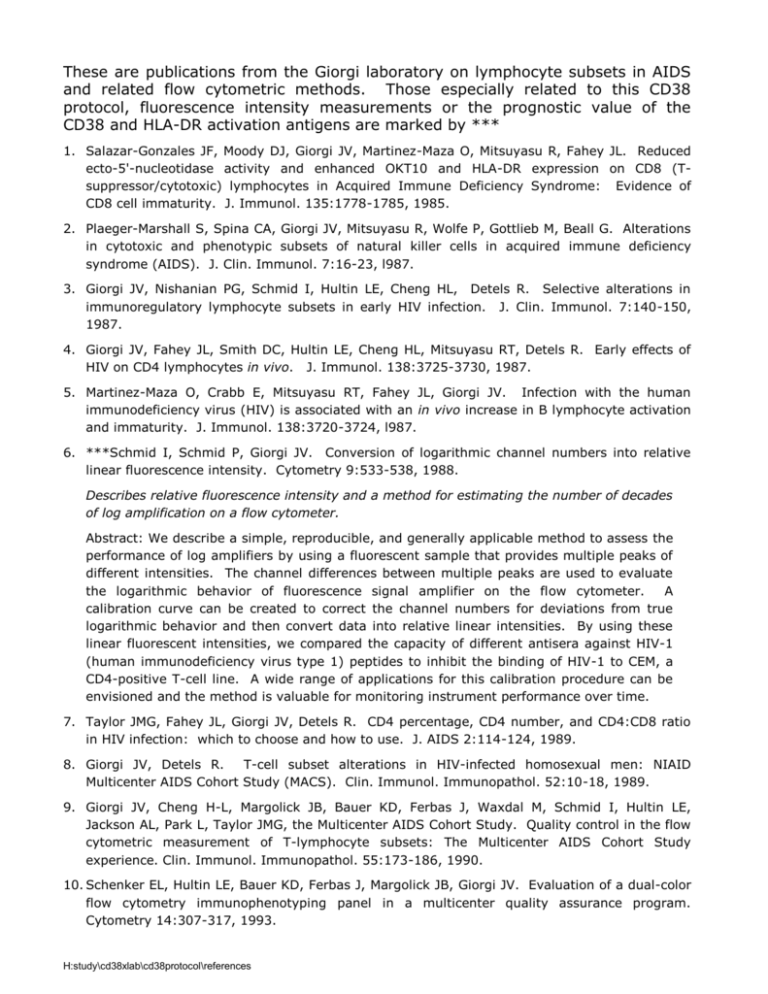
These are publications from the Giorgi laboratory on lymphocyte subsets in AIDS and related flow cytometric methods. Those especially related to this CD38 protocol, fluorescence intensity measurements or the prognostic value of the CD38 and HLA-DR activation antigens are marked by *** 1. Salazar-Gonzales JF, Moody DJ, Giorgi JV, Martinez-Maza O, Mitsuyasu R, Fahey JL. Reduced ecto-5'-nucleotidase activity and enhanced OKT10 and HLA-DR expression on CD8 (Tsuppressor/cytotoxic) lymphocytes in Acquired Immune Deficiency Syndrome: Evidence of CD8 cell immaturity. J. Immunol. 135:1778-1785, 1985. 2. Plaeger-Marshall S, Spina CA, Giorgi JV, Mitsuyasu R, Wolfe P, Gottlieb M, Beall G. Alterations in cytotoxic and phenotypic subsets of natural killer cells in acquired immune deficiency syndrome (AIDS). J. Clin. Immunol. 7:16-23, l987. 3. Giorgi JV, Nishanian PG, Schmid I, Hultin LE, Cheng HL, Detels R. Selective alterations in immunoregulatory lymphocyte subsets in early HIV infection. J. Clin. Immunol. 7:140-150, 1987. 4. Giorgi JV, Fahey JL, Smith DC, Hultin LE, Cheng HL, Mitsuyasu RT, Detels R. Early effects of HIV on CD4 lymphocytes in vivo. J. Immunol. 138:3725-3730, 1987. 5. Martinez-Maza O, Crabb E, Mitsuyasu RT, Fahey JL, Giorgi JV. Infection with the human immunodeficiency virus (HIV) is associated with an in vivo increase in B lymphocyte activation and immaturity. J. Immunol. 138:3720-3724, l987. 6. ***Schmid I, Schmid P, Giorgi JV. Conversion of logarithmic channel numbers into relative linear fluorescence intensity. Cytometry 9:533-538, 1988. Describes relative fluorescence intensity and a method for estimating the number of decades of log amplification on a flow cytometer. Abstract: We describe a simple, reproducible, and generally applicable method to assess the performance of log amplifiers by using a fluorescent sample that provides multiple peaks of different intensities. The channel differences between multiple peaks are used to evaluate the logarithmic behavior of fluorescence signal amplifier on the flow cytometer. A calibration curve can be created to correct the channel numbers for deviations from true logarithmic behavior and then convert data into relative linear intensities. By using these linear fluorescent intensities, we compared the capacity of different antisera against HIV-1 (human immunodeficiency virus type 1) peptides to inhibit the binding of HIV-1 to CEM, a CD4-positive T-cell line. A wide range of applications for this calibration procedure can be envisioned and the method is valuable for monitoring instrument performance over time. 7. Taylor JMG, Fahey JL, Giorgi JV, Detels R. CD4 percentage, CD4 number, and CD4:CD8 ratio in HIV infection: which to choose and how to use. J. AIDS 2:114-124, 1989. 8. Giorgi JV, Detels R. T-cell subset alterations in HIV-infected homosexual men: NIAID Multicenter AIDS Cohort Study (MACS). Clin. Immunol. Immunopathol. 52:10-18, 1989. 9. Giorgi JV, Cheng H-L, Margolick JB, Bauer KD, Ferbas J, Waxdal M, Schmid I, Hultin LE, Jackson AL, Park L, Taylor JMG, the Multicenter AIDS Cohort Study. Quality control in the flow cytometric measurement of T-lymphocyte subsets: The Multicenter AIDS Cohort Study experience. Clin. Immunol. Immunopathol. 55:173-186, 1990. 10. Schenker EL, Hultin LE, Bauer KD, Ferbas J, Margolick JB, Giorgi JV. Evaluation of a dual-color flow cytometry immunophenotyping panel in a multicenter quality assurance program. Cytometry 14:307-317, 1993. H:study\cd38xlab\cd38protocol\references 11. Ho H-N, Hultin LE, Mitsuyasu RT, Matud JL, Hausner MA, Bockstoce D, Chou C-C, O'Rourke S, Taylor JMG, Giorgi JV. Circulating HIV-specific CD8+ cytotoxic T cells express CD38 and HLADR antigens. J. Immunol. 150:3070-3079, 1993. 12. ***Giorgi JV, Liu Z, Hultin LE, Cumberland WG, Hennessey K, Detels R. Elevated levels of CD38+CD8+ T cells in HIV infection add to the prognostic value of low CD4+ T cell levels: Results of 6 years of follow-up. J.AIDS 6:904-912, 1993. Shows prognostic value for poor outcome of CD38+ CD8+ percent in early HIV-1 infection. Abstract: A cohort of 98 HIV-infected initially AIDS-free homosexual men from the Multicenter AIDS Cohort Study (MACS) was followed for six years to investigate whether CD8+ cell subsets have prognostic value for progression to AIDS. In the present study, four subsets of CD8+ T cells that previously have been shown to be selectively elevated in HIVinfected asymptomatic persons, specifically the CD8+ T cell subsets that were CD38+, HLA_ DR+, CD57+ and L-selectin negative (Leu8 ), were measured. Forty-nine of the 98 developed AIDS. Prognostic value of these CD8+ cell subsets was evaluated using the _ proportional hazards model. Levels of both CD38+CD8+ and Leu8 CD8+ cells individually had prognostic value for progression to AIDS. In contrast, CD57 +CD8+ and HLA-DR+CD8+ cell subset levels did not have prognostic value. After adjustment for level of CD4+ T cells, however, only the elevation in the CD38+CD8+ cell subset had additional prognostic value. These results suggest that the level of CD38+CD8+ cells could be used together with the CD4+ T cell level to more accurately predict progression to AIDS among HIV-infected men. These results provide further support for the observation that dramatic and progressive activation of CD8+ T cells in HIV infection occurs. The power of elevated levels of the CD38+CD8+ subset to predict poor prognosis in this cohort suggests these CD8 + T cells reflect an immune stimulation that is ultimately unable to control disease progression. 13. Schmid I, Uittenbogaart CH, Giorgi JV. Sensitive method for measuring apoptosis and cell surface phenotype in human thymocytes by flow cytometry. Cytometry 15:12-20, 1994. 14. Schmid I, Uittenbogaart CH, Keld B, Giorgi JV. A rapid method for measuring apoptosis and dual-color immunofluorescence by single laser flow cytometry. J. Immunol. Methods 170:145157, 1994. 15. Chou C-C, Gudeman V, O'Rourke S, Isacescu V, Detels R, Williams GJ, Mitsuyasu RT, Giorgi JV. Phenotypically defined memory CD4+ cells are not selectively decreased in chronic HIV disease. J. AIDS 7:665-675, 1994. 16. ***Giorgi JV, Ho H-N, Chou C-C, Hultin LE, Matud JL, Hirji K, O'Rourke S, Park L, Margolick J, Ferbas J, Phair JP, and the Multicenter AIDS Cohort Study. CD8 + lymphocyte activation at HIV-1 seroconversion: Development of HLA-DR+CD38-CD8+ cells is associated with subsequent stable CD4+ cell levels. J. Infect. Dis. 170:775-781, 1994. Shows HLA-DR+ CD38– percent is marker of good prognosis in HIV-1 infection. Abstract: Subsets of activated CD8+ T lymphocytes defined by membrane expression of the activation antigens HLA-DR and CD38 were enumerated by three-color flow cytometric analysis in homosexual men who became infected with human immunodeficiency virus type 1 (HIV-1). Profound CD8+ T cell activation was observed in all 15 subjects at seroconversion and at 6 and 12 months thereafter. The HLA-DR+CD38+CD8+ cell population, which has potent direct anti-HIV cytotoxic T cell activity, was markedly elevated at seroconversion. Throughout the first year of infection, some men also had an elevation in the HLA-DR+CD38¯CD8+ T cell population. During the next 5 years, these men had stable H:study\cd38xlab\cd38protocol\references CD4+ T cell levels whereas the others did not. Long-term survivors (9 years seropositive, CD4+ T cells >800/mm3) also had elevated levels of this subset despite few other activated CD8+ T cells. Selective elevation of HLA-DR+CD38¯CD8+ T cells was thus a marker of subsequent stable HIV-1 disease. 17. Giorgi JV, Boumsell L, Autran B. Reactivity of T Cell Section monoclonal antibodies with circulating CD4+ and CD8+ T cells in HIV disease and following in vitro activation. In: Schlossman S, Boumsell L, Gilks W, Harlan J, Kishimoto T, Morimoto C, Ritz J, Shaw S, Silverstein R, Springer T, Tedder T, Todd R, eds. pp. 446-461. Leukocyte Typing V: White Cell Differentiation Antigens. Oxford University Press, Oxford, 1995. 18. Ferbas J, Kaplan AH, Hausner MA, Hultin LE, Matud JL, Liu Z, Panicali DL, Ho, H-N, Detels R, Giorgi JV. Virus burden in long-term survivors of human immunodeficiency virus (HIV) infection is a determinant of anti-HIV CD8+ lymphocyte activity. J. Inf. Dis. 172:329-339, 1995. 19. Hu P-F, Hultin LE, Hultin P, Hausner MA, Hirji K, Jewett A, Bonavida B, Detels R, Giorgi JV. Natural killer cell immunodeficiency in HIV disease is manifest by profoundly decreased numbers of CD16+CD56+ cells and an expansion of a population of CD16dimCD56¯ cells with low lytic activity. J. AIDS 10:331-340, 1995. 20. Giorgi JV. Phenotype and function of T cells in HIV disease. In Gupta, S. ed. Immunology of HIV Infection: pp.181-199, Plenum Press, New York, 1996. 21. ***Liu Z, Hultin LE, Cumberland WG, Hultin P, Schmid I, Matud J, Detels R, Giorgi JV. Elevated relative fluorescence intensity of CD38 antigen expression on CD8 + T cells is a marker of poor prognosis in HIV infection: Results of 6 years of follow-up. Cytometry (Clin. Comm.) 26: 1-7, 1996. Based on reference 12 data; shows CD38 relative fluorescence intensity (RFI) has predictive value similar to CD38+CD8+ cell percent. Abstract: Relative fluorescence intensity measurements from a flow cytometer were used to evaluate expression of CD38 and HLA-DR antigens. These molecules are associated with cellular activation and are present at increased levels on the CD8 + T lymphocytes of HIV-1infected subjects. In the current study, the prognostic value of mean fluorescence intensity measurements of CD38 and HLA-DR on CD8+ T cells was compared to results from our previous study in which we reported prognostic value for an elevated percentage of CD8 + T cells that were positive for expression of the CD38 antigen (Giorgi et al.: JAIDS 6:904–912, 1993). Using the proportional hazards model, elevated mean fluorescence intensity of CD38 expression on CD8+ T cells had prognostic value for development of AIDS that was almost identical to the prognostic value of the percentage of CD8 + T cells that were positive for expression of CD38. This prognostic value was in addition to that provided by the patient’s CD4+ T cell measurement. To our knowledge, this is the first report that a measurement of fluorescence intensity can be used as a prognostic marker in an immunodeficiency disease. Efforts are needed to establish methods that will allow wide-spread application of this observation in the clinical management of HIV-1-infected subjects. 22. Effros RB, Allsopp R, Chiu C-P, Hausner MA, Hirji K, Wang L, Harley CB, Villeponteau B, West MD, Giorgi JV. Shortened telomeres in the expanded CD28¯CD8 + cell subset in HIV disease implicate replicative senescence in HIV pathogenesis. AIDS 10:F17-F22, 1996. 23. Giorgi JV, Hultin LE, Desrosiers RC. The immunopathogenesis of retroviral diseases: No immunophenotypic alterations in T, B and NK cell subsets in SIV mac239-challenged rhesus H:study\cd38xlab\cd38protocol\references macaques protected by SIVnef vaccination. J. Med. Primatol. 25: 186-191, 1996. 24. Schmid I, Nicholson JKA, Giorgi JV, Janossy G, Kunkl A, Lopez PA, Perfetto S, Seamer LC, Dean PN. Biosafety guidelines for sorting of unfixed cells. Cytometry 28:99-117, 1997. 25. ***Liu Z, Cumberland WG, Hultin LE, Prince HE, Detels R, Giorgi JV. Elevated CD38 antigen expression on CD8+ T cells is a stronger marker for the risk of chronic HIV disease progression to AIDS and death in the MACS than CD4+ cell count, soluble immune activation markers or combinations of HLA-DR and CD38 expression. J. Acq. Immune Deficienc. & Hum. Retrovirol. 16:83-92, 1997. Uses CD38 molecules on CD8+ T cells (CD38 on CD8) for the first time; shows high median CD38 on CD8 is more predictive of poor outcome than neopterin, B 2 microglobulin, TNF-, CD4+ cell number and percent and mean CD38 on CD8; study of subjects infected for 8 years when markers were measured and followed to AIDS and death. Abstract: The prognostic value of several immunologic markers were compared in Los Angeles Multicenter AIDS Cohort Study (MACS) participants, most of whom had been HIVinfected for more than 8 years. Markers studied included CD4+ cell number, flow cytometric measurements of CD8+ cell expression of CD38 and HLA-DR antigens, and serum markers of immune activation including neopterin, 2-microglobulin, soluble IL-2 receptor, soluble CD8 and soluble tumor necrosis factor receptor type II. Cox proportional hazards models indicated that elevated CD38 on CD8, a flow cytometric measurement of CD8 + T lymphocyte activation, was the most predictive marker of those studied for development of a clinical AIDS diagnosis and death. As compared with the reference group, who had CD38 on CD8 <2470 molecules per CD8+ cell and in whom 4/99 developed clinical AIDS within 3 years, participants with CD38 on CD8 between 2470–3899, 3900–7250 and >7250 had relative risks (and numbers developing AIDS within 3 years) of 5.0 (15/81), 12.3 (24/60), and 41.4 (36/49), respectively. The strong prognostic value of CD38 on CD8 measurements and the fundamental importance of chronic immune activation in the pathogenesis of HIV disease suggests that this marker might have utility in the clinical management of HIVinfected persons. 26. Lynne JE, Schmid I, Matud JL, Hirji K, Buessow S, Shlian DM, Giorgi JV. Major expansions of select CD8+ subsets in acute Epstein-Barr virus infection: Comparison with chronic human immunodeficiency virus disease. J. Infect. Dis. 177:1083-1087, 1998. 27. ***Liu Z, Cumberland WG, Hultin LE, Kaplan AH, Detels R, Giorgi JV. CD8 + T lymphocyte activation in HIV-1 disease reflects an aspect of pathogenesis distinct from viral burden and immunodeficiency. J. Acq. Immun. Deficienc. & Hum. Retrovirol. 18:332-340, 1998. Shows elevated levels of CD38 expression on CD8+ T cells are even more predictive than plasma HIV-1 RNA levels of progression to AIDS and death in men who have been infected with HIV-1 for 8 years. Abstract: The CD8+ T cell response is central to the control and eventual elimination of persistent virus infections. Although it might be expected that CD8+ T cell activation would be associated with a better clinical outcome during virus infections, in chronic human immunodeficiency virus type 1 (HIV-1)-infection, high levels of CD8+ T cell activation are instead associated with faster disease progression. In this study, cell surface expression of CD38, a flow cytometric marker of T cell activation of CD8+ T cells, had predictive value for HIV-1 disease progression that was in part independent of the predictive value of plasma virus burden and CD4+ T cell number. Measurements of CD38 antigen expression on CD8+ T cells H:study\cd38xlab\cd38protocol\references in HIV-1-infected patients may be of value for assessing prognosis and the impact of therapeutic interventions. The pathogenetic reason why CD8+ T cell activation is associated with poor outcome in HIV-1 disease is unknown. Possibly CD8+ T cell activation contributes to immunologic exhaustion, hyporesponsiveness of T cells to their cognate antigens, or perturbations in the T cell receptor repertoire. 28. Giorgi JV, Majchrowicz MA, Johnson TD, Hultin P, Matud J, Detels R. Immunologic effects of combined protease inhibitor and reverse transcriptase inhibitor therapy in previously treated chronic HIV-1 infection. AIDS 12:1833-1844, 1998. Shows value of monitoring CD38 following initiation of PART; effective anti-retroviral therapy reverses immune activation leading to a decrease in CD38 expression and an increase in resting HLA-DR– CD38– cell levels. Abstract: Our objective was to evaluate the efficacy of combination protease and reverse transcriptase inhibitor therapy in correcting HIV-1-induced lymphocyte subset abnormalities in previously treated adults. For this project, we designed a 48 week observational study of lymphocyte subsets in 12 participants in the Multicenter AIDS Cohort Study (MACS) who were already taking at least one reverse transcriptase inhibitor and added protease inhibitor to their treatment regimen. Comparison groups were seronegative homosexual men (SNHm), seronegative heterosexual men and homosexual HIV-1-infected men who were long-term nonprogressors. Three-color immunofluorescence and monoclonal antibodies were used to assess HIV-1-induced lymphocyte subset alterations related to immune deficiency and immune activation. Plasma HIV-1 RNA levels were monitored to assess suppression of viral replication. We found that CD4+ T cell counts significantly increased and lymphocyte activation measured as CD38 and HLA-DR expression on CD8+ T cells significantly decreased by 48 weeks. CD4+ T cell values remained abnormal even in those who were fully suppressed. Some T cell activation markers decreased to levels observed in LTNP. The increase in CD4+ T cell numbers reached a plateau by week 24, but the increase in resting HLA-DR–CD38– T cells was sustained through week 48. Proportions of CD45RA+CD62L+ and CD28+CD4+ T cell subsets and Fas expression were not abnormal at baseline compared with SNHm controls. The most significant impact of suppression of viral replication was reversal of T cell activation. However, normalization of lymphocyte subset perturbations associated with chronic HIV-1 infection was not achieved after a year of treatment with current combination anti-retroviral regimens. More profound viral suppression, therapy for longer than a year, or immunologic augmentation may be needed to fully reverse the abnormalities. 29. Hultin LE, Matud JL, Giorgi JV. Quantitation of CD38 activation antigen expression on CD8 + T cells in HIV-1 infection using CD4 expression on CD4+ T lymphocytes as a biological calibrator. Cytometry 33:123-132, 1998. This is the publication that describes the method provided at this website. Abstract: For some membrane-associated antigens, the number of molecules expressed per cell carries information about the cell’s differentiation and activation state. Quantitating antigen expression by flow cytometry has immediate application in monitoring CD38 expression on CD8+ T cells in HIV-1 disease, where elevated CD38 antigen expression is a marker of CD8+ T cell activation and a poor prognostic indicator. Reproducible methods are needed in order to quantify such antigens. Here we describe a reproducible method for quantitative fluorescence cytometry (QFCM) that depends on the tightly regulated expression of CD4 antigen on human CD4+ T lymphocytes, which we estimated in a study H:study\cd38xlab\cd38protocol\references of 57 normal donors, to have an inter-person coefficient of variation of 4.9%. Using PEconjugated CD4 mAb with a nominal fluorochrome to protein ratio of 1:1 and a nominal published value of approximately 50,000 CD4 antibody molecules bound per CD4 + T lymphocyte, we estimated the number of PE molecules detected per RFI (relative fluorescence intensity) unit on our flow cytometer to be 41 (19,20). This value is called the “RFI multiplier”. To estimate the number of CD38 antibodies bound per CD8 + T cell (CD38ABC) on patient samples, we multiply the measured CD38 RFI value of CD38 staining using a nominal 1:1 conjugate of CD38-PE by the “RFI multiplier”. The measurements for CD4 and CD38 were stable for two years despite the use of different mAb lots and the potential for drift in instrumentation. We used this approach in a study of nine flow cytometers where the inter-instrument interlaboratory CV’s for CD3-ABC ranged from 3.3% to 5.8% and those for CD38-ABC ranged from 9.8% to 13.8%. These data indicate that CD4 expression can serve as a biological calibrator to standardize fluorescence intensity measurements in longitudinal and multicenter studies. 30. Iyer SB, Hultin LE, Zawadzki JA, Davis KA, Giorgi JV. Quantitation of CD38 expression using QuantiBRITE™ beads. Cytometry 33:206-212, 1998. This publication describes a commercial product that may simplify CD38 quantitation for clinical laboratories. Abstract: The QuantiBRITE™ bead method was compared with the CD4 biological calibration method for quantitation of CD38 expression on CD8+ T-lymphocytes of Multicenter AIDS Cohort Study participants. Results were expressed as CD38 antibodies bound per cell (ABCs) and were the same with the two methods provided two conditions were met. These were the use of repurified (>95% of the monoclonal antibodies [mAbs] have 1 phycoerythrin [PE] molecule per mAb) CD38-PE for both methods and use of repurified CD4PE to calculate the relative fluorescence intensity multiplier for the CD4 biological calibration method. Our results indicate that the prognostic significance of CD38 values obtained using the QuantiBRITE method can be interpreted using previously published reports (Liu et al., J Aquir Immune Defic Syndr Hum Retrovirol 16:83-92, 1997 and 18:332-340, 1998). Sample preparation using NH4Cl and FACS lysing solution gave similar results for CD38 relative fluorescence intensity. Dilution into either phosphate-buffered saline with 2% fetal calf serum and 0.1% sodium azide or fixation in 1% paraformaldehyde for 1 or 24 h also gave similar results. In experiments using Raji cells, which express high levels of CD38, the valence of binding of the intact Leu 17 antibody was ~68% bivalent and ~32% monovalent. This emphasizes the complexity of determining antigen density from ABCs. We conclude that repurified PE conjugates of CD38, which can be consistently made, together with QuantiBRITE PE beads, provide a convenient and reliable method for quantitation of CD38 expression as ABCs. 31. ***Giorgi JV, Hultin LE, McKeating JA, Johnson TD, Owens B, Jacobson LP, Shih R, Lewis J, Wiley D, Phair JP, Wolinsky SM, Detels R. Shorter survival in advanced human immunodeficiency virus type 1 infection is more closely associated with T lymphocyte activation than with plasma virus burden or viral chemokine coreceptor usage. J. Infect. Dis. in press 1999. Shows CD38 is elevated on CD4+ and CD8+ cells in advanced HIV-1 disease and is more predictive of death than plasma HIV-1 RNA, naïve T cell percents, or any other marker. Abstract: To define predictors of survival time in late HIV-1 disease, we studied long and short duration survivors after their CD4+ T cell counts fell to <50 per mm3. Immune H:study\cd38xlab\cd38protocol\references activation of CD4+ and CD8+ T cells, as measured by elevated cell surface expression of CD38 antigen, was strongly associated with shorter subsequent survival (P .002). The naïve CD45RA+ CD62L+ T cell reserve was low in all subjects and did not predict survival (P = .34 for CD4+ and .08 for CD8+ cells). Higher viral burden correlated with CD8+ but not CD4+ cell activation and after correcting for multiple comparisons was not associated with shorter survival (P = .02). All of the patients’ viruses used CCR5, CXCR4 or both and coreceptor usage did not predict survival (P = .27). Through mechanisms apparently unrelated to higher viral burden, immune activation is a major determinant of survival in advanced HIV-1 disease. H:study\cd38xlab\cd38protocol\references
