Nonlinear Optical Imaging - West Virginia University
advertisement

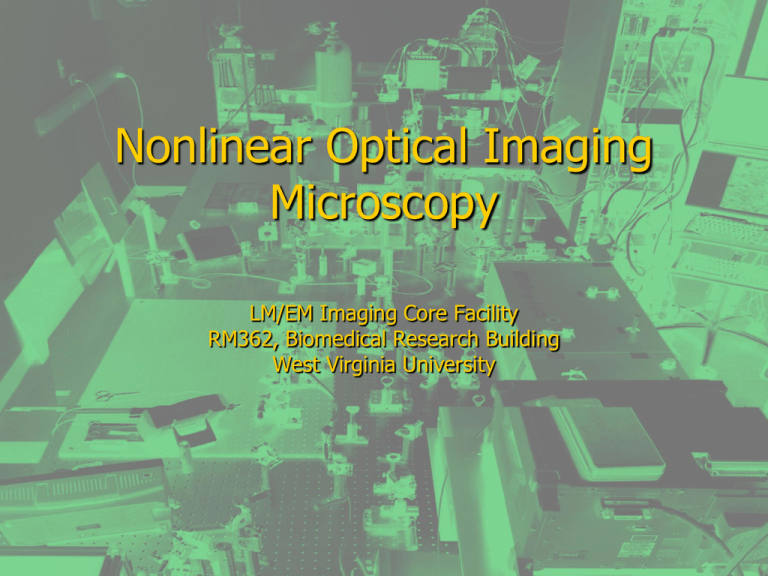
Nonlinear Optical Imaging Microscopy LM/EM Imaging Core Facility RM362, Biomedical Research Building West Virginia University NLOM Bench NLO Facility • Two Photon Microscopy • Second Harmonic Generation Microscopy (SHG) • Third Harmonic Generation Microscopy (THG) • Coherent anti-Stokes Raman Scattering (CARS) Microscopy Advantage of 2-P vs OPF (Confocal) No pinhole necessary meaning more fluorescence light collected. Nondescan setup meaning scanning mirror is out of fluorescence light path, detector closer to specimen. Laser tunable; one wavelength can simultaneously excite many probes Less out-of-focus photobleaching/photodamage good for live cell/tissue imaging Less scattering for deeper tissue penetration TPF Probes 1. Organic dye – immunostaining, colors ranging from Blue to NIR 2. Fluorescent protein (GFP, CFP YFP, RFP, mCherry, tdTomato, etc.) 3. Nanoparticle (Quantum dots, Carbon nanotube) 4. Autofluorescence (NAD(P)H, flavoprotein, elastin, etc.) Two Platforms • Inverted Microscope Configuration • Upright Microscope Configuration IX71 Inverted Microscope Features: Inverted; slow and fast scanner; Equipped with TC incubator/CO2 System Epi-detection: 1. Two-channel 2P/3P fluorescence 2. CARS Forward detection: 1. Second harmonic generation (SHG) 2. Third harmonic generation (THG) 3. CARS MOM Upright Microscope Unique Features of MOM: Upright; objective is xyz motorized; electrophysiology and imaging combined Epi-detection: 4 channel capability, 3 channel simultaneous 2P or 3P imaging, SHG or CARS Forward detection: 1 channel, SHG, THG, or CARS Equiped with perfusion system: equipped for in vitro and in vivo imaging TPF: In Vitro and In Vivo Study Barry Masters and Peter So, Handbook of Biomedical Nonlinear Optical Microscopy TPF Applications Mammalian neurobiology Intracellular imaging of calcium dynamics in dendritic spine Long-term imaging of neurodegeneration measurement of vascular hemodynamics Targeted intracellular recording In Vivo Tumor biology Chronic in vivo models for Dynamic imaging of Tumor-associated blood and lymphatic vessels Tumor Vessel permeability Tumor/host cell interaction in vessels and stroma TPF Applications cont’d Immunology Visualization of lymphocyte mobility, chemotaxis Visualization of antigen recognition in isolated lymphoid organs Visualization of In Vivo lymph nodes in anesthetized mice SHG Applications SHG from Endogenous Tissue (noncentrosymmetric) Collagen, acto-myosin complexes, mitotic spindles, microtubules, and cellulose. Such as in skin, bone, tendon, cartilage, teeth, brain tissue, cornea, etc. SHG Applications cont’d SHG from Exogenous Chromophores Molecular “Flip-Flop” Dynamics in Membranes Intermembrane Separation Measurement Membrane Potential Imaging (using voltage sensitive dyes) Figure. High resolution SHG recording of action potentials deep in hippocampal brain slices. (A) SHG image of a neuron patch clamped and filled with FM464 in slice. (B) SHG line-scan recording of elicited action potentials (55 line scans were averaged). (C) Electrical patch pipette recording of action potentials seen in B. Calibrations: (A) 20 µm; (B) 2.5% ΔSHG/SHG, 50 ms; (C) 20 mV, 50 ms. (W. Webb lab) SHG Applications Figure. Rat hippocampal neurons are imaged by SHG (green pseudocolor) after 5 days (left) and 7 days (right) in culture. At the later development stage, protoprocesses have presumably matured into dendrites whose microtubules are of mixed polarity and cannot produce SHG. Only the axon microtubules, which maintain a uniform polarity, are revealed by SHG (red pseudocolor represents autofluorescence). (W. Web lab) THG Applications Changing the Look of Malaria The optical effect called third harmonic generation causes malaria secretions to glow blue in infected blood cells (left), promising a faster, more efficient diagnosis than traditional microscopy imagery (right) Coherent anti-Stokes Raman Scattering (CARS) Microscopy CARS Applications Tissue imaging (chemical selectivity & noninvasive) CARS: very sensitive to lipids To study dynamic processes in living cells; Lipid rafts on membrane Trafficking and growth of lipid droplets Intracellular water diffusion CARS Images CARS TPF Lipid droplet of A Nile red-stained daf-4 mutant (C. elegans) arrested in its dauer stage for 3 weeks. Hellerer T et al. PNAS 2007; 104:14658-14663 Optical Parametric Oscillator Technology (OPO) Deep Tissue Imaging cont’d Kobat et al. 2009 / Vol. 17, No. 16 / OPTICS EXPRESS 13354 Deep Tissue Imaging Helmchen et al. Nature Methods 2005, 2(12), 932 Data Visualization & Image Analysis 2D image processing Photoshop, MATLAB, ImageJ 3D/4D image visualization & measurement (3D – stack images; 4D – stack images + time lapse) Commercial (Amira, Imaris, MATLAB), Free (ImageJ, Voxx, VisBio) Technology Development Administrative supplement to Neuro CoBRE grant supports: Optical Parametric Amplification Long wavelength imaging Develop new applications for SHG, CARS Neuro CoBRE grant also supports: Develop in vivo imaging capability Set up chamber for time-lapse in vitro imaging Imaging Discovery Grants to facilitate purchase of reagents and use of facility Imaging Discover Grants


