High-Energy Photon Standard Dosimetry Data
advertisement
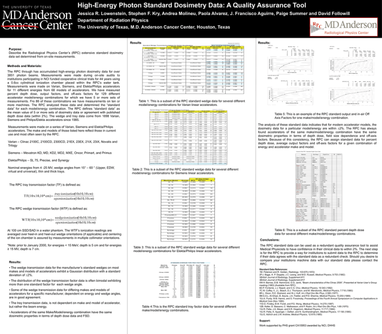
High-Energy Photon Standard Dosimetry Data: A Quality Assurance Tool Jessica R. Lowenstein, Stephen F. Kry, Andrea Molineu, Paola Alvarez, J. Francisco Aguirre, Paige Summer and David Followill Department of Radiation Physics The University of Texas, M.D. Anderson Cancer Center, Houston, Texas Results: Results: Purpose: Describe the Radiological Physics Center’s (RPC) extensive standard dosimetry data set determined from on-site measurements. b. d. Methods and Materials: The RPC through has accumulated high-energy photon dosimetry data for over 3851 photon beams. Measurements were made during on-site audits to institutions participating in NCI funded cooperative clinical trials for 44 years using a 0.6cc cylindrical ionization chamber placed within the RPC’s water tank. Measurements were made on Varian, Siemens, and Elekta/Philips accelerators for 11 different energies from 68 models of accelerators. We have measured percent depth dose, output factors, and off-axis factors for 129 different accelerator model/energy combinations for which we have 5 or more sets of measurements. Fro 86 of these combinations we have measurements on ten or more machines. The RPC analyzed these data and determined the “standard data” for each model/energy combination. The RPC defines “standard data” as the mean value of 5 or more sets of dosimetry data or agreement with published depth dose data (within 2%). The wedge and tray data come from 1898 Varian, Siemens and Philips/Elekta accelerators since 1985. Table 1: This is a subset of the RPC standard wedge data for several different model/energy combinations for Varian linear accelerators. Table 5: This is an example of the RPC standard output and in-air Off Axis Factors for one make/model/energy combination. The analysis of these standard data indicates that for modern accelerator models, the dosimetry data for a particular model/energy are within 2%. The RPC has always found accelerators of the same make/model/energy combination have the same dosimetric properties in terms of depth dose, field size dependence and off-axis factors. Because of this consistency, the RPC can assign standard data for percent depth dose, average output factors and off-axis factors for a given combination of energy and accelerator make and model. Measurements were made on a series of Varian, Siemens and Elekta/Philips accelerators. The make and models of those listed here reflect those in current use and most often seen by the RPC. Varian – Clinac 2100C, 2100CD, 2300CD, 21EX, 23EX, 21iX, 23iX, Novalis and Trilogy Siemens – Mevatron KD, MD, KD2, MD2, MXE, Oncor, Primart, and Primus Elekta/Philips – SL 75, Precise, and Synergy Nominal energies from 4 -25 MV, wedge angles from 15° – 60 ° (Upper, EDW, virtual and universal), thin and thick trays. Table 2: This is a subset of the RPC standard wedge data for several different model/energy combinations for Siemens linear accelerators. The RPC tray transmission factor (TF) is defined as: tray ionization(10x10, 10 cm) T F(10 x 10,10* cm) openionization(10x10, 10 cm) The RPC wedge transmission factor (WTF) is defined as: wedgeionization(10x10, 10 cm) WT F(10 x 10,10* cm) openionization(10x10, 10 cm) At 100 cm SSD/SAD in a water phantom. The WTF’s ionization readings are averaged over heel-in and heel-out wedge orientations (if applicable) and centering of the ion chamber is assured by measurements in multiple collimator orientations. *Note: prior to January 2000, for energies < 15 MeV, depth is 5 cm and for energies ≥ 15 MV, depth is 7 cm. Table 6: This is a subset of the RPC standard percent depth dose data for several different make/model/energy combinations. Conclusions: Table 3: This is a subset of the RPC standard wedge data for several different model/energy combinations for Elekta/Philips linear accelerators. Results: • The wedge transmission data for the manufacturer’s standard wedges on most makes and models of accelerators exhibit a Gaussian distribution with a standard deviation of 2%. • The distribution of the wedge transmission factor data is often bimodal exhibiting more than one standard factor for each wedge angle. • Some of the wedge transmission data for differing makes and models of accelerators for a specific manufacturer, dependent on energy and wedge angles, are in good agreement. • The tray transmission data, is not dependent on make and model of accelerator, but rather the beam energy. • Accelerators of the same Make/Model/energy combination have the same dosimetric properties in terms of depth dose data and FSD. Table 4:This is the RPC standard tray factor data for several different make/model/energy combinations. The RPC standard data can be used as a redundant quality assurance tool to assist Medical Physicists to have confidence in their clinical data to within 2%. The next step is for the RPC to provide a way for institutions to submit data to the RPC to determine if their data agrees with the standard data as a redundant check. Should you desire to compare your institutions machine data with our standard data please contact the RPC. Standard Data References: 1M. Peterson and R. Golden, Radiology, 103:675 (1972) 2P.J. Biggs, K. P. Doppke, J.C. Leong, and M.D. Russell, Medical Physics, 9:753 (1982) 3British Journal of Radiology, Supplement #11 4British Journal of Radiology, Supplement #17 5W.H. Barnes, D.B. Hammond, G.G. Janik, “Beam characteristics of the Clinac 2500”, Presented at Varian User’s Group meeting (1983) (Available from RPC) 6D.P. Fontenla. J.J. Napoli, and C.S. Chui, Medical Physics, 19:343 (1992) 7C.W. Coffey, II, J.L. Beach, D.J. Thompson, and M. Mendiondo, Medical Physics, 7:716 (1980) 8R.L. Dixon, R.E. Ekstrand, and W.J. Huff, Int J Rad Onc Bio Phys, 2:585 (1977) 9M.S.A.L. Al-Ghazi, B. Arjune, J.A. Fiedler, and P.D. Sharma, Medical Physics, 15:250 (1988) 10J.A. Purdy, W.B. Harms, and S. Fivozinsky, Proceedings of the Fourth Annual Symposium on Computer Applications in Medical Care (Nov 1980) 11D.O. Findley, B.W. Forell, and P.S. Wong, Medical Physics, 14:270 (1987) 12B. Keller, D. Bassano, C. Mathewson, and P. Rubin, Int J Rad Onc Bio Phys, 1:69 (1975) 13J.R. Palta, J.A. Meyer, and K.R. Hogstrom, Medical Physics, 11:717 (1984) 14J.R. Palta, K. Ayyanger, I. Daftari, and N. Suntharalingham, Medical Physics, 17:106 (1990) 15J.E. Aldrich and J.W. Andrew, Medical Physics, 12:619 (1985) Support: Work supported by PHS grant CA10953 awarded by NCI, DHHS
