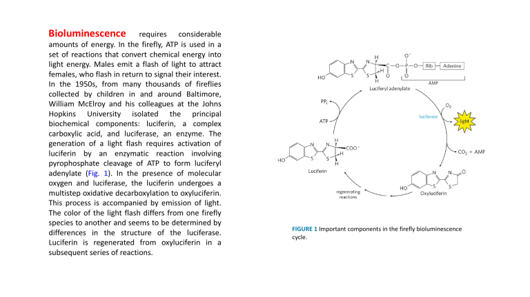
Bioluminescence requires considerable amounts of energy. In the firefly, ATP is used in a set of reactions that convert chemical energy into light energy. Males emit a flash of light to attract females, who flash in return to signal their interest. In the 1950s, from many thousands of fireflies collected by children in and around Baltimore, William McElroy and his colleagues at the Johns Hopkins University isolated the principal biochemical components: luciferin, a complex carboxylic acid, and luciferase, an enzyme. The generation of a light flash requires activation of luciferin by an enzymatic reaction involving pyrophosphate cleavage of ATP to form luciferyl adenylate (Fig. 1). In the presence of molecular oxygen and luciferase, the luciferin undergoes a multistep oxidative decarboxylation to oxyluciferin. This process is accompanied by emission of light. The color of the light flash differs from one firefly species to another and seems to be determined by differences in the structure of the luciferase. Luciferin is regenerated from oxyluciferin in a subsequent series of reactions. FIGURE 1 Important components in the firefly bioluminescence cycle. Bacteriorhodopsin, with only 247 amino acid residues, is the simplest light-driven proton pump known. • It is found in the plasma membrane of H. salinarum as the light- absorbing pigment, which contains retinal (the aldehyde derivative of vitamin A) as a light-harvesting prosthetic group. • Halobacterium salinarum lives only in brine ponds and salt where the high salt concentration—which can exceed 4 M—results from water loss by evaporation. These organisms are aerobes and normally use O2 to oxidize organic fuel molecules. However, the solubility of O2 is so low in brine ponds that sometimes oxidative metabolism must be supplemented by sunlight as an alternative source of energy. • When the cells are illuminated, all-trans-retinal bound to the bacteriorhodopsin absorbs a photon and undergoes photoisomerization to 13-cisretinal, forcing a conformational change in the protein. The restoration of all-trans-retinal is accompanied by outward movement of protons through the plasma membrane. • The chromophore retinal is bound through a Schiff-base linkage to the ε-amino group of a Lys residue. In the dark, the nitrogen of this Schiff base is protonated; in the light,photoisomerization of retinal lowers the pKa of this group and it releases its proton to a nearby Asp residue, triggering a series of proton hops that ultimately result in release of a proton at the outer surface of the membrane. • The electrochemical potential across the membrane drives protons back into the cell through a membrane ATP synthase complex very similar to that of mitochondria and chloroplasts. Thus, when O2 is limited, halobacteria can use light to supplement the ATP synthesized by oxidative phosphorylation. Halobacteria do not evolve O2, nor do they carry out photoreduction of NADP+; their phototransducing machinery is therefore much simpler than that of cyanobacteria or plants. FIGURE 20-29 A different mechanism for light-driven proton pumping evolved independently in a halophilic archaeon. (a) Bacteriorhodopsin (Mr 26,000) of Halobacterium halobium has seven membrane-spanning α helices. The chromophore all-trans-retinal (purple) is covalently attached via a Schiff base to the ε-amino group of a Lys residue deep in the membrane interior. Running through the protein are a series of Asp and Glu residues and a series of closely associated water molecules that together provide the transmembrane path for protons (red arrows). Steps through indicate proton movements, described below. (b) In the dark (left panel), the Schiff base is protonated. Illumination (right panel) photoisomerizes the retinal, forcing subtle conformational changes in the protein that alter the distance between the Schiff base and its neighboring amino acid residues. Interaction with these neighbors (Leu93 and Val49) lowers the pKa of the protonated Schiff base, and the base gives up its proton to a nearby carboxyl group on Asp85 (step 1 in (a)). This initiates a series of concerted proton hops between water molecules in the interior of the protein, which ends with 2 release of a proton that was shared by Glu194 and Glu204 near the extracellular surface. (Tyr83 forms a hydrogen bond with Glu194 that facilitates this proton release.) 3 The Schiff base reacquires a proton from Asp96, which 4 takes up a proton from the cytosol. Finally, Asp85 gives up its proton, leading to reprotonation of the Glu204– Glu194 pair. The system is now ready for another round of proton pumping. Efficiency of photophosphorylation As electrons move from water to NADP+ in plant chloroplasts, about 12 protons move from the stroma into the thylakoid lumen per four electrons passed (that is, per O2 formed). In illuminated chloroplasts, the energy stored in the proton gradient per mole of protons is so the movement of 12 mol of protons across the thylakoid membrane represents conservation of about 200 kJ of energy—enough energy to drive the synthesis of several moles of ATP (ΔGʹ° = 30.5 kJ/mol). Experimental measurements yield values of about 3 ATP per O2 produced. At least eight photons must be absorbed to drive four electrons from H2O to NADPH (one photon per electron at each reaction center). The energy in eight photons of visible light is more than enough for the synthesis of three molecules of ATP. ATP synthesis is not the only energy-conserving reaction of photosynthesis in plants; the NADPH formed in the final electron transfer is also energetically rich. The overall equation for noncyclic photophosphorylation (a term explained below) is
