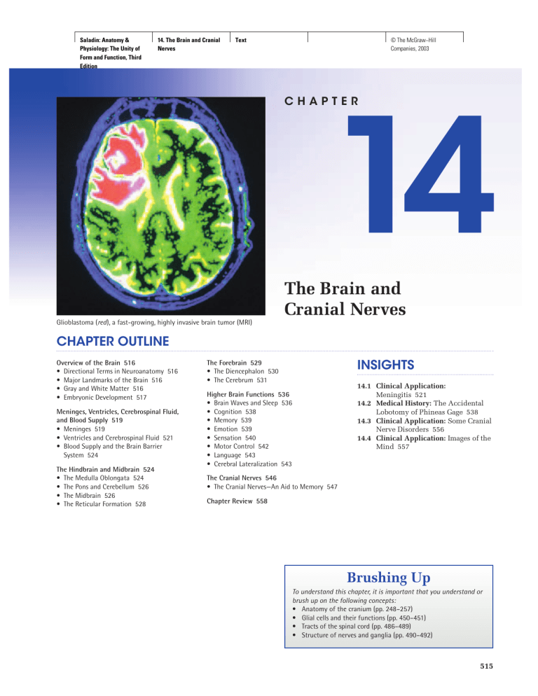
Saladin: Anatomy & Physiology: The Unity of Form and Function, Third Edition 14. The Brain and Cranial Nerves © The McGraw−Hill Companies, 2003 Text CHAPTER 14 The Brain and Cranial Nerves Glioblastoma (red), a fast-growing, highly invasive brain tumor (MRI) CHAPTER OUTLINE Overview of the Brain 516 • Directional Terms in Neuroanatomy 516 • Major Landmarks of the Brain 516 • Gray and White Matter 516 • Embryonic Development 517 Meninges, Ventricles, Cerebrospinal Fluid, and Blood Supply 519 • Meninges 519 • Ventricles and Cerebrospinal Fluid 521 • Blood Supply and the Brain Barrier System 524 The Hindbrain and Midbrain 524 • The Medulla Oblongata 524 • The Pons and Cerebellum 526 • The Midbrain 526 • The Reticular Formation 528 INSIGHTS The Forebrain 529 • The Diencephalon 530 • The Cerebrum 531 14.1 Clinical Application: Meningitis 521 14.2 Medical History: The Accidental Lobotomy of Phineas Gage 538 14.3 Clinical Application: Some Cranial Nerve Disorders 556 14.4 Clinical Application: Images of the Mind 557 Higher Brain Functions 536 • Brain Waves and Sleep 536 • Cognition 538 • Memory 539 • Emotion 539 • Sensation 540 • Motor Control 542 • Language 543 • Cerebral Lateralization 543 The Cranial Nerves 546 • The Cranial Nerves—An Aid to Memory 547 Chapter Review 558 Brushing Up To understand this chapter, it is important that you understand or brush up on the following concepts: • Anatomy of the cranium (pp. 248–257) • Glial cells and their functions (pp. 450–451) • Tracts of the spinal cord (pp. 486–489) • Structure of nerves and ganglia (pp. 490–492) 515 Saladin: Anatomy & Physiology: The Unity of Form and Function, Third Edition 14. The Brain and Cranial Nerves © The McGraw−Hill Companies, 2003 Text 516 Part Three Integration and Control T When you have completed this section, you should be able to • describe the major subdivisions and anatomical landmarks of the brain; and • describe the embryonic development of the CNS and relate this to adult brain anatomy. of its major landmarks (figs. 14.1 and 14.2). These will provide important points of reference as we progress through a more detailed study. The average adult brain weighs about 1,600 g (3.5 lb) in men and 1,450 g in women. Its size is proportional to body size, not intelligence—the Neanderthal people had larger brains than modern humans. The brain is divided into three major portions—the cerebrum, cerebellum, and brainstem. The cerebrum (SER-eh-brum or seh-REE-brum) constitutes about 83% of its volume and consists of a pair of cerebral hemispheres. Each hemisphere is marked by thick folds called gyri3 (JYrye; singular, gyrus) separated by shallow grooves called sulci4 (SUL-sye; singular, sulcus). A very deep groove, the longitudinal fissure, separates the right and left hemispheres from each other. At the bottom of this fissure, the hemispheres are connected by a thick bundle of nerve fibers called the corpus callosum5—a prominent landmark for anatomical description (fig. 14.1d). The cerebellum6 (SER-eh-BEL-um) lies inferior to the cerebrum and occupies the posterior cranial fossa. It is also marked by gyri, sulci, and fissures. The cerebellum is the second-largest region of the brain; it constitutes about 10% of its volume but contains over 50% of its neurons. The brainstem is what remains if the cerebrum and cerebellum are removed. In a living person, it is oriented like a vertical stalk with the cerebrum perched on top of it like a mushroom cap. Postmortem changes give it a more oblique angle in the cadaver and consequently in many medical illustrations. The major components of the brainstem, from rostral to caudal, are the diencephalon, midbrain, pons, and medulla oblongata. Some authorities, however, regard only the last three of these as the brainstem. The brainstem ends at the foramen magnum of the skull and the central nervous system (CNS) continues below this as the spinal cord. Directional Terms in Neuroanatomy Gray and White Matter Two directional terms often used to describe brain anatomy are rostral and caudal. Rostral1 means “toward the nose” and caudal2 means “toward the tail,” descriptions especially appropriate to rats and other mammals on which so much neuroanatomy has been done. In human brain anatomy, one structure is rostral to another if it is closer to the forehead and caudal to another if it is closer to the spinal cord. The brain, like the spinal cord, is composed of gray and white matter. Gray matter—the seat of the neuron cell bodies, dendrites, and synapses—forms a surface layer called the cortex over the cerebrum and cerebellum, and deeper masses called nuclei surrounded by white matter. The white matter thus lies deep to the cortical gray matter of the brain, opposite from the relationship of gray and white matter in the spinal cord. As in the spinal cord, the white matter is composed of tracts, or bundles of axons, which here connect one part of the brain to another. he mystique of the brain continues to intrigue modern biologists and psychologists even as it did the philosophers of antiquity. Aristotle interpreted the brain as a radiator for cooling the blood, but generations earlier, Hippocrates had expressed a more accurate view of its functions. “Men ought to know,” he said, “that from the brain, and from the brain only, arise our pleasures, joy, laughter and jests, as well as our sorrows, pains, griefs and tears. Through it, in particular, we think, see, hear, and distinguish the ugly from the beautiful, the bad from the good, the pleasant from the unpleasant.” Brain function is so strongly associated with what it means to be alive and human that the cessation of brain activity is taken as a clinical criterion of death even when other organs of the body are still functioning. With its hundreds of neuronal pools and trillions of synapses, the brain performs sophisticated tasks beyond our present understanding. Still, all of our mental functions, no matter how complex, are ultimately based on the cellular activities described in chapter 12. The relationship of the mind or personality to the cellular function of the brain is a question that will provide fertile ground for scientific study and philosophical debate long into the future. This chapter is a study of the brain and the cranial nerves directly connected to it. Here we will plumb some of the mysteries of motor control, sensation, emotion, thought, language, personality, memory, dreams, and plans. Your study of this chapter is one brain’s attempt to understand itself. Chapter 14 Overview of the Brain Objectives Major Landmarks of the Brain Before we consider the form and function of specific regions of the brain, it will help to get a general overview gy ⫽ turn, twist sulc ⫽ furrow, groove 5 corpus ⫽ body ⫹ call ⫽ thick 6 cereb ⫽ brain ⫹ ellum ⫽ little 3 4 rostr ⫽ nose caud ⫽ tail 1 2 Saladin: Anatomy & Physiology: The Unity of Form and Function, Third Edition 14. The Brain and Cranial Nerves © The McGraw−Hill Companies, 2003 Text Chapter 14 The Brain and Cranial Nerves 517 Cerebral hemispheres Rostral Caudal Central sulcus Frontal lobe Gyrus Cerebrum Lateral sulcus Central sulcus Longitudinal fissure Temporal lobe Parietal lobe Brainstem Cerebellum Occipital lobe Spinal cord (a) (c) Cerebral hemispheres Olfactory tracts Parietal lobe Frontal lobe Optic chiasm Parietooccipital sulcus Pituitary gland Corpus callosum Temporal lobe Thalamus Pons Hypothalamus Cranial nerves Midbrain Medulla oblongata Pons Occipital lobe Cerebellum Medulla oblongata Cerebellum Spinal cord (b) (d) Figure 14.1 Four Views of the Brain. (a) Superior; (b) inferior (base of the brain); (c) left lateral; (d ) median section (color indicates brainstem). Embryonic Development To understand the terminology of mature brain anatomy, it is necessary to be aware of the embryonic development of the CNS. The nervous system develops from ectoderm, the outermost germ layer of an embryo. By the third week of development, a dorsal streak called the neuroectoderm appears along the length of the embryo and thickens to form a neural plate (fig. 14.3). This is destined to give rise to all neurons and glial cells except microglia, which come from mesoderm. As development progresses, the neural plate sinks and forms a neural groove with a raised neural fold along each side. The neural folds then fuse along the midline, somewhat like a closing zipper. By 4 weeks, this process creates a hollow channel called the neural tube. The neural tube now separates from the overlying ectoderm, sinks a little deeper, and grows lateral processes that later form motor nerve fibers. The lumen of the neural tube develops into fluid-filled spaces called the central canal of the spinal cord and ventricles of the brain. Chapter 14 Central sulcus Frontal lobe Saladin: Anatomy & Physiology: The Unity of Form and Function, Third Edition 14. The Brain and Cranial Nerves © The McGraw−Hill Companies, 2003 Text 518 Part Three Integration and Control Precentral gyrus Central sulcus Postcentral gyrus Parietal lobe Frontal lobe Insula Occipital lobe Temporal lobe Cerebellum Posterior Medulla oblongata Chapter 14 (a) Corpus callosum Lateral ventricle Parieto-occipital sulcus Thalamus Hypothalamus Midbrain Cerebellum Fourth ventricle Pons Medulla oblongata (b) Figure 14.2 Dissections of the Brain. (a) Left lateral view with part of the left hemisphere cut away to expose the insula. The arachnoid mater is visible as a delicate translucent membrane over many of the sulci of the occipital lobe. (b) Median section, left lateral view. After reading about the ventricles of the brain, locate the cerebral aqueduct in figure b. Saladin: Anatomy & Physiology: The Unity of Form and Function, Third Edition 14. The Brain and Cranial Nerves © The McGraw−Hill Companies, 2003 Text Chapter 14 The Brain and Cranial Nerves 519 22 days 19 days Neural plate Level of section Neural crest Level of section Surface ectoderm 20 days 26 days Neural groove Level of section Neural folds Level of section Neural tube Surface ectoderm Figure 14.3 Embryonic Development of the CNS to the Neural Tube Stage. The left-hand figure in each box represents a dorsal view of the embryo and the right-hand figure shows a three-dimensional reconstruction at the indicated level. Before You Go On Answer the following questions to test your understanding of the preceding section: 1. List the three major parts of the brain and describe their locations. 2. Define gyrus and sulcus. 3. Explain how the five secondary brain vesicles arise from the neural tube. Meninges,Ventricles, Cerebrospinal Fluid, and Blood Supply Objectives When you have completed this section, you should be able to • describe the meninges of the brain; • describe the system of fluid-filled chambers within the brain; • discuss the production, circulation, and function of the cerebrospinal fluid that fills these chambers; and • explain the significance of the brain barrier system. Meninges pros ⫽ before, in front ⫹ encephal ⫽ brain 8 mes ⫽ middle 9 rhomb ⫽ rhombus 10 tele ⫽ end, remote 11 di ⫽ through, between 12 met ⫽ behind, beyond, distal to 13 myel ⫽ spinal cord 7 The meninges of the brain are basically the same as those of the spinal cord: dura mater, arachnoid mater, and pia mater (fig. 14.5). The dura mater, however, shows some significant differences. In the cranial cavity, it consists of two layers—an outer periosteal layer, equivalent to the periosteum of the cranial bone, and an inner meningeal layer. Only the meningeal layer continues into the vertebral Chapter 14 As the neural tube develops, some ectodermal cells that originally lay along the margin of the groove separate from the rest and form a longitudinal column on each side called the neural crest. Some neural crest cells become sensory neurons, while others migrate to other locations and become sympathetic neurons, Schwann cells, and other cell types. By the fourth week, the neural tube exhibits three anterior dilations, or primary vesicles, called the forebrain (prosencephalon7) (PROSS-en-SEF-uh-lon), midbrain (mesencephalon8) (MEZ-en-SEF-uh-lon), and hindbrain (rhombencephalon9) (ROM-ben-SEF-uh-lon) (fig. 14.4). By the fifth week, the neural tube undergoes further flexion and subdivides into five secondary vesicles. The forebrain divides into two of them, the telencephalon10 (TELen-SEF-uh-lon) and diencephalon11 (DY-en-SEF-uh-lon); the midbrain remains undivided and retains the name mesencephalon; and the hindbrain divides into two vesicles, the metencephalon12 (MET-en-SEF-uh-lon) and myelencephalon13 (MY-el-en-SEF-uh-lon). The telencephalon has a pair of lateral outgrowths that later become the cerebral hemispheres, and the diencephalon exhibits a pair of small cuplike optic vesicles that become the retinas of the eyes. Figure 14.4c shows structures of the fully developed brain that arise from each of the secondary vesicles. Saladin: Anatomy & Physiology: The Unity of Form and Function, Third Edition 14. The Brain and Cranial Nerves © The McGraw−Hill Companies, 2003 Text 520 Part Three Integration and Control Telencephalon Prosencephalon Optic vesicle Diencephalon Mesencephalon Metencephalon Rhombencephalon Myelencephalon Spinal cord 4 weeks 5 weeks (a) (b) Telencephalon Diencephalon Forebrain Mesencephalon (midbrain) Pons Cerebellum Metencephalon Hindbrain Myelencephalon (medulla oblongata) Chapter 14 (c) Spinal cord Figure 14.4 Primary and Secondary Vesicles of the Embryonic Brain. (a) The primary vesicles at 4 weeks; (b) the secondary vesicles at 5 weeks; (c) the fully developed brain, color-coded to relate its structures to the secondary embryonic vesicles. Periosteal Dura layer mater Meningeal layer Subdural space Skull Arachnoid mater Arachnoid villus Subarachnoid space Superior sagittal sinus Pia mater Blood vessel Falx cerebri (in longitudinal fissure only) Figure 14.5 The Meninges of the Brain. Frontal section of the head. Gray matter Brain White matter Saladin: Anatomy & Physiology: The Unity of Form and Function, Third Edition 14. The Brain and Cranial Nerves Text © The McGraw−Hill Companies, 2003 Chapter 14 The Brain and Cranial Nerves 521 canal. The cranial dura mater lies closely against the cranial bone, with no intervening epidural space like that of the spinal cord. In some places, the two layers are separated by dural sinuses, spaces that collect blood that has circulated through the brain. Two major dural sinuses are the superior sagittal sinus, found just under the cranium along the midsagittal line, and the transverse sinus, which runs horizontally from the rear of the head toward each ear. These sinuses meet like an inverted T at the back of the brain and ultimately empty into the internal jugular veins of the neck. In certain places, the meningeal layer of the dura mater folds inward to separate major parts of the brain: the falx14 cerebri (falks SER-eh-bry) extends into the longitudinal fissure between the right and left cerebral hemispheres; the tentorium15 (ten-TOE-ree-um) cerebelli stretches like a roof over the posterior cranial fossa and separates the cerebellum from the overlying cerebrum; and the falx cerebelli partially separates the right and left halves of the cerebellum on the inferior side. The arachnoid mater and pia mater are similar to those of the spinal cord. A subarachnoid space separates the arachnoid from the pia, and in some places, a subdural space separates the dura from the arachnoid. 15 Insight 14.1 Clinical Application Meningitis Meningitis—inflammation of the meninges—is one of the most serious diseases of infancy and childhood. It occurs especially between 3 months and 2 years of age. Meningitis is caused by a variety of bacteria and viruses that invade the CNS by way of the nose and throat, often following respiratory, throat, or ear infections. The pia mater and arachnoid are most likely to be affected, and from here the infection can spread to the adjacent nervous tissue. In bacterial meningitis, the brain swells, the ventricles enlarge, and the brainstem may have hemorrhages. Signs include a high fever, stiff neck, drowsiness, and intense headache and may progress to vomiting, loss of sensory and motor functions, and coma. Death can occur within hours of the onset. Infants and toddlers with a high fever should therefore receive immediate medical attention. Meningitis is diagnosed partly by examining the cerebrospinal fluid (CSF) for bacteria and white blood cells. The CSF is obtained by making a lumbar puncture (spinal tap) between two lumbar vertebrae and drawing fluid from the subarachnoid space. This site is chosen because it has an abundance of CSF and there is no risk of injury to the spinal cord, which does not extend into the lower lumbar vertebrae. Ventricles and Cerebrospinal Fluid The brain has four internal chambers called ventricles (fig. 14.6). The most rostral ones are the lateral ventricles, 1. Buoyancy. Because the brain and CSF are very similar in density, the brain neither sinks nor floats in the CSF but remains suspended in it—that is, the brain has neutral buoyancy. A human brain removed from the body weighs about 1,500 g, but when suspended in CSF its effective weight is only about 50 g. By analogy, consider how much easier it is to lift another person when you are standing in a lake than it is on land. Neutral buoyancy allows the brain to attain considerable size without being impaired by its own weight. If the brain rested heavily on the floor of the cranium, the pressure would kill the nervous tissue. 2. Protection. CSF also protects the brain from striking the cranium when the head is jolted. If the jolt is severe, however, the brain still may strike the inside of the cranium or suffer shearing injury from contact with the angular surfaces of the cranial floor. This is one of the common findings in child abuse (shaken child syndrome) and in head injuries (concussions) from auto accidents, boxing, and the like. 3. Chemical stability. CSF is secreted into each ventricle of the brain and is ultimately absorbed into the bloodstream. It provides a means of rinsing metabolic Chapter 14 falx ⫽ sickle tentorium ⫽ tent 14 which form a large arc in each cerebral hemisphere. Through a tiny passage called the interventricular foramen, each lateral ventricle is connected to the third ventricle, a narrow medial space inferior to the corpus callosum. From here, a canal called the cerebral aqueduct passes down the core of the midbrain and leads to the fourth ventricle, a small chamber between the pons and cerebellum. Caudally, this space narrows and forms a central canal that extends through the medulla oblongata into the spinal cord. These ventricles and canals are lined with ependymal cells, a type of neuroglia that resembles a simple cuboidal epithelium. Each ventricle contains a choroid (CO-royd) plexus, named for its histological resemblance to a fetal membrane called the chorion. The choroid plexus is a network of blood capillaries anchored to the floor or wall of the ventricle and covered by ependymal cells. A clear, colorless liquid called cerebrospinal fluid (CSF) fills the ventricles and canals of the CNS and bathes its external surface. The brain produces about 500 mL of CSF per day, but the fluid is constantly reabsorbed at the same rate and only 100 to 160 mL are present at one time. About 40% of it is formed in the subarachnoid space external to the brain, 30% by the general ependymal lining of the brain ventricles, and 30% by the choroid plexuses. CSF forms partly by the filtration of blood plasma through the choroid plexuses and other capillaries of the brain. The ependymal cells modify this filtrate, however, so the CSF has more sodium and chloride than the blood plasma, but less potassium, calcium, and glucose and very little protein. Cerebrospinal fluid serves three purposes: Saladin: Anatomy & Physiology: The Unity of Form and Function, Third Edition 14. The Brain and Cranial Nerves © The McGraw−Hill Companies, 2003 Text 522 Part Three Integration and Control Caudal Rostral Lateral ventricles Cerebrum Lateral ventricle Interventricular foramen Interventricular foramen Third ventricle Cerebral aqueduct Lateral aperture Third ventricle Cerebral aqueduct Fourth ventricle Median aperture Lateral aperture Fourth ventricle Median aperture Central canal (a) (b) Rostral (anterior) Longitudinal fissure Frontal lobe Gray matter (cortex) Chapter 14 White matter Corpus callosum (anterior part) Lateral ventricle Caudate nucleus Septum pellucidum Sulcus Gyrus Temporal lobe Third ventricle Lateral sulcus Insula Thalamus Choroid plexus Lateral ventricle Corpus callosum (posterior part) Longitudinal fissure Occipital lobe (c) Caudal (posterior) Figure 14.6 Ventricles of the Brain. (a) Right lateral aspect; (b) anterior aspect; (c) superior aspect of a horizontal section of the brain, showing the lateral ventricles and other features of the cerebrum. Saladin: Anatomy & Physiology: The Unity of Form and Function, Third Edition 14. The Brain and Cranial Nerves © The McGraw−Hill Companies, 2003 Text Chapter 14 The Brain and Cranial Nerves 523 wastes from the CNS and homeostatically regulating its chemical environment. Slight changes in its composition can cause malfunctions of the nervous system. For example, a high glycine concentration disrupts temperature and blood pressure control, and a high pH causes dizziness and fainting. along the way. A small amount of CSF fills the central canal of the spinal cord, but ultimately, all of it escapes through three pores in the walls of the fourth ventricle—a median aperture and two lateral apertures. These lead into the subarachnoid space on the brain surface. From this space, the CSF is absorbed by arachnoid villi, cauliflower-like extensions of the arachnoid meninx that protrude through the dura mater into the superior sagittal sinus. CSF penetrates the walls of the arachnoid villi and mixes with the blood in the sinus. Hydrocephalus16 is the abnormal accumulation of CSF in the brain, usually resulting from a blockage in its The CSF is not a stationary fluid but continually flows through and around the CNS, driven partly by its own pressure and partly by rhythmic pulsations of the brain produced by each heartbeat. The CSF secreted in the lateral ventricles flows through the interventricular foramina into the third ventricle (fig. 14.7) and then down the cerebral aqueduct to the fourth ventricle. The third and fourth ventricles and their choroid plexuses add more CSF hydro ⫽ water ⫹ cephal ⫽ head 16 Superior sagittal sinus 8 Arachnoid villus 1 Subarachnoid space 1 2 CSF flows through interventricular foramina into third ventricle. 2 Choroid plexus Third ventricle 3 3 4 5 Choroid plexus in third ventricle adds more CSF. 4 7 CSF flows down cerebral aqueduct to fourth ventricle. Cerebral aqueduct Choroid plexus in fourth ventricle adds more CSF. Lateral aperture 6 6 CSF flows out two lateral apertures and one median aperture. 7 CSF fills subarachnoid space and bathes external surfaces of brain and spinal cord. 8 At arachnoid villi, CSF is resorbed into venous blood of dural venous sinuses. 5 Median aperture Central canal of spinal cord Subarachnoid space of spinal cord Figure 14.7 The Flow of Cerebrospinal Fluid. Locate the sites of the obstructions that cause hydrocephalus. Chapter 14 CSF is secreted by choroid plexus in each lateral ventricle. Saladin: Anatomy & Physiology: The Unity of Form and Function, Third Edition 14. The Brain and Cranial Nerves © The McGraw−Hill Companies, 2003 Text 524 Part Three Integration and Control route of flow and reabsorption. Such obstructions occur most often in the interventricular foramen, cerebral aqueduct, and the apertures of the fourth ventricle. The accumulated CSF makes the ventricles expand and compress the nervous tissue, with potentially fatal consequences. In a fetus or infant, it can cause the entire head to enlarge because the cranial bones are not yet fused. Good recovery can be achieved if a tube (shunt) is inserted to drain fluid from the ventricles into a vein of the neck. Blood Supply and the Brain Barrier System Chapter 14 Although the brain constitutes only 2% of the adult body weight, it receives 15% of the blood (about 750 mL/min) and consumes 20% of the body’s oxygen and glucose. Because neurons have such a high demand for ATP, and therefore glucose and oxygen, the constancy of blood supply is especially critical to the nervous system. A mere 10second interruption in blood flow can cause loss of consciousness; an interruption of 1 to 2 minutes can significantly impair neural function; and 4 minutes without blood causes irreversible brain damage. Despite its critical importance to the brain, the blood is also a source of antibodies, macrophages, and other potentially harmful agents. Consequently, there is a brain barrier system that strictly regulates what substances can get from the bloodstream into the tissue fluid of the brain. There are two potential points of entry that must be guarded: the blood capillaries throughout the brain tissue and the capillaries of the choroid plexuses. At the former site, the brain is well protected by the blood-brain barrier (BBB), which consists mainly of the tightly joined endothelial cells that form the blood capillaries and partly of the basement membrane surrounding them. In the developing brain, astrocytes reach out and contact the capillaries with their perivascular feet. They stimulate the endothelial cells to form tight junctions, which seal off the capillaries and ensure that anything leaving the blood must pass through the cells and not between them. At the choroid plexuses, the brain is protected by a similar blood-CSF barrier, composed of ependymal cells joined by tight junctions. Tight junctions are absent from ependymal cells elsewhere, because it is important to allow exchanges between the brain tissue and CSF. That is, there is no brain-CSF barrier. The brain barrier system (BBS) is highly permeable to water, glucose, and lipid-soluble substances such as oxygen, carbon dioxide, alcohol, caffeine, nicotine, and anesthetics. It is slightly permeable to sodium, potassium, chloride, and the waste products urea and creatinine. While the BBS is an important protective device, it is an obstacle to the delivery of drugs to treat brain diseases. Trauma and inflammation sometimes damage the BBS and allow pathogens to enter the brain tissue. Furthermore, there are places called circumventricular organs (CVOs) in the third and fourth ventricles where the barrier system is absent and the blood does have direct access to the brain. These enable the brain to monitor and respond to fluctuations in blood glucose, pH, osmolarity, and other variables. Unfortunately, the CVOs also afford a route for the human immunodeficiency virus (HIV, the AIDS virus) to invade the brain. Before You Go On Answer the following questions to test your understanding of the preceding section: 4. Name the three meninges from superficial to deep. 5. Describe three functions of the cerebrospinal fluid. 6. Where does the CSF originate and what route does it take through and around the CNS? 7. Name the two components of the brain barrier system and explain the importance of this system. The Hindbrain and Midbrain Objectives When you have completed this section, you should be able to • list the components of the hindbrain and midbrain and their functions; • discuss the role of the cerebellum in movement and equilibrium; • define the term brainstem and describe its anatomical relationship to the cerebellum and forebrain; and • describe the location and functions of the reticular formation. Our study of the brain is organized around the five secondary vesicles of the embryonic brain and their mature derivatives. We proceed in a caudal to rostral direction, beginning with the hindbrain and its relatively simple functions and progressing to the forebrain, the seat of such complex functions as thought, memory, and emotion. The Medulla Oblongata As noted earlier, the embryonic hindbrain differentiates into two subdivisions, the myelencephalon and metencephalon (see fig. 14.4). The myelencephalon develops into just one structure, the medulla oblongata (meh-DULLuh OB-long-GAH-ta). The medulla is about 3 cm long and superficially looks like an extension of the spinal cord, but slightly wider. Significant differences are seen, however, on closer inspection of its gross and microscopic anatomy. The anterior surface bears a pair of clublike ridges, the pyramids. Resembling two side-by-side baseball bats, the pyramids are wider at the rostral end, taper caudally, and are separated by an anterior median fissure continuous with that of the spinal cord (fig. 14.8). The pyramids Saladin: Anatomy & Physiology: The Unity of Form and Function, Third Edition 14. The Brain and Cranial Nerves © The McGraw−Hill Companies, 2003 Text Chapter 14 The Brain and Cranial Nerves 525 Diencephalon Thalamus Infundibulum Optic tract Cranial nerves Optic nerve (II) Cerebral peduncle Oculomotor nerve (III) Trochlear nerve (IV) Mammillary body Pons Trigeminal nerve (V) Abducens nerve (VI) Facial nerve (VII) Medulla oblongata Pyramid Anterior median fissure Pyramidal decussation Vestibulocochlear nerve (VIII) Glossopharyngeal nerve (IX) Vagus nerve (X) Accessory nerve (XI) Hypoglossal nerve (XII) Spinal nerves Spinal cord (a) Chapter 14 Diencephalon Thalamus Pineal gland Lateral geniculate body Midbrain Superior colliculus Medial geniculate body Inferior colliculus Optic tract Cerebral peduncle Pons Hindbrain Fourth ventricle Medulla oblongata Superior cerebellar peduncle Middle cerebellar peduncle Inferior cerebellar peduncle Olive Cuneate fasciculus Gracile fasciculus Spinal cord (b) Figure 14.8 The Brainstem. (a) Ventral aspect; (b) right dorsolateral aspect. Some authorities do not include the diencephalon in the brainstem. Saladin: Anatomy & Physiology: The Unity of Form and Function, Third Edition 14. The Brain and Cranial Nerves © The McGraw−Hill Companies, 2003 Text 526 Part Three Integration and Control contain corticospinal tracts of nerve fibers that carry motor signals from the cerebrum to the spinal cord, ultimately to stimulate the skeletal muscles. Most of these motor nerve fibers decussate at a visible point near the caudal end of the pyramids. As a result, each side of the brain controls muscles on the contralateral side of the body. (This pertains only to muscles below the head.) Lateral to each pyramid is an elevated area called the olive. It contains a wavy layer of gray matter, the inferior olivary nucleus, which is a center that receives information from many levels of the brain and spinal cord and relays it mainly to the cerebellum. Dorsally, the medulla exhibits ridges called the gracile fasciculus and cuneate fasciculus. These are continuations of the spinal cord tracts of the same names that carry sensory signals to the brain. In addition to ascending and descending nerve tracts, the medulla contains sensory nuclei that receive input from the taste buds, pharynx, and viscera of the thoracic and abdominal cavities, and motor nuclei that control several primitive visceral and somatic functions. Some motor nuclei of the medulla discussed later in the book are: tion of the pons and medulla. The pons contains nuclei that relay signals from the cerebrum to the cerebellum, and nuclei concerned with sleep, hearing, equilibrium, taste, eye movements, facial expressions, facial sensation, respiration, swallowing, bladder control, and posture. The Cerebellum The embryonic metencephalon develops into two structures, the pons and cerebellum. The cerebellum is the largest part of the hindbrain (fig. 14.9). It consists of right and left cerebellar hemispheres connected by a narrow bridgelike vermis.18 Three pairs of stalklike cerebellar peduncles19 (peh-DUN-culs) connect the cerebellum to the brainstem: the inferior peduncles to the medulla oblongata, the middle peduncles to the pons, and the superior peduncles to the midbrain. These are composed of the nerve fibers that carry all signals between the cerebellum and the rest of the brain. The cerebellum receives most of its input from the pons, via the middle peduncles, but the spinocerebellar tracts enter through the inferior peduncles. Motor output leaves the cerebellum by way of the superior peduncles. Each hemisphere exhibits slender, parallel folds called folia20 (gyri) separated by shallow sulci. The cerebellum has a surface cortex of gray matter and a deeper layer of white matter. In a sagittal section, the white matter, called the arbor vitae,21 exhibits a branching, fernlike pattern. Each hemisphere has four pairs of deep nuclei, masses of gray matter embedded in the white matter. All input to the cerebellum goes to the cortex and all of its output comes from the deep nuclei. The cerebellum contains about 100 billion neurons. The most distinctive of these are the Purkinje22 (pur-KINjee) cells—unusually large, globose neurons with a tremendous profusion of dendrites (see fig. 12.5, p. 448), arranged in a single row in the cortex. Their axons travel to the deep nuclei, where they synapse on output neurons that issue fibers to the brainstem. The cerebellum is mostly concerned with muscular coordination and will be discussed in a later section on motor control. Evidence is also emerging that the cerebellum plays a role in judging the passage of time, in some other cognitive processes (awareness, judgment, and memory), and in emotion. The Pons The Midbrain • the cardiac center, which adjusts the rate and force of the heartbeat; Chapter 14 • the vasomotor center, which adjusts blood vessel diameter to regulate blood pressure and reroute blood from one part of the body to another; and • two respiratory centers, which control the rate and depth of breathing. Other nuclei of the medulla are concerned with speech, coughing, sneezing, salivation, swallowing, gagging, vomiting, gastrointestinal secretion, sweating, and movements of the tongue and head. Many of the medulla’s sensory and motor functions are mediated through the last four cranial nerves, which begin or end here: cranial nerves IX (glossopharyngeal), X (vagus), XI (accessory), and XII (hypoglossal). The Pons and Cerebellum 17 The pons appears as an anterior bulge in the brainstem rostral to the medulla (fig. 14.8). Its white matter includes tracts that conduct signals from the cerebrum down to the cerebellum and medulla, and tracts that carry sensory signals up to the thalamus. Cranial nerve V (trigeminal) arises from the pons, and cranial nerves VI (abducens), VII (facial), and VIII (vestibulocochlear) arise from the juncpons ⫽ bridge 17 The embryonic mesencephalon develops into just one mature brain structure, the midbrain—a short segment of the brainstem that connects the hindbrain and fore- verm ⫽ worm ped ⫽ foot ⫹ uncle ⫽ little 20 foli ⫽ leaf 21 “tree of life” 22 Johannes E. von Purkinje (1787–1869), Bohemian anatomist 18 19 Saladin: Anatomy & Physiology: The Unity of Form and Function, Third Edition 14. The Brain and Cranial Nerves © The McGraw−Hill Companies, 2003 Text Chapter 14 The Brain and Cranial Nerves 527 Pineal gland Posterior commissure Superior colliculus Inferior colliculus Cerebral aqueduct Mammillary body Midbrain White matter (arbor vitae) Oculomotor nerve Gray matter Fourth ventricle Pons Medulla oblongata (a) Anterior Anterior lobe Vermis Posterior lobe Folia Posterior (b) Figure 14.9 The Cerebellum. (a) Midsagittal section, showing relationship to the brainstem; (b) superior aspect. brain (see figs. 14.1d, 14.2b, and 14.8). It contains the cerebral aqueduct and gives rise to two cranial nerves that control eye movements: cranial nerve III (oculomotor) and IV (trochlear). Some major regions of the midbrain are (fig. 14.10): • The cerebral peduncles, which help to anchor the cerebrum to the brainstem. The corticospinal tracts pass through the peduncles on their way to the medulla. • The tegmentum,23 the main mass of the midbrain, located between the cerebral peduncles and cerebral aqueduct. It contains the red nucleus, which has a pink color in life because of its high density of blood vessels. Fibers from the red nucleus form the tegmen ⫽ cover 23 rubrospinal tract in most mammals, but in humans its connections go mainly to and from the cerebellum, with which it collaborates in fine motor control. • The substantia nigra24 (sub-STAN-she-uh NY-gruh), a dark gray to black nucleus pigmented with melanin, located between the peduncles and the tegmentum. This is a motor center that relays inhibitory signals to the thalamus and basal nuclei (both of which are discussed later). Degeneration of the neurons in the substantia nigra leads to the muscle tremors of Parkinson disease (see insight 12.4, p. 475). • The central (periaqueductal) gray matter, a large arrowhead-shaped region of gray matter surrounding the cerebral aqueduct. It is involved with the substantia ⫽ substance ⫹ nigra ⫽ black 24 Chapter 14 Cerebellar hemisphere Saladin: Anatomy & Physiology: The Unity of Form and Function, Third Edition 14. The Brain and Cranial Nerves © The McGraw−Hill Companies, 2003 Text 528 Part Three Integration and Control Plane of section Posterior Superior colliculus Cerebral aqueduct Tectum Medial geniculate nucleus Reticular formation Central gray matter Oculomotor nucleus Tegmentum Medial lemniscus Red nucleus Substantia nigra Cerebral peduncle Oculomotor nerve (III) Chapter 14 Anterior Figure 14.10 Cross Section of the Midbrain. reticulospinal tracts in controlling our awareness of pain (see chapter 16). • The tectum,25 a rooflike region dorsal to the aqueduct. It consists of four nuclei, the corpora quadrigemina,26 which bulge from the midbrain roof. The two superior nuclei, called the superior colliculi27 (col-LIC-youlye), function in visual attention, visually tracking moving objects, and such reflexes as turning the eyes and head in response to a visual stimulus, for example to look at something that you catch sight of in your peripheral vision. The two inferior colliculi receive afferent signals from the inner ear and relay them to other parts of the brain, especially the thalamus. Among other functions, they mediate the reflexive turning of the head in response to a sound. • The medial lemniscus, a continuation of the gracile and cuneate tracts of the spinal cord and brainstem. The reticular formation, discussed next, is a prominent feature of the midbrain but is not limited to this region. tectum ⫽ roof, cover corpora ⫽ bodies ⫹ quadrigemina ⫽ quadruplets 27 colli ⫽ hill ⫹ cul ⫽ little 25 26 Think About It Why are the inferior colliculi shown in figure 14.9 but not in figure 14.10? How are these two figures related? The Reticular Formation Running vertically through the core of the midbrain, pons, and medulla is a loosely organized core of gray matter called the reticular formation, composed of more than 100 small nuclei, including several of those already discussed, mingled with bundles of nerve fibers (fig. 14.11). The functions of the reticular formation include the following: • Somatic motor control. Some motor neurons of the cerebral cortex send their axons to reticular formation nuclei, which then give rise to the reticulospinal tracts of the spinal cord. These tracts modulate (adjust) muscle contraction to maintain tone, balance, and posture. The reticular formation also relays signals from the eyes and ears to the cerebellum so the cerebellum can integrate visual, auditory, and vestibular (balance and motion) stimuli into its role in motor coordination. Other reticular formation motor nuclei include gaze centers, which Saladin: Anatomy & Physiology: The Unity of Form and Function, Third Edition 14. The Brain and Cranial Nerves © The McGraw−Hill Companies, 2003 Text Chapter 14 The Brain and Cranial Nerves 529 Radiations to cerebral cortex Reticular formation Auditory input Visual input Descending motor fibers Ascending general sensory fibers Figure 14.11 The Reticular Formation. The formation consists of over 100 nuclei scattered through the brainstem region indicated in red. Arrows represent the breadth of its projections to and from the cerebral cortex and other CNS regions. • Cardiovascular control. The reticular formation includes the cardiac center and vasomotor center of the medulla oblongata. Before You Go On Answer the following questions to test your understanding of the preceding section: 8. Name the visceral functions controlled by nuclei of the medulla. 9. Describe the general functions of the cerebellum. 10. What are some functions of the midbrain nuclei? 11. Describe the reticular formation and list several of its functions. • Pain modulation. The reticular formation is the origin of the descending analgesic pathways mentioned in the earlier description of the reticulospinal tracts. • Sleep and consciousness. The reticular formation has projections to the cerebral cortex and thalamus that allow it some control over what sensory signals reach the cerebrum and come to our conscious attention. It plays a central role in states of consciousness such as alertness and sleep. Injury to the reticular formation can result in irreversible coma. General anesthetics work by blocking signal transmission through the reticular formation. The reticular formation also is involved in habituation—a process in which the brain learns to ignore repetitive, inconsequential stimuli while remaining sensitive to others. In a noisy city, for example, a person can sleep through traffic sounds but wake promptly to the sound of an alarm clock or a crying baby. Reticular formation nuclei that modulate activity of the cerebral cortex are called the reticular activating system or extrathalamic cortical modulatory system. The Forebrain Objectives When you have completed this section, you should be able to • name the three major components of the diencephalon and describe their locations and functions; • identify the five lobes of the cerebrum; • describe the three types of tracts in the cerebral white matter; • describe the distinctive cell types and histological arrangement of the cerebral cortex; and • describe the location and functions of the basal nuclei and limbic system. The forebrain consists of the diencephalon and telencephalon. The diencephalon encloses the third ventricle and is the most rostral part of the brainstem. The telencephalon develops chiefly into the cerebrum. Chapter 14 enable the eyes to track and fixate on objects, and central pattern generators—neuronal pools that produce rhythmic signals to the muscles of breathing and swallowing. Saladin: Anatomy & Physiology: The Unity of Form and Function, Third Edition 14. The Brain and Cranial Nerves © The McGraw−Hill Companies, 2003 Text 530 Part Three Integration and Control The Diencephalon The embryonic diencephalon has three major derivatives: the thalamus, hypothalamus, and epithalamus. The Thalamus The thalamus28 (fig. 14.12) is about four-fifths of the diencephalon. It consists of an oval mass of gray matter that underlies each cerebral hemisphere, protrudes into the lateral ventricle, and medially protrudes into the third ventricle. In about 70% of people, a narrow intermediate mass connects the right and left halves to each other. The thalamus is the “gateway to the cerebral cortex.” Nearly all sensory input and other information going to thalamus ⫽ chamber, inner room 28 Thalamus Intermediate mass the cerebrum passes by way of synapses in the thalamus, and the thalamus receives input from several areas of the cerebrum involved in motor control. The thalamus has several nuclei that integrate sensory information originating throughout the body and direct it to the appropriate processing centers in the cerebrum. It is heavily interconnected with the limbic system (discussed later) and thus involved in its emotional and memory functions. It is also involved in arousal, eye movements, taste, smell, hearing, equilibrium, and the somesthetic senses. Its involvement in motor and sensory circuits is further discussed later in this chapter and in chapter 16. The Hypothalamus The hypothalamus (fig. 14.12) forms part of the walls and floor of the third ventricle. It extends anteriorly to the optic chiasm (ky-AZ-um), where the optic nerves meet, and posteriorly to a pair of humps called the mammil- Chapter 14 Lateral group Somesthetic input to association areas; contributes to limbic system Medial group Emotional input to prefrontal cortex; awareness of emotions Ventral group Somesthetic input to postcentral gyrus; signals from cerebellum and basal nuclei to primary motor and motor association areas Anterior group Part of limbic system Lateral geniculate nucleus Visual signals to occipital lobe Mammillary body Relays signals from limbic system to thalamus Paraventricular nucleus Produces oxytocin (involved in childbirth, lactation, orgasm); autonomic motor effects; control of posterior pituitary Anterior nucleus Thirst center Ventromedial nucleus Satiety center (supresses hunger); emotion Preoptic nucleus Thermoregulation; control of female reproductive cycle; sexual behavior Supraoptic nucleus Produces antidiuretic hormone (involved in water conservation); control of posterior pituitary Suprachiasmatic nucleus Biological clock; regulates circardian rhythms and female reproductive cycle Hypothalamus Optic chiasm Optic nerve Pituitary gland Figure 14.12 The Diencephalon. Only some of the nuclei of the thalamus and hypothalamus are shown, and only some of their functions listed. These lists are by no means complete. Saladin: Anatomy & Physiology: The Unity of Form and Function, Third Edition 14. The Brain and Cranial Nerves © The McGraw−Hill Companies, 2003 Text Chapter 14 The Brain and Cranial Nerves 531 lary29 bodies. The mammillary bodies relay signals from the limbic system to the thalamus. The pituitary gland is attached to the hypothalamus by a stalk (infundibulum) between the optic chiasm and mammillary bodies. The hypothalamus is the major control center of the autonomic nervous system and endocrine system and plays an essential role in the homeostatic regulation of nearly all organs of the body. Its nuclei include centers concerned with a wide variety of visceral functions. • Hormone secretion. The hypothalamus secretes hormones that control the anterior pituitary gland. Acting through the pituitary, the hypothalamus regulates growth, metabolism, reproduction, and stress responses. The hypothalamus also produces two hormones that are stored in the posterior pituitary gland, concerned with labor contractions, lactation, and water conservation. These relationships are explored especially in chapter 17. • Autonomic effects. The hypothalamus is a major integrating center for the autonomic nervous system. It sends descending fibers to nuclei lower in the brainstem that influence heart rate, blood pressure, gastrointestinal secretion and motility, and pupillary diameter, among other functions. • Food and water intake. Neurons of the hunger and satiety centers monitor blood glucose and amino acid levels and produce sensations of hunger and satiety. Hypothalamic neurons called osmoreceptors monitor the osmolarity of the blood and stimulate the hypothalamic thirst center when we are dehydrated. Dehydration also stimulates the hypothalamus to produce antidiuretic hormone, which conserves water by reducing urine output. • Sleep and circadian rhythms. The caudal part of the hypothalamus is part of the reticular formation. It contains nuclei that regulate falling asleep and waking. Superior to the optic chiasm, the hypothalamus contains a suprachiasmatic nucleus that controls our 24-hour (circadian) rhythm of activity. • Memory. The mammillary bodies of the hypothalamus lie in the pathway of signals traveling from the hippocampus, an important memory center of the mammill ⫽ nipple 29 • Emotional behavior. Hypothalamic centers are involved in a variety of emotional responses including anger, aggression, fear, pleasure, and contentment; and in sexual drive, copulation, and orgasm. The Epithalamus The epithalamus consists mainly of the pineal gland (an endocrine gland discussed in chapter 17), the habenula (a relay from the limbic system to the midbrain), and a thin roof over the third ventricle. The Cerebrum The embryonic telencephalon becomes the cerebrum, the largest and most conspicuous part of the human brain. Your cerebrum enables you to turn these pages, read and comprehend the words, remember ideas, talk about them with your peers, and take an examination. It is the seat of your sensory perception, memory, thought, judgment, and voluntary motor actions. It is the most challenging frontier of neurobiology. Gross Anatomy The surface of the cerebrum, including its gray matter (cerebral cortex) and part of the white matter, is folded into gyri that allow a greater amount of cortex to fit in the cranial cavity. These folds give the cerebrum a surface area of about 2,500 cm2, comparable to 41⁄2 pages of this book. If the cerebrum were smooth-surfaced, it would have only onethird as much area and proportionately less informationprocessing capability. This extensive folding is one of the greatest differences between the human brain and the relatively smooth-surfaced brains of most other mammals. Some gyri have consistent and predictable anatomy, while others vary from brain to brain and from the right hemisphere to the left. Certain unusually prominent sulci divide each hemisphere into five anatomically and functionally distinct lobes, listed next. The first four lobes are visible superficially and are named for the cranial bones overlying them (fig. 14.13); the fifth lobe is not visible from the surface. 1. The frontal lobe lies immediately behind the frontal bone, superior to the orbits. Its posterior boundary is the central sulcus. The frontal lobe is chiefly concerned with voluntary motor functions, motivation, foresight, planning, memory, mood, emotion, social judgment, and aggression. 2. The parietal lobe forms the uppermost part of the brain and underlies the parietal bone. Its rostral Chapter 14 • Thermoregulation. The hypothalamic thermostat is a nucleus that monitors blood temperature. When the temperature becomes too high or too low, the thermostat signals other hypothalamic nuclei—the heat-losing center or heat-producing center, respectively, which control cutaneous vasodilation and vasoconstriction, sweating, shivering, and piloerection. brain, to the thalamus. Thus they are important in memory, and lesions to the mammillary bodies result in memory deficits. (Memory is discussed more fully later in this chapter.) Saladin: Anatomy & Physiology: The Unity of Form and Function, Third Edition 14. The Brain and Cranial Nerves © The McGraw−Hill Companies, 2003 Text 532 Part Three Integration and Control Frontal lobe Parietal lobe Precentral gyrus Postcentral gyrus Central sulcus Occipital lobe Lateral sulcus Cerebellum Temporal lobe Figure 14.13 Lobes of the Cerebrum. The insula is not visible from the surface (see fig. 14.2a). Chapter 14 boundary is the central sulcus and its caudal boundary is the parieto-occipital sulcus, visible on the medial surface of each hemisphere (see fig. 14.1d ). This lobe is concerned with the sensory reception and integration of somesthetic, taste, and some visual information. 3. The occipital lobe is at the rear of the head underlying the occipital bone. It is the principal visual center of the brain. 4. The temporal lobe is a lateral, horizontal lobe deep to the temporal bone, separated from the parietal lobe above it by a deep lateral sulcus. It is concerned with hearing, smell, learning, memory, visual recognition, and emotional behavior. 5. The insula30 is a small mass of cortex deep to the lateral sulcus, made visible only by retracting or cutting away some of the overlying cerebrum (see figs. 14.2a, 14.6c, and 14.16). It is not as accessible to study in living people as other parts of the cortex and is still little-known territory. It apparently plays roles in understanding spoken language, in the sense of taste, and in integrating sensory information from visceral receptors. between the cerebrum and lower brain centers. These fibers are arranged in three types of tracts (fig. 14.14): 1. Projection tracts extend vertically from higher to lower brain or spinal cord centers and carry information between the cerebrum and the rest of the body. The corticospinal tracts, for example, carry motor signals from the cerebrum to the brainstem and spinal cord. Other projection tracts carry signals upward to the cerebral cortex. Superior to the brainstem, such tracts form a dense band called the internal capsule between the thalamus and basal nuclei (described shortly), then radiate in a diverging, fanlike array (the corona radiata31) to specific areas of the cortex. 2. Commissural tracts cross from one cerebral hemisphere to the other through bridges called commissures (COM-ih-shurs). The great majority of commissural fibers pass through the large C-shaped corpus callosum (see fig. 14.1d), which forms the floor of the longitudinal fissure. A few tracts pass through the much smaller anterior and posterior commissures. Commissural tracts enable the two sides of the cerebrum to communicate with each other. 3. Association tracts connect different regions of the same hemisphere. Long association fibers connect different lobes of a cerebral hemisphere to each other, whereas short association fibers connect The Cerebral White Matter Most of the volume of the cerebrum is white matter. This is composed of glia and myelinated nerve fibers that transmit signals from one region of the cerebrum to another and insula ⫽ island 30 31 corona ⫽ crown ⫹ radiata ⫽ radiating Saladin: Anatomy & Physiology: The Unity of Form and Function, Third Edition 14. The Brain and Cranial Nerves © The McGraw−Hill Companies, 2003 Text Chapter 14 The Brain and Cranial Nerves 533 Association tracts Parietal lobe Frontal lobe Corpus callosum Temporal lobe Occipital lobe (a) Longitudinal fissure Corpus callosum Commissural tracts Lateral ventricle Thalamus Third ventricle Basal nuclei Mammillary body Cerebral peduncle Pons Pyramid Decussation in pyramids Medulla oblongata (b) Figure 14.14 Tracts of Cerebral White Matter. (a) Left lateral aspect, showing association tracts; (b) frontal section, showing commissural and projection tracts. What route can commissural tracts take other than the one shown here? different gyri within a single lobe. Among their roles, association tracts link perceptual and memory centers of the brain; for example, they enable you to smell a rose, name it, and picture what it looks like. The Cerebral Cortex Neural integration is carried out in the gray matter of the cerebrum, which is found in three places—the cerebral cortex, basal nuclei, and limbic system. We begin with the cerebral cortex,32 a layer covering the surface of the hemispheres. Even though it is only 2 to 3 mm thick, the cortex constitutes about 40% of the mass of the brain and contains 14 to 16 billion neurons. It is composed of two principal types of neurons (fig. 14.15): (1) Stellate cells have spheroidal somas with dendrites projecting for short distances in all directions. They are concerned largely with receiving cortex ⫽ bark, rind 32 sensory input and processing information on a local level. (2) Pyramidal cells are tall and conical (triangular in tissue sections). Their apex points toward the brain surface and has a thick dendrite with many branches and small, knobby dendritic spines. The base gives rise to horizontally oriented dendrites and an axon that passes into the white matter. Pyramidal cells are the output neurons of the cerebrum— they transmit signals to other parts of the CNS. Their axons have collaterals that synapse with other neurons in the cortex or in deeper regions of the brain. About 90% of the human cerebral cortex is a sixlayered tissue called neocortex33 because of its relatively recent evolutionary origin. Although vertebrates have existed for about 600 million years, the neocortex did not develop significantly until about 60 million years ago, when there was a sharp increase in the diversity of mammals. It attained its highest development by neo ⫽ new 33 Chapter 14 Projection tracts Saladin: Anatomy & Physiology: The Unity of Form and Function, Third Edition 14. The Brain and Cranial Nerves © The McGraw−Hill Companies, 2003 Text 534 Part Three Integration and Control The Basal Nuclei Cortical surface I Small pyramidal cells II III Stellate cells IV Large pyramidal cells Chapter 14 V VI White matter Figure 14.15 Histology of the Neocortex. Neurons are arranged in six layers. Are the long processes leading upward from each pyramidal cell body dendrites or axons? far in the primates. The six layers of neocortex, numbered in figure 14.15, vary from one part of the cerebrum to another in relative thickness, cellular composition, synaptic connections, size of the neurons, and destination of their axons. Layer IV is thickest in sensory regions and layer V in motor regions, for example. All axons that leave the cortex and enter the white matter arise from layers III, V, and VI. Some regions of cerebral cortex have fewer than six layers. The earliest type of cerebral cortex to appear in vertebrate evolution was a one- to five-layered tissue called paleocortex (PALE-ee-oh-cor-tex), limited in humans to part of the insula and certain areas of the temporal lobe concerned with smell. The next to evolve was a three-layered archicortex (AR-kee-cor-tex), found in the human hippocampus. The neocortex was the last to evolve. The basal nuclei are masses of cerebral gray matter buried deep in the white matter, lateral to the thalamus (fig. 14.16). They are often called basal ganglia, but the word ganglion is best restricted to clusters of neurons outside the CNS. Neuroanatomists disagree on how many brain centers to classify as basal nuclei, but agree on at least three: the caudate34 nucleus, putamen,35 and globus pallidus.36 The putamen and globus pallidus are also collectively called the lentiform37 nucleus, while the putamen and caudate nucleus are collectively called the corpus striatum after their striped appearance. The basal nuclei receive input from the substantia nigra of the midbrain and motor areas of the cerebral cortex and send signals back to both of these locations. They are involved in motor control and are further discussed in a later section on that topic. The Limbic System The limbic38 system, named for the medial border of the temporal lobe, is a loop of cortical structures surrounding the corpus callosum and thalamus (fig. 14.17). It includes nuclei called the amygdala and hippocampus on the medial side of the temporal lobe, a tract called the fornix leading to the mammillary bodies of the hypothalamus, and a fold of cortex called the cingulate gyrus that arches over the corpus callosum. From the earliest investigations of this system in the 1930s, it was thought to be involved in emotion and smell; later, memory was added to the list of functions. Recently, some neuroanatomists have argued that the components of this system have so little in common that there is no point to calling it the limbic system. But whether or not this term is abandoned, it is still agreed that the amygdala is important in emotion and the hippocampus in memory. These functions are explored in later sections of this chapter. Before You Go On Answer the following questions to test your understanding of the preceding section: 12. What are the three major components of the diencephalon? Which ventricle does it enclose? 13. What is the role of the thalamus in sensory function? 14. List at least six functions of the hypothalamus. 15. Name the five lobes of the cerebrum and describe their locations and boundaries. caudate ⫽ tailed, tail-like putam ⫽ pod, husk 36 glob ⫽ globe, ball ⫹ pall ⫽ pale 37 lenti ⫽ lens ⫹ form ⫽ shape 38 limbus ⫽ border 34 35 Saladin: Anatomy & Physiology: The Unity of Form and Function, Third Edition 14. The Brain and Cranial Nerves © The McGraw−Hill Companies, 2003 Text Chapter 14 The Brain and Cranial Nerves 535 Superior Cerebrum Corpus callosum Lateral ventricle Anterior Thalamus Internal capsule Caudate nucleus Corpus striatum Putamen Insula Lentiform nucleus Third ventricle Globus pallidus Hypothalamus Subthalamic nucleus Optic tract Pituitary gland Figure 14.16 The Basal Nuclei (frontal section of the brain). Chapter 14 Corpus callosum Cingulate gyrus Olfactory bulb Olfactory tract Hypothalamus Amygdala Temporal lobe Figure 14.17 The Limbic System. Fornix Third ventricle Mammillary body Hippocampus Saladin: Anatomy & Physiology: The Unity of Form and Function, Third Edition 14. The Brain and Cranial Nerves © The McGraw−Hill Companies, 2003 Text 536 Part Three Integration and Control Brain Waves and Sleep 16. Distinguish between commissural, association, and projection tracts of the cerebrum. 17. Where are the basal nuclei located? What is their general function? 18. Where is the limbic system located? What component of it is involved in emotion? What component is involved in memory? Higher Brain Functions Objectives When you have completed this section, you should be able to • list the types of brain waves and discuss their relationship to sleep and other mental states; • explain how the brain controls the skeletal muscles; • identify the parts of the cerebrum that receive and interpret somatic sensory signals; • identify the parts of the cerebrum that receive and interpret signals from the special senses; • describe the locations and functions of the language centers; • discuss the brain regions involved in memory; and • discuss the functional relationship between the right and left cerebral hemispheres. Chapter 14 This section concerns such brain functions as sleep, memory, cognition, emotion, sensation, motor control, and language. These are associated especially with the cerebral cortex, but not exclusively; they involve interactions between the cerebral cortex and such areas as the cerebellum, basal nuclei, limbic system, hypothalamus, and reticular formation. Some of these functions present the most difficult challenges for neurobiology, but they are the most intriguing functions of the brain and involve its largest areas. Brain waves are rhythmic voltage changes resulting predominantly from synchronized postsynaptic potentials in the superficial layers of the cerebral cortex. They are recorded from electrodes on the scalp. The recording, called an electroencephalogram39 (EEG), is useful in studying normal brain functions such as sleep and consciousness and in diagnosing degenerative brain diseases, metabolic abnormalities, brain tumors, sites of trauma, and so forth. States of consciousness ranging from high alert to deep sleep are correlated with changes in the EEG. The complete and persistent absence of brain waves is often used as a clinical and legal criterion of brain death. There are four types of brain waves, distinguished by differences in amplitude (mV) and frequency. Frequency is expressed in hertz (Hz), or cycles per second (fig. 14.18): 1. Alpha (␣) waves have a frequency of 8 to 13 Hz and are recorded especially in the parieto-occipital area. They occur when a person is awake and resting, with the eyes closed and the mind wandering. They disappear when a person opens the eyes, receives specific sensory stimulation, or engages in a mental task such as performing mathematical calculations. They are also absent during deep sleep. 2. Beta () waves have a frequency of 14 to 30 Hz and occur in the frontal to parietal region. They are seen during mental activity and sensory stimulation. 3. Theta () waves have a frequency of 4 to 7 Hz. They are normal in children and sleeping adults, but in electro ⫽ electricity ⫹ encephalo ⫽ brain ⫹ gram ⫽ record 39 Alpha (α) Beta (β) Theta (θ) Delta (δ) 1 second (a) (b) Figure 14.18 The Electroencephalogram (EEG). (a) An EEG is recorded from electrodes on the forehead and scalp. (b) Four classes of brain waves are seen in EEGs. Saladin: Anatomy & Physiology: The Unity of Form and Function, Third Edition 14. The Brain and Cranial Nerves © The McGraw−Hill Companies, 2003 Text Chapter 14 The Brain and Cranial Nerves 537 • In stage 1 we close our eyes and begin to relax; thoughts come and go, and we often have a drifting sensation. We awaken easily if stimulated. The EEG is dominated by alpha waves. • In stage 2 we are less easily aroused. The EEG is more irregular, with short bursts of 12- to 14-Hz brain waves called sleep spindles. • In stage 3, about 20 minutes after stage 1, sleep deepens, the muscles relax, and the vital signs (body temperature, blood pressure, pulse, and respiratory rate) decline. Theta and delta waves appear in the EEG. • In stage 4 the vital signs are at their lowest levels, the muscles are very relaxed, and it is difficult to arouse the sleeper. Stage 4 is also called slow-wave sleep (SWS) because the EEG is dominated by delta waves. About five times a night, a sleeper backtracks from stage 4 to stage 1 and enters episodes of rapid eye movement (REM) sleep. The eyes flicker under the eyelids as if watching a movie. Vital signs increase and the brain consumes more oxygen than when a person is awake. In males, REM sleep is usually accompanied by penile erection. REM sleep is also called paradoxical sleep because the EEG circa ⫽ approximately ⫹ dia ⫽ a day, 24 hours 40 β waves (fully awake, eyes open) Deepening sleep α waves Stage 1 (drowsy) Sleep spindles Stage 2 (light sleep) θ and δ waves Stages 3–4 (deep sleep) REM sleep Non-REM sleep Sleep-stage cycles Chapter 14 Sleep is a temporary state of unconsciousness from which a person can awaken when stimulated. Coma, by contrast, is a state of unconsciousness from which no amount of stimulation arouses a person. The cycle of sleep and waking is controlled by nuclei in the hypothalamus and brainstem. In the anterior hypothalamus, just above the optic chiasm, is the suprachiasmatic (SOO-pra-KY-azMAT-ic) nucleus (SCN), which acts as a biological clock to regulate our daily circadian40 rhythm of sleep and waking. Some nerve fibers lead from the eyes to the SCN and serve to synchronize our body rhythms with the external rhythm of night and day. If an animal’s SCN is destroyed, it sleeps at random times with no daily rhythm. The reticular formation and hypothalamus have some nuclei that produce wakefulness and alertness and others that bring on sleep. Reticular formation nuclei issue fibers to the thalamus and cerebral cortex and seem to regulate consciousness by modulating communication between those two centers. In the first 30 to 45 minutes of sleep, our brain waves decline in frequency but increase in amplitude as we pass through four sleep stages (fig. 14.19): Waking awake adults they suggest emotional stress or brain disorders. 4. Delta (␦) waves are high-amplitude “slow waves” with a frequency of less than 3.5 Hz. Infants exhibit delta waves when awake and adults exhibit them in deep sleep. When present in awake adults, they indicate serious brain damage. REM 3–4 2 1 Awake 0 1 2 3 4 5 6 7 8 Hours of sleep Figure 14.19 Sleep Stages and Brain Activity. (a) Correlation of the brain waves with the stages of non-REM sleep. (b) Stages of sleep over an 8-hour night in an average young adult. Stages 3 to 4 dominate the first half of the night and REM sleep dominates the second half. Most dreaming occurs during REM sleep. resembles that of the waking state, yet the sleeper is harder to arouse than in any other stage. Most dreams occur during REM sleep, although nightmares are usually associated with stages 3 and 4. Another characteristic of REM sleep is sleep paralysis—inhibition of the skeletal muscles (except for those of eye movement). Sleep paralysis prevents us Saladin: Anatomy & Physiology: The Unity of Form and Function, Third Edition 14. The Brain and Cranial Nerves © The McGraw−Hill Companies, 2003 Text 538 Part Three Integration and Control from acting out our dreams, and it may have prevented our tree-dwelling ancestors from falling in their sleep. The first period of REM sleep occurs about 90 minutes after we fall asleep. In the second half of the night, REM sleep becomes longer and more frequent. We still know very little about the function of sleep. Non-REM sleep seems to have a restorative effect on the body, and prolonged sleep deprivation is fatal to experimental animals. Yet it is unclear why quiet bed rest is not sufficient—that is, why we must lose consciousness. Unlike total sleep deprivation, selective deprivation of REM sleep (waking a person up whenever he or she begins REM sleep) has no adverse effects. Some have suggested that REM sleep is a period in which the brain either “consolidates” and strengthens memories or purges unwanted information from memory, but there is little evidence for either view. Cognition Chapter 14 Cognition41 refers to mental processes such as awareness, perception, thinking, knowledge, and memory. It is an integration of information between the points of sensory input and motor output. Seventy-five percent of our brain tissue consists of cognitive association areas of the cerebrum. We know the functions of these areas largely through studies of people with brain lesions—local injuries resulting from such causes as infection, trauma, cancer, and stroke. A few examples of the effects of cerebral lesions reveal some functions of the association areas: of the world, enabling us to think about it and to plan and execute appropriate behavior. It is responsible for giving appropriate expression to our emotions. As a broad generalization, we can conclude that the parietal association cortex is responsible for perceiving and attending to stimuli, the temporal association cortex for identifying them, and the frontal association cortex for planning our responses. Insight 14.2 Medical History The Accidental Lobotomy of Phineas Gage Accidental but nonfatal destruction of parts of the brain has afforded many clues to the function of various regions. One of the most famous incidents occurred in 1848 to Phineas Gage, a laborer on a railroad construction project in Vermont. Gage was packing blasting powder into a hole with a 31⁄2 ft tamping iron when the powder prematurely exploded. The tamping rod was blown out of the hole and passed through Gage’s maxilla, orbit, and the frontal lobe of his brain before emerging from his skull near the hairline and landing 50 ft away (fig. 14.20). Gage went into convulsions, but later sat up and conversed with his crewmates as they drove him to a physician in an oxcart. On arrival, he stepped out on his own and told the physician, “Doctor, here is business enough for you.” His doctor, John Harlow, reported that he could insert his index finger all the way into Gage’s wound. Yet 2 months later, Gage was walking around town carrying on his normal business. He was not, however, the Phineas Gage people had known. Before the accident, he had been a competent, responsible, financially pru- • Parietal lobe lesions can cause people to become unaware of objects on the other side of the body or even of their own limbs on that side—a condition called contralateral neglect syndrome. In typical cases, men shave only one half of the face, women apply makeup to only one side, patients dress only half of the body, and some people deny that one arm or leg belongs to them. • Temporal lobe lesions often result in agnosia42 (ag-NO-zee-ah), the inability to recognize, identify, and name familiar objects. In prosopagnosia,43 a person cannot remember familiar faces, even his or her own reflection in a mirror. • Frontal lobe lesions are especially devastating to the qualities we think of as the personality (see insight 14.2). The frontal association area (prefrontal cortex) is well developed only in primates, especially humans. It integrates information from sensory and motor regions of the cortex and from other association areas. It gives us a sense of our relationship to the rest cognit ⫽ to know a ⫽ without ⫹ gnos ⫽ knowledge 43 prosopo ⫽ face, person 41 42 Figure 14.20 Phineas Gage’s 1848 Accident. This is a computer-generated image made in 1994 to show the path taken by the tamping bar through Gage’s skull and brain. Saladin: Anatomy & Physiology: The Unity of Form and Function, Third Edition 14. The Brain and Cranial Nerves © The McGraw−Hill Companies, 2003 Text Chapter 14 The Brain and Cranial Nerves 539 dent man, well liked by his associates. In an 1868 publication on the incident, Harlow said that following the accident, Gage was “fitful, irreverent, indulging at times in the grossest profanity.” He became irresponsible, lost his job, worked for a while as a circus sideshow attraction, and died a vagrant 12 years later. A 1994 computer analysis of Gage’s skull indicated that the brain injury was primarily to the ventromedial region of both frontal lobes. In Gage’s time, scientists were reluctant to attribute social behavior and moral judgment to any region of the brain. These functions were strongly tied to issues of religion and ethics and were considered inaccessible to scientific analysis. Based partly on Phineas Gage and other brain injury patients like him, neuroscientists today recognize that planning, moral judgment, and emotional control are among the functions of the prefrontal cortex. Memory hippocampus ⫽ sea horse, named for its shape 44 Emotion The prefrontal cortex is the seat of judgment, intent, and control over the expression of our emotions. However we may feel, it is here that we decide the appropriate way to show our feelings. But the feelings themselves, and our emotional memories, form in deeper regions of the brain—the hypothalamus and amygdala45 (ah-MIG-da-la). Here are the nuclei that stimulate us to recoil in fear from a rattlesnake or yearn for a lost love. (The amygdala also seems to be involved in a broad range of functions including food intake, sexual activity, and drawing our attention to novel stimuli.) Emotional control centers of the brain have been identified not only by studying people with brain lesions, but also by such techniques as surgical removal, ablation (destruction) of small regions with electrodes, and stimulation with electrodes and chemical implants, especially in experimental animals. Changes in behavior following removal, ablation, or stimulation give clues to the functions that a region performs. However, interpretation of the results is difficult and controversial because of the complex connections between the emotional brain and other regions. Many important aspects of personality depend on an intact, functional amygdala and hypothalamus. When specific regions of the amygdala or hypothalamus are destroyed or artificially stimulated, humans and other animals exhibit blunted or exaggerated expressions of anger, fear, aggression, self-defense, pleasure, pain, love, sexuality, and parental affection, as well as abnormalities in learning, memory, and motivation. Much of our behavior is shaped by learned associations between stimuli, our responses to them, and the rewards or punishments that result. Nuclei involved in the senses of reward and punishment have been identified in the hypothalamus of cats, rats, monkeys, and other animals. In a representative experiment, an electrode is amygdal ⫽ almond 45 Chapter 14 We studied the neuronal and molecular mechanisms of memory in chapter 12. Now that you have been introduced to the gross anatomy of the brain, we can consider the anatomical sites of those processes. Our subject is really a little broader than memory per se. Information management by the brain entails learning (acquiring new information), memory proper (information storage and retrieval), and forgetting (eliminating trivial information). Forgetting is as important as remembering. People with a pathological inability to forget trivial information have great difficulty in reading comprehension and other functions that require us to separate what is important from what is not. More often, though, braininjured people are either unable to store new information (anterograde amnesia) or to recall things they knew before the injury (retrograde amnesia). Amnesia refers to defects in declarative memory (such as the ability to describe past events), not procedural memory (such as the ability to tie your shoes) (see definitions in chapter 12). The hippocampus44 of the limbic system (see fig. 14.17) does not store memories but organizes sensory and cognitive experiences into a unified long-term memory. The hippocampus learns from sensory input while an experience is happening, but it has a short memory. Later, perhaps when one is sleeping, it plays this memory repeatedly to the cerebral cortex, which is a “slow learner” but forms longer-lasting memories through the processes of long-term memory described in chapter 12. This process of “teaching the cerebral cortex” until a long-term memory is established is called memory consolidation. Long-term memories are stored in various areas of cerebral cortex. Our vocabulary and memory of faces and familiar objects, for example, are stored in the superior temporal lobe, and memories of our plans and social roles are stored in the prefrontal cortex. Lesions of the hippocampus can cause profound anterograde amnesia. For example, in 1953, a famous patient known as H. M. underwent surgical removal of a large portion of both temporal lobes, including both hippocampi, in an effort to treat his severe epilepsy. The operation had no adverse effect on his intelligence, procedural memory, or declarative memory for things that had happened early in his life, but it left him with an inability to establish new memories. He could hold a conversation with his psychologist, but a few minutes later deny that it had taken place. He worked with the same psychologist for more than 40 years after his operation, yet had no idea of who she was from day to day. Other parts of the brain involved in memory include the cerebellum, with a role in learning motor skills, and the amygdala, with a role in emotional memory—both examined shortly. Saladin: Anatomy & Physiology: The Unity of Form and Function, Third Edition 14. The Brain and Cranial Nerves © The McGraw−Hill Companies, 2003 Text 540 Part Three Integration and Control the homunculus is that the amount of cerebral tissue devoted to a given body region is proportional to how richly innervated and sensitive that part of the body is. Thus, the hands and face are represented by a much larger area of cortex than the trunk. Such a point-for-point correspondence between an area of the body and an area of the CNS is called somatotopy.48 Sensation • vision—posterior occipital lobe; Sensory functions fall into two categories: • hearing—superior temporal lobe and nearby insula; and Chapter 14 implanted in an area of an animal’s hypothalamus called the median forebrain bundle (MFB). The animal is placed in a chamber with a foot pedal wired to that electrode. When the animal steps on the pedal, it receives a mild electrical stimulus to the MFB. The sensation is apparently strongly rewarding, because the animal soon learns to press the pedal over and over and may spend most of its time doing so—even to the point of neglecting food and water. Rats have been known to bar-press 5,000 to 12,000 times an hour, and monkeys up to 17,000 times an hour, to stimulate their MFBs. These animals cannot tell us what they are feeling, but electrode implants have also been used to treat people who suffer otherwise incurable schizophrenia, pain, or epilepsy. These patients also repeatedly press a button to stimulate the MFB, but they do not report feelings of joy or ecstasy. Some are unable to explain why they enjoy the stimulus, and others report “relief from tension” or “a quiet, relaxed feeling.” With electrodes misplaced in other areas of the hypothalamus, subjects report feelings of fear or terror when stimulated. 1. somesthetic46 (somatosensory, somatic) sensation, which comes from receptors widely distributed over the body for such stimuli as touch, pressure, stretch, movement, heat, cold, and pain; and 2. special senses, which originate in sense organs of the head and include vision, hearing, equilibrium, taste, and smell. Somesthetic Sensation Somesthetic nerve signals travel up the spinal cord and brainstem to the thalamus, which routes them to the postcentral gyrus. This is the most anterior gyrus of the parietal lobe, immediately posterior to the central sulcus. The cortex of this gyrus is the primary somesthetic cortex (somatosensory area) (fig. 14.21). Somesthetic fibers decussate on their way to the thalamus, so the right postcentral gyrus receives signals from the left side of the body and the left gyrus receives signals from the right. Each gyrus is like an upside-down sensory map of the contralateral side of the body, traditionally diagrammed as a sensory homunculus47 (fig. 14.21b). As the diagram shows, receptors in the lower limb project to the superior and medial parts of the gyrus and receptors in the face project to the inferior and lateral parts of the gyrus. The reason for the bizarre, distorted appearance of som ⫽ body ⫹ esthet ⫽ feeling homunculus ⫽ little man The Special Senses The receptors of taste also project to the postcentral gyrus, but the organs of smell, vision, hearing, and equilibrium project to other specialized regions of the brain. Their pathways are described in chapter 16. For now, we will simply identify the primary sensory areas where the signals are ultimately received (fig. 14.22)—that is, what parts of the brain initially process the input: • taste (gustation)—near the inferior lateral end of the postcentral gyrus and part of the insula; • smell (olfaction)—medial surface of the temporal lobe and inferior surface of the frontal lobe; • equilibrium—mainly the cerebellum, but also via the thalamus to unknown destinations in the cerebral cortex. Sensory Association Areas Each primary sensory area of the cerebral cortex lies adjacent to an association area (secondary sensory cortex) that interprets sensory information and makes it identifiable and useful (fig. 14.22). The largest and best-known association areas are: • The somesthetic association area located in the parietal lobe immediately posterior to the postcentral gyrus. It makes us aware of the positions of our limbs, the location of a touch or pain, and the shape, weight, and texture of an object in our hand, for example. • The visual association area is located mainly in the occipital lobe between the somesthetic association area and primary visual cortex. It enables us to identify the things we see. Recognition of faces, however, resides in the inferior temporal lobe. • The auditory association area is located in the superior temporal lobe deep within the lateral sulcus. It enables us to remember the name of a piece of music and to identify a person by his or her voice. 46 47 somato ⫽ body ⫹ topy ⫽ place 48 Saladin: Anatomy & Physiology: The Unity of Form and Function, Third Edition 14. The Brain and Cranial Nerves © The McGraw−Hill Companies, 2003 Text Chapter 14 The Brain and Cranial Nerves 541 Anterior Right hemisphere Precentral gyrus Central sulcus s er ng Fi i In ddle de x um b Eye Nose Th Head Shoulder Arm Elbow Forearm M Wrist nd Ha tle Lit g n Ri Frontal lobe Neck Tru nk Left hemisphere p Hi Leg Foot Toes Genitalia Face Upper lip Lips Postcentral gyrus Lower lip Teeth, gums, and jaw e Tongu l nx ina om Phary abd a r Int ans org Parietal lobe Occipital lobe Posterior (a) (b) Primary motor area Primary somesthetic area Primary gustatory (taste) area Motor association (premotor) area Somesthetic association area Visual association area Frontal association area Primary visual area Primary olfactory (smell) area Primary auditory area Auditory association area Figure 14.22 Some Functional Regions of the Cerebral Cortex. Left hemisphere. Chapter 14 Figure 14.21 The Primary Somesthetic Cortex (postcentral gyrus). (a) Location, superior view. (b) Sensory homunculus, drawn so that body parts are in proportion to the amount of cortex dedicated to their sensation. Saladin: Anatomy & Physiology: The Unity of Form and Function, Third Edition 14. The Brain and Cranial Nerves © The McGraw−Hill Companies, 2003 Text 542 Part Three Integration and Control Motor Control Anterior t Wris nd Ha tle Lit ng Ri M In iddl d e Th ex u Ne mb ck Bro w Eye lid a nd e yeba ll Face Frontal lobe s er ng Fi Precentral gyrus ee Kn Ankle Toes Lips Postcentral gyrus Tongue S t M as a ti Parietal lobe Jaw S a li v V o c a l i z a ti o n Central sulcus Elbow Right hemisphere Shoulder Left hemisphere Trunk Hi p Chapter 14 The intention to contract a skeletal muscle begins in the motor association (premotor) area of the frontal lobes (fig. 14.22). This is where we plan our behavior—where neurons compile a program for the degree and sequence of muscle contractions required for an action such as dancing, typing, or speaking. The program is then transmitted to neurons of the precentral gyrus (primary motor area), which is the most posterior gyrus of the frontal lobe, immediately anterior to the central sulcus (fig. 14.23a). Neurons here send signals to the brainstem and spinal cord that ultimately result in muscle contractions. The precentral gyrus, like the postcentral one, exhibits somatotopy. The neurons for toe movements, for example, are deep in the longitudinal fissure on the medial side of the gyrus. The summit of the gyrus controls the trunk, shoulder, and arm, and the inferolateral region controls the facial muscles. This map is diagrammed as a motor homunculus (fig. 14.23b). Like the sensory homunculus, it has a distorted look because the amount of cortex devoted to a given body region is proportional to the number of muscles and motor units in that region, not to the size of the region. Areas of fine control, such as the hands, have more muscles than such areas as the trunk and thigh, more motor units per muscle, and larger areas of motor cortex to control them. The pyramidal cells of the precentral gyrus are called upper motor neurons. Their fibers project caudally, with about 19 million fibers ending in nuclei of the brainstem and 1 million forming the corticospinal tracts, which decussate in the pyramids of the medulla oblongata on their way to the spinal cord. Below the neck, each precentral gyrus thus controls muscles on the contralateral side of the body. In the brainstem or spinal cord, the fibers from the upper motor neurons synapse with lower motor neurons whose axons innervate the skeletal muscles. Other areas of the brain important to muscle control are the basal nuclei and cerebellum. The basal nuclei receive input from many sensory and motor regions of cerebral cortex—most importantly the prefrontal cortex. Their output goes by way of the thalamus back to the prefrontal cortex, motor association area, and precentral gyrus. The basal nuclei are part of a feedback circuit involved in the planning and execution of movement. They assume responsibility for controlling highly practiced behaviors such as tying your shoes or driving a car—skilled movements that you carry out with little thought. People with lesions of the basal nuclei tend to move slowly and have difficulty initiating movement, such as getting up from a chair. They show changes in muscle tone and posture, the tremors of Parkinson disease, or involuntary movements such as flailing of the limbs (ballismus). The cerebellum smooths muscle contractions, maintains muscle tone and posture, coordinates the motions of different joints with each other (such as the shoulder and wa ll o wing i o n c a ti o n Occipital lobe Posterior (a) (b) Figure 14.23 The Primary Motor Cortex (precentral gyrus). (a) Location, superior view. (b) Motor homunculus, drawn so that body parts are in proportion to the amount of primary motor cortex dedicated to their control. Saladin: Anatomy & Physiology: The Unity of Form and Function, Third Edition 14. The Brain and Cranial Nerves © The McGraw−Hill Companies, 2003 Text Input to cerebellum Output from cerebellum Primary motor area Primary motor area Motor association area Motor association area Primary somesthetic area Cerebral cortex Cerebral cortex Thalamus Pons Vestibular pathways (relatively few fibers) Visual pathways Sensory input Chapter 14 Auditory pathways Cerebellum Pons Medulla Red nucleus of midbrain Rubrospinal tract (relatively insignificant in humans) Reticular formation Pons and medulla Muscle and joint proprioceptors Spinocerebellar tracts Reticulospinal and vestibulospinal tracts Limb and postural muscles Figure 14.24 Motor Pathways Involving the Cerebellum. The cerebellum receives its input from the afferent pathways (red ) on the left and sends its output through the efferent pathways (green) on the right. 543 Saladin: Anatomy & Physiology: The Unity of Form and Function, Third Edition 14. The Brain and Cranial Nerves Text © The McGraw−Hill Companies, 2003 544 Part Three Integration and Control elbow joints in pitching a baseball), coordinates eye and body movements, and aids in learning motor skills. It receives signals from the upper motor neurons about intended movements and gets feedback about the actual performance from proprioceptors in the muscles and joints, via the spinocerebellar tracts of the spinal cord (fig. 14.24). The Purkinje cells of the cerebellum compare the performance of the muscles with stored information on learned skills and with the motor command issued from the cerebral cortex. When there is a discrepancy between the intent and performance, they send signals to the deep cerebellar nuclei, which in turn relay them to the brainstem and cerebral cortex. Motor centers here use information from the cerebellum to correct muscle performance to match the intent. Lesions in the cerebellum can result in a clumsy, awkward gait (ataxia) and make some tasks such as climbing a flight of stairs virtually impossible. Language Chapter 14 Language includes several abilities—reading, writing, speaking, and understanding words—assigned to different regions of cerebral cortex (fig. 14.25). Wernicke’s49 (WURni-keez) area is responsible for the recognition of spoken and written language. It lies just posterior to the lateral sulcus, usually in the left hemisphere. It is a sensory association area that receives visual and auditory input from the respective regions of primary sensory cortex. The angular gyrus just posterior to Wernicke’s area processes the words we read into a form that we can speak. Wernicke’s area formulates phrases according to learned rules of grammar and transmits a plan of speech to Broca’s50 area, located in the inferior prefrontal cortex in the same hemisphere. Broca’s area generates a motor program for the muscles of the larynx, tongue, cheeks, and lips to produce speech. This program is then transmitted to the primary motor cortex, which executes it—that is, it issues commands to the lower motor neurons that supply the relevant muscles. PET scans show a rise in the metabolic activity of Broca’s area as we prepare to speak. The emotional aspect of language is controlled by regions in the opposite hemisphere that mirror Wernicke’s and Broca’s areas. Opposite Broca’s area is the affective language area. Lesions to this area result in aprosodia— flat, emotionless speech. The cortex opposite Wernicke’s area is concerned with recognizing the emotional content of another person’s speech. Lesions here can result in such problems as the inability to understand a joke. Aphasia51 (ah-FAY-zee-uh) is any language deficit resulting from lesions in the hemisphere (usually the left) containing Wernicke’s and Broca’s areas. The many forms 49 Karl Wernicke (1848–1904), German neurologist Pierre Paul Broca (1824–80), French surgeon and anthropologist 51 a ⫽ without ⫹ phas ⫽ speech 50 of aphasia are difficult to classify. Nonfluent (Broca’s) aphasia, due to a lesion to Broca’s area, results in slow speech, difficulty in choosing words, or use of words that only approximate the correct word. For example, a person may say “tssair” when asked to identify a picture of a chair. In extreme cases, the person’s entire vocabulary consists of two or three words, sometimes those that were being spoken when a stroke occurred. Patients feel very frustrated with themselves and often maintain a tightlipped reluctance to talk. A lesion to Wernicke’s area may cause fluent (Wernicke’s) aphasia, in which a person speaks normally and sometimes excessively, but uses jargon and invented words that make little sense (for example, “choss” for chair). Such people also cannot comprehend written and spoken words. In anomic aphasia, a person can speak normally and understand speech but cannot identify written words or pictures. Shown a picture of a chair, the person may say, “I know what it is. . . . I have a lot of them,” but be unable to name the object. This represents only a small sample of the complex and puzzling linguistic effects of brain lesions. Other lesions to small areas of cortex can cause impaired mathematical ability, a tendency to write only consonants, or difficulty understanding the second half of each word a person reads. Cerebral Lateralization The two cerebral hemispheres look identical at a glance, but close examination reveals a number of differences. For example, in women the left temporal lobe is longer than the right. In left-handed people, the left frontal, parietal, and occipital lobes are usually wider than those on the right. The two hemispheres also differ in some of their functions (fig. 14.26). Neither hemisphere is “dominant,” but each is specialized for certain tasks. This difference in function is called cerebral lateralization. One hemisphere, usually the left, is called the categorical hemisphere. It is specialized for spoken and written language and for the sequential and analytical reasoning employed in such fields as science and mathematics. This hemisphere seems to break information into fragments and analyze it in a linear way. The other hemisphere, usually the right, is called the representational hemisphere. It perceives information in a more integrated, holistic way. It is the seat of imagination and insight, musical and artistic skill, perception of patterns and spatial relationships, and comparison of sights, sounds, smells, and tastes. Cerebral lateralization is highly correlated with handedness. The left hemisphere is the categorical one in 96% of right-handed people, and the right hemisphere in 4%. Among left-handed people, the right hemisphere is categorical in 15%, the left in 70%, and in the remaining 15% neither hemisphere is distinctly specialized. Lateralization develops with age. In young children, if one cerebral hemisphere is damaged or removed (for Saladin: Anatomy & Physiology: The Unity of Form and Function, Third Edition 14. The Brain and Cranial Nerves © The McGraw−Hill Companies, 2003 Text Chapter 14 The Brain and Cranial Nerves 545 Anterior Posterior Precentral gyrus Postcentral gyrus Speech center of primary motor cortex Angular gyrus Primary auditory cortex (in lateral sulcus) Primary visual cortex Broca's area Wernicke's area Figure 14.25 Language Centers of the Left Hemisphere. Olfaction, left nostril Verbal memory Memory for shapes (Limited language comprehension, mute) Frontal Speech Left hand motor control Right hand motor control Feeling shapes with right hand Left hemisphere Right hemisphere Hearing nonvocal sounds (left ear advantage) Hearing vocal sounds (right ear advantage) Rational, symbolic thought Feeling shapes with left hand Musical ability Intuitive, nonverbal thought Parietal Superior language comprehension Superior recognition of faces and spatial relationships Vision, right field Vision, left field Figure 14.26 Lateralization of Cerebral Functions. example, because of brain cancer), the other hemisphere can often take over its functions. Adult males exhibit more lateralization than females and suffer more functional loss when one hemisphere is damaged. When the left hemisphere is damaged, men are three times as likely as women to become aphasic. The reason for this difference is not yet clear, but it may be related to the corpus callosum. In men, the corpus callosum has a fairly uniform thickness, but in women its caudal portion is thickened by additional commissural fibers, suggesting that women have a more extensive communication between their hemispheres. Not surprisingly, the brain is the most structurally complex organ of the body. We have barely scratched the surface in this chapter. Table 14.1 summarizes the gross anatomy of the brain and will help you put its structures into context. Chapter 14 Olfaction, right nostril Saladin: Anatomy & Physiology: The Unity of Form and Function, Third Edition 14. The Brain and Cranial Nerves © The McGraw−Hill Companies, 2003 Text 546 Part Three Integration and Control Table 14.1 Anatomical Checklist for the Brain Meninges Reticular Formation Dura mater Prosencephalon (forebrain) Falx cerebri Diencephalon Falx cerebelli Thalamus Tentorium cerebelli Hypothalamus Arachnoid mater Arachnoid villi Pia mater Ventricle System Lateral ventricles Epithalamus Pineal gland Habenula Telencephalon (cerebrum) Cerebral hemispheres Major sulci and fissure Interventricular foramen Longitudinal fissure Third ventricle Central sulcus Cerebral aqueduct Parieto-occipital sulcus Fourth ventricle Median and lateral apertures Lateral sulcus Lobes Chapter 14 Central canal Frontal lobe Choroid plexuses Parietal lobe Rhombencephalon (hindbrain) Occipital lobe Myelencephalon Medulla oblongata Pyramids Olive Inferior olivary nucleus Metencephalon Pons Cerebellum Cerebellar hemispheres Vermis Cerebellar peduncles Folia Arbor vitae Deep nuclei Mesencephalon (midbrain) Temporal lobe Insula Gray matter (cerebral cortex) White matter Projection tracts Commissural tracts Corpus callosum Anterior commissure Posterior commissure Association tracts Major gyri Precentral gyrus Postcentral gyrus Cingulate gyrus Basal nuclei Caudate nucleus Cerebral peduncles Putamen Tegmentum Globus pallidus Red nucleus Limbic system Substantia nigra Hippocampus Central gray matter Amygdala Tectum Fornix Corpora quadrigemina Superior colliculi Inferior colliculi Saladin: Anatomy & Physiology: The Unity of Form and Function, Third Edition 14. The Brain and Cranial Nerves © The McGraw−Hill Companies, 2003 Text Chapter 14 The Brain and Cranial Nerves 547 Before You Go On Answer the following questions to test your understanding of the preceding section: 19. Suppose you are reading a novel and gradually fall asleep and begin to dream. How would your brain waves change during this sequence of events? 20. Describe the locations and functions of the somesthetic, visual, auditory, and frontal association areas. 21. Describe the somatotopy of the primary motor area and primary sensory area. 22. What are the roles of Wernicke’s area, Broca’s area, and the precentral gyrus in language? The Cranial Nerves Objectives When you have completed this section, you should be able to • list the 12 cranial nerves by name and number; • identify where each cranial nerve originates and terminates; and • state the functions of each cranial nerve. The Cranial Nerves— An Aid to Memory Generations of biology and medical students have relied on mnemonic (memory-aiding) phrases and ditties, ranging from the sublimely silly to the unprintably ribald, to help them remember the cranial nerves and other anatomy. An old classic began, “On old Olympus’ towering tops . . . ,” with the first letter of each word matching the first letter of each cranial nerve (olfactory, optic, oculomotor, etc.). Some cranial nerves have changed names, however, since that passage was devised. One of the author’s former students* devised a better mnemonic that can remind you of the first two to four letters of most cranial nerves: Old olfactory (I) feels facial (VII) Opie optic (II) very vestibulocochlear (VIII) occasionally oculomotor (III) gloomy, glossopharyngeal (IX) tries trochlear (IV) vague, vagus (X) trigonometry trigeminal (V) and accessory (XI) and hypoactive hypoglossal (XII) abducens (VI) Another mnemonic, but using only the first letter of each nerve’s name, is “Oh, once one takes the anatomy final, very good vacation ahead” (author unknown). The first two letters of ahead represent nerves XI and XII. *Courtesy of Marti Haykin, M.D. Chapter 14 To be functional, the brain must communicate with the rest of the body. Most of its input and output travels by way of the spinal cord, but it also communicates by way of the cranial nerves, which arise from the base of the brain, exit the cranium through its foramina, and lead to muscles and sense organs primarily in the head and neck. There are 12 pairs of cranial nerves, numbered I to XII starting with the most rostral (fig. 14.27). Each nerve also has a descriptive name such as optic nerve and vagus nerve. The cranial nerves are illustrated and described in table 14.2. Cranial nerves are traditionally classified as sensory (I, II, and VIII), motor (III, IV, VI, XI, and XII), and mixed (V, VII, IX, and X). In reality, only cranial nerves I and II (for smell and vision) are purely sensory, whereas all the rest contain both afferent and efferent fibers and are therefore mixed nerves. Those traditionally classified as motor not only stimulate muscle contractions but also contain afferent fibers of proprioception, which provide your brain with unconscious feedback for controlling muscle contraction and make you consciously aware of such things as the position of your tongue and orientation of your head. Cranial nerve VIII, concerned with hearing and equilibrium, is traditionally classified as sensory, but it has motor fibers that return signals to the inner ear and “tune” it to sharpen our sense of hearing. The nerves traditionally classified as mixed have sensory functions quite unrelated to their motor functions—for example, the facial nerve (VII) has a sensory role in taste and a motor role in controlling facial expressions. In order to teach the traditional classification (which is relevant for such purposes as board examinations and comparison to other books), yet remind you that all but two of these nerves are mixed, table 14.2 describes many of the nerves as predominantly sensory or motor. The motor fibers of the cranial nerves begin in nuclei of the brainstem and lead to glands and muscles. The sensory fibers begin in receptors located mainly in the head and neck and lead mainly to the brainstem. Pathways for the special senses are described in chapter 16. Sensory fibers for proprioception begin in the muscles innervated by the motor fibers of the cranial nerves, but they often travel to the brain in a different nerve than the one which supplies the motor innervation. Most cranial nerves carry fibers between the brainstem and ipsilateral receptors and effectors. Thus, a lesion to one side of the brainstem causes a sensory or motor deficit on the same side of the head. This contrasts with lesions to the motor and somesthetic cortex of the cerebrum, which, as we saw earlier, cause sensory and motor deficits on the contralateral side of the body. The exceptions are the optic nerve (cranial nerve II), where half the fibers decussate to the opposite side of the brain (see chapter 16), and trochlear nerve (cranial nerve IV), in which all efferent fibers go to a muscle of the contralateral eye. Saladin: Anatomy & Physiology: The Unity of Form and Function, Third Edition 14. The Brain and Cranial Nerves © The McGraw−Hill Companies, 2003 Text 548 Part Three Integration and Control Frontal lobe Cranial nerves Olfactory bulb Fibers of olfactory nerve (I) Olfactory tract Optic nerve (II) Optic chiasma Oculomotor nerve (III) Temporal lobe Infundibulum Trochlear nerve (IV) Trigeminal nerve (V) Abducens nerve (VI) Facial nerve (VII) Vestibulocochlear nerve (VIII) Medulla Glossopharyngeal nerve (IX) Vagus nerve (X) Accessory nerve (XI) Cerebellum Hypoglossal nerve (XII) (a) Chapter 14 Longitudinal fissure Frontal lobe Olfactory tract Cranial nerves Olfactory bulb (from olfactory n., I) Optic n. (II) Optic chiasma Optic tract Temporal lobe Pons Medulla oblongata Cerebellum Spinal cord Oculomotor n. (III) Trochlear n. (IV) Trigeminal n. (V) Abducens n. (VI) Facial n. (VII) Vestibulocochlear n. (VIII) Glossopharyngeal n. (IX) Vagus n. (X) Accessory n. (XI) Hypoglossal n. (XII) (b) Figure 14.27 The Cranial Nerves. (a) Base of the brain, showing the 12 cranial nerves. (b) Photograph of the cranial nerves. Saladin: Anatomy & Physiology: The Unity of Form and Function, Third Edition 14. The Brain and Cranial Nerves © The McGraw−Hill Companies, 2003 Text Table 14.2 The Cranial Nerves Origins of proprioceptive fibers are not tabulated; they are the muscles innervated by the motor fibers. Nerves listed as mixed or sensory are agreed by all authorities to be either mixed or purely sensory nerves. Nerves classified as predominantly motor or sensory are traditionally classified that way but contain some fibers of the other type. I. Olfactory Nerve Composition: Sensory Cranial passage: Cribriform plate of ethmoid bone Function: Smell Effects of damage: Impaired sense of smell Origin: Olfactory mucosa in nasal cavity Clinical test: Determine whether subject can smell (not necessarily identify) aromatic substances such as coffee, vanilla, clove oil, or soap Termination: Olfactory bulbs beneath frontal lobe of brain Olfactory bulb Olfactory tract Cribriform plate of ethmoid bone Fascicles of olfactory nerve (I) Nasal mucosa Figure 14.28 The Olfactory Nerve. Composition: Sensory Cranial passage: Optic foramen Function: Vision Effects of damage: Blindness in part or all of the visual field Origin: Retina Clinical test: Inspect retina with ophthalmoscope; test peripheral vision and visual acuity Termination: Thalamus Chapter 14 II. Optic Nerve Eyeball Optic nerve (II) Optic chiasm Pituitary gland Mammillary body Optic tract Figure 14.29 The Optic Nerve. (continued) 549 Saladin: Anatomy & Physiology: The Unity of Form and Function, Third Edition 14. The Brain and Cranial Nerves © The McGraw−Hill Companies, 2003 Text 550 Part Three Integration and Control Table 14.2 The Cranial Nerves (continued) III. Oculomotor (OC-you-lo-MO-tur) Nerve Composition: Predominantly motor Cranial passage: Superior orbital fissure Function: Eye movements, opening of eyelid, constriction of pupil, focusing, proprioception Effects of damage: Drooping eyelid, dilated pupil, inability to move eye in certain directions, tendency of eye to rotate laterally at rest, double vision, and difficulty focusing Origin: Midbrain Termination: Somatic fibers lead to levator palpebrae superioris, superior, medial, and inferior rectus, and inferior oblique muscles; parasympathetic fibers enter eyeball and lead to constrictor of iris and ciliary muscle of lens Clinical test: Look for differences in size and shape of right and left pupil; test pupillary response to light; test ability to track moving objects Oculomotor nerve (III) Superior branch Inferior branch Ciliary ganglion Superior orbital fissure Figure 14.30 The Oculomotor Nerve. Chapter 14 IV. Trochlear (TROCK-lee-ur) Nerve Composition: Predominantly motor Cranial passage: Superior orbital fissure Function: Eye movements and proprioception Origin: Midbrain Effects of damage: Double vision and inability to rotate eye inferolaterally. Eye points superolaterally, and patient often tilts head toward affected side. Termination: Superior oblique muscle of eye Clinical test: Test ability to rotate eye inferolaterally. Superior oblique muscle Superior orbital fissure Trochlear nerve (IV) Figure 14.31 The Trochlear Nerve. (continued) Saladin: Anatomy & Physiology: The Unity of Form and Function, Third Edition 14. The Brain and Cranial Nerves © The McGraw−Hill Companies, 2003 Text Chapter 14 The Brain and Cranial Nerves 551 Table 14.2 The Cranial Nerves (continued) V. Trigeminal52 (tri-JEM-ih-nul) Nerve Largest of the cranial nerves; consists of three divisions designated V1 to V3 V3 , Mandibular Division V1, Ophthalmic Division Composition: Mixed Composition: Sensory Function: Same sensations as V1–V2 lower on face; mastication Function: Main sensory nerve of upper face (touch, temperature, pain) Origin: Superior region of face as illustrated, surface of eyeball, tear gland, superior nasal mucosa, frontal and ethmoid sinuses Sensory origin: Inferior region of face as illustrated, anterior two-thirds of tongue (but not taste buds), lower teeth and gums, floor of mouth, dura mater Termination: Pons Sensory termination: Pons Cranial passage: Superior orbital fissure Motor origin: Pons Effects of damage: Loss of sensation Motor termination: Anterior belly of digastric, masseter, temporalis, mylohyoid, pterygoids, and tensor tympani of middle ear Clinical test: Test corneal reflex—blinking in response to light touch to eyeball Cranial passage: Foramen ovale V2 , Maxillary Division Effects of damage: Loss of sensation; impaired chewing Composition: Sensory Clinical test: Assess motor functions by palpating masseter and temporalis muscles while subject clenches teeth; test ability of subject to move mandible from side to side and to open mouth against resistance Function: Same sensations as V1 lower on face Origin: Middle region of face as illustrated, nasal mucosa, maxillary sinus, palate, upper teeth and gums Termination: Pons Cranial passage: Foramen rotundum and infraorbital foramen Effects of damage: Loss of sensation Chapter 14 Clinical test: Test sense of touch, pain, and temperature with light touch, pinpricks, and hot and cold objects Superior orbital fissure Ophthalmic division (V1) Trigeminal ganglion Trigeminal nerve (V) Mandibular division (V3) Foramen ovale Maxillary division (V2) Foramen rotundum Infraorbital nerve Superior alveolar nerves Lingual nerve Anterior trunk to chewing muscles Inferior alveolar nerve Temporalis muscle Lateral pterygoid muscle Medial pterygoid muscle Distribution of sensory fibers of each division Masseter muscle V1 Anterior belly of digastric muscle V2 V3 Inset shows motor branches of the mandibular division (V3) Figure 14.32 The Trigeminal Nerve. (continued) tri ⫽ three ⫹ gemin ⫽ twin 52 Saladin: Anatomy & Physiology: The Unity of Form and Function, Third Edition 14. The Brain and Cranial Nerves © The McGraw−Hill Companies, 2003 Text Table 14.2 The Cranial Nerves (continued) VI. Abducens (ab-DOO-senz) Nerve Composition: Predominantly motor Function: Eye movements Origin: Inferior pons Lateral rectus muscle Termination: Lateral rectus muscle of eye Cranial passage: Superior orbital fissure Superior orbital fissure Effects of damage: Inability to rotate eye laterally; at rest, eye rotates medially because of action of antagonistic muscles Abducens nerve (VI) Clinical test: Test lateral eye movements Figure 14.33 The Abducens Nerve. VII. Facial Nerve Composition: Mixed Function: Major motor nerve of facial expression; autonomic control of tear glands, nasal and palatine glands, submandibular and sublingual salivary glands; sense of taste Clinical test: Test anterior two-thirds of tongue with substances such as sugar, salt, vinegar (sour), and quinine (bitter); test response of tear glands to ammonia fumes; test motor functions by asking subject to close eyes, smile, whistle, frown, raise eyebrows, etc. Sensory origin: Taste buds on anterior two-thirds of tongue Sensory termination: Thalamus Motor origin: Pons Chapter 14 Motor termination: Divides into temporal, zygomatic, buccal, mandibular, and cervical branches. Somatic motor fibers end on digastric muscle, stapedius muscle of middle ear, and muscles of facial expression; autonomic fibers end on submandibular and sublingual salivary glands Temporal Cranial passage: Stylomastoid foramen Effects of damage: Inability to control facial muscles; sagging resulting from loss of muscle tone; distorted sense of taste, especially for sweets Zygomatic Buccal Mandibular Cervical Facial nerve (VII) Internal acoustic meatus Stylomastoid foramen (b) Geniculate ganglion Sphenopalatine ganglion Tear gland Chorda tympani branch (taste) Tem p Submandibular ganglion Parasympathetic fibers Sublingual gland oral Zygom atic Motor branch to muscles of facial expression Buccal Submandibular gland Mandibular Stylomastoid foramen (a) ical Cerv Figure 14.34 The Facial Nerve. (a) The facial nerve and associated organs. (b) The five major branches of the facial nerve. (c) A way to remember the distribution of the five major branches. (c) (continued) Saladin: Anatomy & Physiology: The Unity of Form and Function, Third Edition 14. The Brain and Cranial Nerves © The McGraw−Hill Companies, 2003 Text Table 14.2 The Cranial Nerves (continued) VIII. Vestibulocochlear (vess-TIB-you-lo-COC-lee-ur) Nerve Composition: Predominantly sensory Motor termination: Outer hair cells of cochlea of inner ear (see chapter 16) Function: Hearing and equilibrium Cranial passage: Internal acoustic meatus Sensory origin: Inner ear Effects of damage: Nerve deafness, dizziness, nausea, loss of balance, and nystagmus (involuntary oscillation of the eyes from side to side) Sensory termination: Fibers for equilibrium end at junction of pons and medulla; fibers for hearing end in medulla Clinical test: Test hearing, balance, and ability to walk a straight line Motor origin: Pons Vestibular ganglia Vestibular nerve Cochlear nerve Vestibulocochlear nerve (VIII) Internal auditory meatus Cochlea Vestibule Semicircular ducts Figure 14.35 The Vestibulocochlear Nerve. Composition: Mixed Function: Swallowing, salivation, gagging; regulation of blood pressure and respiration; touch, pressure, taste, and pain sensations from tongue and pharynx; touch, pain, and temperature sensations from outer ear Sensory origin: Pharynx, middle and outer ear, posterior one-third of tongue (including taste buds), internal carotid arteries Sensory termination: Medulla oblongata Motor termination: Parotid salivary gland, glands of posterior tongue, stylopharyngeal muscle (which dilates the pharynx during swallowing) Cranial passage: Jugular foramen Effects of damage: Loss of bitter and sour taste; impaired swallowing Clinical test: Test gag reflex, swallowing, and coughing; note speech impediments; test posterior one-third of tongue using bitter and sour substances Motor origin: Medulla oblongata Glossopharyngeal nerve (IX) Parotid salivary gland Parasympathetic fibers Superior ganglion Jugular foramen Inferior ganglion Otic ganglion Carotid sinus Pharyngeal muscles Figure 14.36 The Glossopharyngeal Nerve. (continued) 553 Chapter 14 IX. Glossopharyngeal (GLOSS-oh-fah-RIN-jee-ul) Nerve Saladin: Anatomy & Physiology: The Unity of Form and Function, Third Edition 14. The Brain and Cranial Nerves © The McGraw−Hill Companies, 2003 Text 554 Part Three Integration and Control Table 14.2 The Cranial Nerves (continued) X. Vagus53 (VAY-gus) Nerve Composition: Mixed Motor origin: Medulla oblongata Function: Swallowing; taste; speech; pulmonary, cardiovascular, and gastrointestinal regulation; sensations of hunger, fullness, and intestinal discomfort Motor termination: Tongue, palate, pharynx, larynx, thoracic and abdominal viscera Cranial passage: Jugular foramen Sensory origin: Thoracic and abdominal viscera, root of tongue, epiglottis, pharynx, larynx, outer ear, dura mater Effects of damage: Hoarseness or loss of voice; impaired swallowing and gastrointestinal motility; fatal if both vagus nerves are damaged Sensory termination: Medulla oblongata Clinical test: Test with cranial nerve IX Vagus nerve (X) Jugular foramen Pharyngeal nerve branches Laryngeal branches Carotid sinus Chapter 14 Lung Heart Spleen Liver Stomach Kidney Colon (proximal portion) Small intestine Figure 14.37 The Vagus Nerve. vag ⫽ wandering 53 (continued) Saladin: Anatomy & Physiology: The Unity of Form and Function, Third Edition 14. The Brain and Cranial Nerves Text © The McGraw−Hill Companies, 2003 Chapter 14 The Brain and Cranial Nerves 555 Table 14.2 The Cranial Nerves (continued) XI. Accessory Nerve Composition: Predominantly motor Function: Swallowing; head, neck, and shoulder movements Origin: Medulla oblongata and segments C1 through C5 or C6 of spinal cord Termination: Palate, pharynx, sternocleidomastoid and trapezius muscles Effects of damage: Impaired movement of head, neck, and shoulders; difficulty in shrugging shoulders on damaged side; paralysis of sternocleidomastoid, causing head to turn toward injured side Clinical test: Test ability to rotate head and shrug shoulders against resistance Cranial passage: Jugular foramen Jugular foramen Vagus nerve (X) Accessory nerve (XI) Sternocleidomastoid muscle Cranial root Spinal root Foramen magnum Cervical region of spinal cord (C1 – C5) Trapezius muscle Chapter 14 Figure 14.38 The Accessory Nerve. XII. Hypoglossal (HY-po-GLOSS-ul) Nerve Composition: Predominantly motor Function: Tongue movements of speech, food manipulation, and swallowing Origin: Medulla oblongata Cranial passage: Hypoglossal canal Termination: Intrinsic and extrinsic muscles of tongue, thyrohyoid and geniohyoid muscles Hypoglossal canal Intrinsic muscles of the tongue Extrinsic muscles of the tongue Hypoglossal nerve (XII) Figure 14.39 The Hypoglossal Nerve. Effects of damage: Difficulty in speech and swallowing; inability to protrude tongue if both right and left nerves are injured; deviation toward injured side, and atrophy of tongue on that side, if only one nerve is damaged Clinical test: Note deviations of tongue as subject protrudes and retracts it; test ability to protrude tongue against resistance Saladin: Anatomy & Physiology: The Unity of Form and Function, Third Edition 14. The Brain and Cranial Nerves © The McGraw−Hill Companies, 2003 Text 556 Part Three Integration and Control Insight 14.3 Clinical Application Some Cranial Nerve Disorders Trigeminal neuralgia54 (tic douloureux55) is a syndrome characterized by recurring episodes of intense stabbing pain in the trigeminal nerve. The cause is unknown; there is no visible change in the nerve. It usually occurs after the age of 50 and mostly in women. The pain lasts only a few seconds to a minute or two, but it strikes at unpredictable intervals and sometimes up to a hundred times a day. The pain usually occurs in a specific zone of the face, such as around the mouth and nose. It may be triggered by touch, drinking, tooth brushing, or washing the face. Analgesics (pain relievers) give only limited relief. Severe cases are treated by cutting the nerve, but this also deadens most other sensation in that side of the face. Bell 56 palsy is a degenerative disorder of the facial nerve, probably due to a virus. It is characterized by paralysis of the facial muscles on one side with resulting distortion of the facial features, such as sagging of the mouth or lower eyelid. The paralysis may interfere with speech, prevent closure of the eye, and cause excessive tear secretion. There may also be a partial loss of the sense of taste. Bell palsy may appear abruptly, sometimes overnight, and often disappears spontaneously within 3 to 5 weeks. Like a machine with a great number of moving parts, the nervous system is highly subject to malfunctions. Table 14.3 lists a few well-known brain and cranial nerve dysfunctions. The effects of aging on the CNS are described on page 1110. Before You Go On Answer the following questions to test your understanding of the preceding section: 23. List the purely sensory cranial nerves and state the function of each. 24. What is the only cranial nerve to extend beyond the head-neck region? 25. If the oculomotor, trochlear, or abducens nerve were damaged, the effect would be similar in all three cases. What would that effect be? 26. Which cranial nerve carries sensory signals from the greatest area of the face? 27. Name two cranial nerves involved in the sense of taste and describe where their sensory fibers originate. neur ⫽ nerve ⫹ algia ⫽ pain douloureux ⫽ painful Sir Charles Bell (1774–1842), Scottish physician 54 55 56 Chapter 14 Table 14.3 Some Disorders Associated with the Brain and Cranial Nerves Cerebral palsy Muscular incoordination resulting from damage to the motor areas of the brain during fetal development, birth, or infancy; causes include prenatal rubella infection, drugs, or radiation exposure; oxygen deficiency during birth; and hydrocephalus Concussion Damage to the brain typically resulting from a blow, often with loss of consciousness, disturbances of vision or equilibrium, and short-term amnesia Encephalitis Inflammation of the brain, accompanied by fever, usually caused by mosquito-borne viruses or herpes simplex virus; causes neuronal degeneration and necrosis; can lead to delirium, seizures, and death Epilepsy Disorder causing sudden, massive discharge of neurons (seizures) resulting in motor convulsions, sensory and psychic disturbances, and often impaired consciousness; may result from birth trauma, tumors, infections, drug or alcohol abuse, or congenital brain malformation Migraine headache Recurring headaches often accompanied by nausea, vomiting, dizziness, and aversion to light, often triggered by such factors as weather changes, stress, hunger, red wine, or noise; more common in women and sometimes running in families Schizophrenia A thought disorder involving delusions, hallucinations, inappropriate emotional responses to situations, incoherent speech, and withdrawal from society, resulting from hereditary or developmental abnormalities in neuronal networks Disorders described elsewhere Alzheimer disease p. 475 Brain tumors p. 451 Multiple sclerosis p. 453 Amnesia p. 539 Cerebellar ataxia p. 543 Parkinson disease p. 475 Aphasia p. 543 Cranial nerve injuries p. 556 Poliomyelitis p. 490 Aprosodia p. 543 Hydrocephalus pp. 262, 523 Tay-Sachs disease p. 453 Bell palsy p. 556 Meningitis p. 521 Trigeminal neuralgia p. 556 Saladin: Anatomy & Physiology: The Unity of Form and Function, Third Edition 14. The Brain and Cranial Nerves © The McGraw−Hill Companies, 2003 Text Chapter 14 The Brain and Cranial Nerves 557 Insight 14.4 Clinical Application Images of the Mind Primary auditory cortex Visual cortex 1. The word car is seen in the visual cortex. Wernicke's area 2. Wernicke’s area conceives of the verb drive to go with it. Premotor area Primary motor cortex Broca's area 3. Broca’s area compiles a motor program to speak the word drive. 4. The primary motor cortex executes the program and the word is spoken. Figure 14.40 PET Scans of the Brain Made During the Performance of a Language Task. These images show the cortical regions that are active when a person reads words and then speaks them. The most active areas are shown in red and the least active areas are shown in blue. Chapter 14 Enclosed as it is in the cranium, there is no easy way to observe a living brain directly. This has long frustrated neurobiologists, who once had to content themselves with glimpses of brain function afforded by electroencephalograms, patients with brain lesions, and patients who remained awake and conversant during brain surgery and who consented to experimentation while the brain was exposed. New imaging methods, however, are yielding dramatic perspectives on brain function. Two of these—positron emission tomography (PET) and magnetic resonance imaging (MRI)—were explained in insight 1.5 at the end of chapter 1. Both techniques rely on transient increases in blood flow to parts of the brain called into action to perform specific tasks. By monitoring these changes, neuroscientists can identify which parts of the brain are involved in specific tasks. To produce a PET scan of the brain, the subject is given an injection of radioactively labeled glucose and a scan is made in a control state before any specific mental task is begun. Then the subject is given a specific task to perform. For example, the subject may be instructed to read the word car and speak a verb related to it, such as drive. New PET scans are made in the task state while the subject performs this task. Neither control- nor task-state images are very revealing by themselves, but the computer subtracts the controlstate data from the task-state data and presents a color-coded image of the difference. To compensate for chance events and individual variation, the computer also produces an image that is either averaged from several trials with one person or from trials with several different people. In such averaged images, the busiest areas of the brain seem to “light up” from moment to moment as the task is performed (fig. 14.40). This identifies the regions used for various stages of the task, such as reading the word, thinking of a verb to go with it, planning to say drive, and actually saying it. Among other things, such experiments demon- strate that Broca’s and Wernicke’s areas are not involved in simply repeating words; they are active, however, when a subject must evaluate a word and choose an appropriate response—that is, they function in formulating the new word the subject is going to say. PET scans also show that different neuronal pools take over a task as we practice and become more proficient at it. Functional magnetic resonance imaging (fMRI) depends on the role of astrocytes in brain metabolism. The main excitatory neurotransmitter secreted by cerebral neurons is glutamate. After a neuron releases glutamate and glutamate stimulates the next neuron, astrocytes quickly remove it from the synapse and convert it to glutamine. Astrocytes acquire the energy for this from the anaerobic fermentation of glucose. High activity in an area of cortex thus requires an increased blood flow to supply this glucose, but it does not consume the oxygen in that blood. Thus, the oxygen supply exceeds the demand for it in that part of the brain, and blood leaving that region contains more oxygen than the blood leaving less active regions. Since the magnetic properties of hemoglobin depend on how much oxygen is bound to it, fMRI can detect changes in brain circulation. fMRI is more precise than PET and pinpoints regions of brain activity with a precision of 1 to 2 mm. It also has the advantages of requiring no injected substances and no exposure to radioisotopes. While it takes about 1 minute to produce a PET scan, fMRI produces images much more quickly, which makes it more useful for determining how the brain responds immediately to sensory input or mental tasks. PET and fMRI scanning have enhanced our knowledge of neurobiology by identifying shifting patterns of brain activity associated with attention and consciousness, sensory perception, memory, emotion, motor control, reading, speaking, musical judgment, planning a chess strategy, and so forth. In addition to their contribution to basic neuroscience, these techniques show promise as an aid to neurosurgery and psychopharmacology. They also are enhancing our understanding of brain dysfunctions such as depression, schizophrenia, and attention deficit–hyperactivity disorder (ADHD). We have entered an exciting era in the safe visualization of normal brain function, virtually producing pictures of the mind at work. Saladin: Anatomy & Physiology: The Unity of Form and Function, Third Edition 14. The Brain and Cranial Nerves Text © The McGraw−Hill Companies, 2003 558 Part Three Integration and Control Chapter Review Review of Key Concepts Chapter 14 Overview of the Brain (p. 516) 1. The adult brain weighs 1,450 to 1,600 g. It is divided into the cerebrum, cerebellum, and brainstem. 2. The cerebrum and cerebellum exhibit folds called gyri separated by grooves called sulci. The groove between the cerebral hemispheres is the longitudinal fissure. 3. The cerebrum and cerebellum have gray matter in their surface cortex and deeper nuclei, and white matter deep to the cortex. 4. Embryonic development of the brain progresses through neural plate and neural tube stages in the first 4 weeks. The anterior neural tube then begins to bulge and differentiate into forebrain, midbrain, and hindbrain. By the fifth week, the forebrain and hindbrain show further subdivision into two secondary vesicles each. Meninges, Ventricles, Cerebrospinal Fluid, and Blood Supply (p. 519) 1. Like the spinal cord, the brain is surrounded by a dura mater, arachnoid mater, and pia mater. The dura mater is divided into two layers, periosteal and meningeal, which in some places are separated by a bloodfilled dural sinus. In some places, a subdural space also separates the dura from the arachnoid. 2. The brain has four internal, interconnected cavities: two lateral ventricles in the cerebral hemispheres, a third ventricle between the hemispheres, and a fourth ventricle between the pons and cerebellum. 3. The ventricles and canals of the CNS are lined with ependymal cells, and each ventricle contains a choroid plexus of blood capillaries. 4. These spaces are filled with cerebrospinal fluid (CSF), which is produced by the ependyma and choroid plexuses and in the subarachnoid space around the brain. The CSF of the ventricles flows from the lateral to the third and then fourth ventricle, out through foramina in the fourth, into the subarachnoid space around the brain and spinal cord, and finally returns to the blood by way of arachnoid villi. 5. CSF provides buoyancy, physical protection, and chemical stability for the CNS. 6. The brain has a high demand for glucose and oxygen and thus receives a copious blood supply. 7. The blood-brain barrier and bloodCSF barrier tightly regulate what substances can escape the blood and reach the nervous tissue. The Hindbrain and Midbrain (p. 524) 1. The medulla oblongata is the most caudal part of the brain, just inside the foramen magnum. It conducts signals up and down the brainstem and between the brainstem and cerebellum, and contains nuclei involved in vasomotion, respiration, coughing, sneezing, salivation, swallowing, gagging, vomiting, gastrointestinal secretion, sweating, and muscles of tongue and head movement. Cranial nerves IX through XII arise from the medulla. 2. The pons is immediately rostral to the medulla. It conducts signals up and down the brainstem and between the brainstem and cerebellum, and contains nuclei involved in sleep, hearing, equilibrium, taste, eye movements, facial expression and sensation, respiration, swallowing, bladder control, and posture. Cranial nerve V arises from the pons, and nerves VI through VIII arise between the pons and medulla. 3. The cerebellum is the largest part of the hindbrain. It is composed of two hemispheres joined by a vermis, and has three pairs of cerebellar peduncles that attach it to the medulla, pons, and midbrain and carry signals between the brainstem and cerebellum. 4. Histologically, the cerebellum exhibits a fernlike pattern of white matter called the arbor vitae, deep nuclei of gray matter embedded in the white matter, and unusually large neurons called Purkinje cells. 5. The cerebellum is concerned with motor coordination and judging the passage of time, and plays lessunderstood roles in awareness, judgment, memory, and emotion. 6. The midbrain is rostral to the pons. It conducts signals up and down the brainstem and between the brainstem and cerebellum, and contains nuclei involved in motor control, pain, visual attention, and auditory reflexes. It gives rise to cranial nerves III and IV. 7. The reticular formation is an elongated cluster of nuclei extending throughout the brainstem, including some of the nuclei already mentioned. It is involved in the control of skeletal muscles, the visual gaze, breathing, swallowing, cardiac and vasomotor control, pain, sleep, consciousness, and sensory awareness. The Forebrain (p. 529) 1. The forebrain consists of the diencephalon and cerebrum. 2. The diencephalon is composed of the thalamus, hypothalamus, and epithalamus. 3. The thalamus receives sensory input from the brainstem and first two cranial nerves, integrates sensory data, and relays sensory information to appropriate areas of the cerebrum. It is also involved in emotion, memory, arousal, and eye movements. 4. The hypothalamus is inferior to the thalamus and forms the walls and floor of the third ventricle. It is a major homeostatic control center. It synthesizes some pituitary hormones and controls the timing of pituitary secretion, and it has nuclei concerned with heart rate, blood pressure, gastrointestinal secretion and motility, pupillary diameter, Saladin: Anatomy & Physiology: The Unity of Form and Function, Third Edition 14. The Brain and Cranial Nerves © The McGraw−Hill Companies, 2003 Text Chapter 14 The Brain and Cranial Nerves 559 Higher Brain Functions (p. 536) 1. The cerebral cortex generates brain waves that can be recorded as an 2. 3. 4. 5. 6. 7. electroencephalogram (EEG). Different types of brain waves (alpha, beta, theta, delta) predominate in various states of consciousness and certain brain disorders. The cycle of sleep and waking is controlled by the suprachiasmatic nucleus of the hypothalamus and the reticular formation of the lower brainstem. Sleep progresses from stage 1 to stage 4 with characteristic changes in the EEG and other physiological values. Most dreaming occurs during a fifth type of sleep called rapid eye movement (REM) sleep. Cognition (consciousness, thought, etc.) involves several association areas of the cerebral cortex, especially in the parietal, temporal, and frontal lobes. The hippocampus of the limbic system processes information and organizes it into long-term memories (memory consolidation). These memories are then stored in other regions of the cerebral cortex, including the prefrontal cortex and the temporal lobe. The cerebellum is also involved in procedural memory (learning motor skills) and the amygdala in emotional memory. The amygdala, hippocampus, and hypothalamus are important emotional centers of the brain, involved in such feelings as love, fear, anger, pleasure, and pain, and in learning to associate behaviors with reward and punishment. Somesthetic sensation is controlled by the postcentral gyrus, where there is a point-for-point correspondence (somatotopy) with specific regions on the contralateral side of the body. Special senses other than taste and equilibrium are controlled by other areas of primary sensory cortex: smell in the temporal and frontal lobes, vision in the occipital lobe, and hearing in the temporal lobe and insula. Taste signals go with somesthetic senses to the postcentral gyrus and equilibrium signals to the cerebellum. 8. The primary sensory areas are surrounded with sensory association areas that process sensory input, relate it to memory, and identify the stimuli. 9. Motor control resides in the motor association area and precentral gyrus of the frontal lobe. The precentral gyrus shows a somatotopic correspondence with muscles on the contralateral side of the body. 10. The basal nuclei and cerebellum play important roles in motor coordination and the conduct of learned motor skills. 11. Language is coordinated largely by Wernicke’s and Broca’s areas. Recognizing language and formulating what one will say or write occur in Wernicke’s area; compiling the motor program of speech resides in Broca’s area; and commands to the muscles of speech originate in the precentral gyrus. 12. The brain exhibits cerebral lateralization: Some functions are coordinated mainly by the left hemisphere and others by the right. The categorical hemisphere (in most people, the left) is responsible for verbal and mathematical skills and logical, linear thinking. The representational hemisphere (usually the right) is a seat of imagination, insight, spatial perception, musical skill, and other “holistic” functions. The Cranial Nerves (p. 546) 1. Twelve pairs of cranial nerves arise from the floor of the brain, pass through foramina of the skull, and lead primarily to structures in the head and neck. 2. Cranial nerves (CN) I and II are purely sensory. All the rest are mixed, although the sensory components of some are only proprioceptive and aid in motor control, so they are often regarded as motor nerves (CN III, IV, VI, XI, and XII). 3. The functions and other characteristics of the cranial nerves are described in table 14.2. Chapter 14 thermoregulation, hunger and thirst, sleep and circadian rhythms, memory, and emotion. 5. The epithalamus lies above the thalamus and includes the pineal gland (an endocrine gland) and habenula (a relay from limbic system to midbrain). 6. The cerebrum is the largest part of the brain. It is divided into two hemispheres, and each hemisphere into five lobes: frontal, parietal, occipital, and temporal lobes and the insula. 7. Nerve fibers of the cerebral white matter are bundled in tracts of three kinds: projection tracts that extend between higher and lower brain centers, commissural tracts that cross between the right and left cerebral hemispheres through the corpus callosum and the anterior and posterior commissures; and association tracts that connect different lobes and gyri within a single hemisphere. 8. The cerebral cortex is gray matter with two types of neurons: stellate cells and pyramidal cells. All output from the cortex travels by way of axons of the pyramidal cells. Most of the cortex is neocortex, in which there are six layers of nervous tissue. Evolutionarily older parts of the cerebrum have one- to five-layered paleocortex and archicortex. 9. The basal nuclei are masses of cerebral gray matter lateral to the thalamus, concerned with motor control. They include the caudate nucleus, putamen, and globus pallidus. 10. The limbic system is a loop of specialized cerebral cortex on the medial border of the temporal lobe. Some of its parts are the hippocampus, amygdala, fornix, and cingulate gyrus. It is important in smell, emotion, and memory. Saladin: Anatomy & Physiology: The Unity of Form and Function, Third Edition 14. The Brain and Cranial Nerves © The McGraw−Hill Companies, 2003 Text 560 Part Three Integration and Control Selected Vocabulary rostral 516 caudal 516 cerebrum 516 cerebral hemispheres 516 gyrus 516 sulcus 516 cerebellum 516 brainstem 516 cortex 516 nucleus 516 tract 516 ventricle 521 cerebrospinal fluid 521 blood-brain barrier 524 medulla oblongata 524 pons 526 midbrain 526 reticular formation 528 thalamus 530 hypothalamus 530 basal nuclei 534 limbic system 534 cognition 538 somesthetic sensation 540 somatotopy 540 primary sensory area 540 association area 540 primary motor area 542 cranial nerves 546 Testing Your Recall 1. Which of these is caudal to the hypothalamus? a. the thalamus b. the optic chiasm c. the cerebral aqueduct d. the pituitary gland e. the corpus callosum Chapter 14 2. If the telencephalon were removed from a 5-week-old embryo, which of the following structures would fail to develop in the fetus? a. cerebral hemispheres b. the thalamus c. the midbrain d. the medulla oblongata e. the spinal cord 3. The blood-CSF barrier is formed by a. blood capillaries. b. endothelial cells. c. protoplasmic astrocytes. d. oligodendrocytes. e. ependymal cells. 4. The pyramids of the medulla oblongata contain a. descending corticospinal fibers. b. commissural fibers. c. ascending spinocerebellar fibers. d. fibers going to and from the cerebellum. e. ascending spinothalamic fibers. 5. Which of the following does not receive any visual input? a. the hypothalamus b. the frontal lobe c. the thalamus d. the occipital lobe e. the midbrain 6. While studying in a noisy cafeteria, you get sleepy and doze off for a few minutes. You awaken with a start and realize that all the cafeteria sounds have just “come back.” While you were dozing, this auditory input was blocked from reaching your auditory cortex by a. the temporal lobe. b. the thalamus. c. the reticular activating system. d. the medulla oblongata. e. the vestibulocochlear nerve. c. trochlear d. abducens e. accessory 11. The right and left cerebral hemispheres are connected to each other by a thick C-shaped bundle of fibers called the ______. 12. The brain has four chambers called ______ filled with ______ fluid. 7. Because of a brain lesion, a certain patient never feels full, but eats so excessively that she now weighs nearly 600 pounds. The lesion is most likely in her a. hypothalamus. b. amygdala. c. hippocampus. d. basal nuclei. e. pons. 13. On a sagittal plane, the cerebellar white matter exhibits a branching pattern called the ______. 8. The ______ is most closely associated with the cerebellum in embryonic development and remains its primary source of input fibers throughout life. a. telencephalon b. thalamus c. midbrain d. pons e. medulla 16. The primary motor area of the cerebrum is the ______ gyrus of the frontal lobe. 9. Damage to the ______ nerve could result in defects of eye movement. a. optic b. vagus c. trigeminal d. facial e. abducens 10. All of the following except the ______ nerve begin or end in the orbit. a. optic b. oculomotor 14. Abnormal accumulation of cerebrospinal fluid in the ventricles can cause a condition called ______ . 15. Cerebrospinal fluid is secreted partly by a mass of blood capillaries called the ______ in each ventricle. 17. Your personality is determined mainly by which lobe of the cerebrum? 18. Areas of cerebral cortex that identify or interpret sensory information are called ______. 19. Linear, analytical, and verbal thinking occurs in the ______ hemisphere of the cerebrum, which is on the left in most people. 20. The motor pattern for speech is generated in an area of cortex called ______ and then transmitted to the primary motor cortex to be carried out. Answers in Appendix B Saladin: Anatomy & Physiology: The Unity of Form and Function, Third Edition 14. The Brain and Cranial Nerves © The McGraw−Hill Companies, 2003 Text Chapter 14 The Brain and Cranial Nerves 561 True or False 4. Broca’s area is ipsilateral to Wernicke’s area. Determine which five of the following statements are false, and briefly explain why. 5. Most of the cerebrospinal fluid is produced by the choroid plexuses. 1. The two hemispheres of the cerebellum are separated by the longitudinal fissure. 6. Hearing is a function of the occipital lobe. 2. The cerebral hemispheres would fail to develop if the neural crests of the embryo were destroyed. 7. Respiration is controlled by nuclei in both the pons and medulla oblongata. 3. The midbrain is caudal to the thalamus. 8. The trigeminal nerve carries sensory signals from a larger area of the face than the facial nerve does. 9. Unlike other cranial nerves, the vagus nerve extends far beyond the headneck region. 10. The optic nerve controls movements of the eye. Answers in Appendix B Testing Your Comprehension 2. How would a lesion in the cerebellum affect skeletal muscle function differently than a lesion in the basal nuclei? 4. A person can survive destruction of an entire cerebral hemisphere but cannot survive destruction of the hypothalamus, which is a much 3. Suppose that a neuroanatomist performed two experiments on an animal with the same basic spinal smaller mass of brain tissue. Explain this difference and describe some ways that destruction of a cerebral hemisphere would affect one’s quality of life. 5. What would be the most obvious effects of lesions that destroyed each of the following: (a) the hippocampus, (b) the amygdala, (c) Broca’s area, (d) the occipital lobe, and (e) the hypoglossal nerve? Answers at the Online Learning Center Answers to Figure Legend Questions 14.2 14.7 It is the narrow channel between the lead lines from the hypothalamus and midbrain labels. The most common points of obstruction are the interventricular foramen at label 2, the cerebral aqueduct near label 4, and the lateral and median apertures near label 5. 14.14 The anterior and posterior commissures. (The posterior commissure is labeled in figure 14.9.) 14.15 Dendrites. The axons project downward into the white matter. www.mhhe.com/saladin3 The Online Learning Center provides a wealth of information fully organized and integrated by chapter. You will find practice quizzes, interactive activities, labeling exercises, flashcards, and much more that will complement your learning and understanding of anatomy and physiology. Chapter 14 brainstem structure as a human’s: In experiment 1, he selectively transected (cut across) the pyramids on the ventral side of the medulla oblongata, and in experiment 2, he selectively transected the gracile and cuneate fasciculi on the dorsal side. How would the outcomes of the two experiments differ? 1. Which cranial nerve conveys pain signals to the brain in each of the following situations: (a) sand blows into your eye; (b) you bite the rear of your tongue; and (c) your stomach hurts from eating too much?
