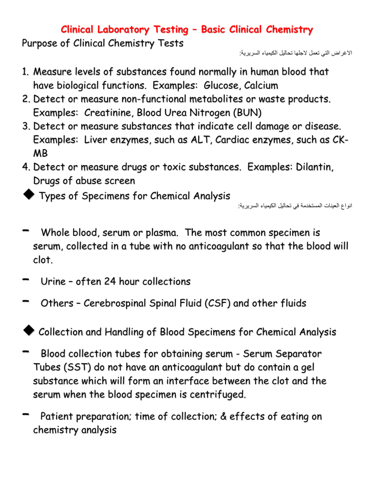
Clinical Laboratory Testing – Basic Clinical Chemistry Purpose of Clinical Chemistry Tests :االغراض التي تعمل الجلها تحاليل الكيمياء السريرية 1. Measure levels of substances found normally in human blood that have biological functions. Examples: Glucose, Calcium 2. Detect or measure non-functional metabolites or waste products. Examples: Creatinine, Blood Urea Nitrogen (BUN) 3. Detect or measure substances that indicate cell damage or disease. Examples: Liver enzymes, such as ALT, Cardiac enzymes, such as CKMB 4. Detect or measure drugs or toxic substances. Examples: Dilantin, Drugs of abuse screen Types of Specimens for Chemical Analysis – – – :انواع العينات المستخدمة في تحاليل الكيمياء السريرية Whole blood, serum or plasma. The most common specimen is serum, collected in a tube with no anticoagulant so that the blood will clot. Urine – often 24 hour collections Others – Cerebrospinal Spinal Fluid (CSF) and other fluids Collection and Handling of Blood Specimens for Chemical Analysis – – Blood collection tubes for obtaining serum - Serum Separator Tubes (SST) do not have an anticoagulant but do contain a gel substance which will form an interface between the clot and the serum when the blood specimen is centrifuged. Patient preparation; time of collection; & effects of eating on chemistry analysis Some specimens are increased or decreased after eating (ex. Glucose, triglycerides), so it is important to know what the test and collection method call for. Specimens for these tests are usually collected in a fasting state. Sometimes serum or plasma appears lipemia (milky) after a patient has eaten a fatty meal. Lipemia affects most chemistry analyses. The blood must be recollected when the patient is fasting. Clinical Chemistry Tests – – Normal or Reference Values – range of values for a particular chemistry test from healthy individuals Chemistry Panel grouping – some tests are “bundled” according to the system or organ targeted. Examples: thyroid panel, liver panel, cardiac panel, kidney panel, basic metabolic panel, etc. Commonly Performed Chemistry Tests or Analytes بعض التحاليل الكيميائية المهمة والشائعة – – Proteins – essential components of cells and body fluids. Some made by body, others acquired from diet. Provides information about state of hydration, nutrition and liver function, since most serum proteins are made in the liver. Electrolytes – sometimes called “lytes” Includes sodium (Na), potassium (K), chloride (Cl) and bicarbonate (HCO3-) Collectively these have a great effect on hydration, acidbase balance and osmotic pressure as well as pH and heart and muscle contraction – Levels differ depending on if inside vs. outside cells Important in transport of substances into and out of cells Minerals المعادن Calcium – – – Used in coagulation and muscle contraction – Hypercalcemia – occurs in parathyroidism, bone malignancies, hormone disorders, excessive vitamin D, and acidosis; may cause kidney stones – 99% is in skeleton and is not metabolically active Influenced by vitamin D, parathyroid hormone, estrogen and calcitonin Hypocalcemia – can cause tetany; occurs in hypoparathyroidism, vitamin D deficiency, poor dietary absorption and kidney disease Phosphorus – – 80% in bone and rest in energy compounds such as ATP – – Essential for hemoglobin – Increased in hemolytic anemia, increased iron intake or blocked synthesis of iron-containing compounds, such as in lead poisoning I r on – Influenced by calcium and certain hormones Deficiency results in anemia; may be caused by lack of iron in diet, poor absorption, poor release of stored iron or loss due to bleeding Kidney Function Tests Serum Creatinine – Best test for overall kidney function; not affected by – – diet or hormone levels Waste product of muscle metabolism Serum creatinine rises when kidney function is impaired BUN (Blood Urea Nitrogen) – – – – BUN is surplus amino acids that are converted to urea and excreted by kidneys as a waste product BUN influenced by diet and hormones, so it is NOT as good an indicator of renal function as serum creatinine levels BUN increased in kidney disease, high protein diet, and after administration of steroids BUN decreased in starvation, pregnancy and in persons on a low protein diet Uric Acid – – – – Formed from breakdown of nucleic acids and excreted as a waste product by kidneys Increased in kidney disease, but most often used to diagnosis gout (pain in joints, mainly big toe, due to precipitated uric acid crystals) Also increased in increased cell destruction, such as after massive radiation or chemotherapy Liver Function Tests Liver functions: – – Synthesizes glycogen from glucose – Forms cholesterol and degrades it into bile acids, Makes plasma proteins (albumin, lipoproteins, coagulation proteins) – – which emulsifies fats for absorption Stores iron, glycogen, vitamins and other substances Destroys old blood cells and recycles components of hemoglobin Total Bilirubin – – Waste production of hemoglobin breakdown – Alkaline Phosphatase (ALP or AP) - Greatly increased in liver tumors and lesions; moderately increased in diseases such as hepatitis Increased in excessive RBC breakdown, such as hemolytic anemia, or impaired liver function or some sort of obstruction, such as a tumor or gall stone Liver Enzymes – levels increase following damage to liver tissues – – – – Alanine Aminotransferase (ALT; formerly called SGPT) - Increases up to 10x in cirrhosis, infections or tumors and up to 100x in viral or toxic hepatitis Asparate Aminotransferase (AST; formerly called SGOT) - Increased in liver disease, but also in heart attacks Gamma Glutamyl Transferase (GGT) - Often used to monitor patients recovering from hepatitis and cirrhosis Lactate Dehydrogenase (LD) - Increased in liver disease and following heart attacks Cardiac Function Tests – – Creatine Kinase (CK) - Widely used to diagnosis and monitor heart attacks Troponins Only present in heart muscle, making it a more accurate indicator of heart attack than CK Cardiac Troponin T (cTnT) Cardiac Troponin I (cTnI) Lipid Metabolism Tests – – Cholesterol Present in all tissues Serves as the skeleton for many hormones Recommended to be less than 200 mg/dL in adults) LDL = “bad” cholesterol; HDL = “good” cholesterol Triglycerides Main storage form of lipids, comprising 95% of fat tissue Hyperlipidemia – having high blood levels of triglycerides – may increase risk of heart attack Carbohydrate Metabolism Tests – Glucose - Largely regulated by insulin Thyroid Function Tests – – – – Thyroid Stimulating Hormone (TSH) - Inverse relationship to thyroid function (the higher the TSH, the lower the thyroid function and vice versa) Other less common thyroid tests include T3 and T4 Hypothyroidism – underactive thyroid gland Hyperthyroidism – overactive thyroid gland انتهت محاضرة التحاليل الكيميائية االساسية Spectrophotometer Spectrophotometer (Spec) An instrument that measures the amount of light that passes through (is transmitted through) a sample. Uses a type of light to detect molecules in a solution Light is a type of energy, and the energy is reported as wavelengths, in nanometers (nm). Two different types of Spectrophotometer: Ultraviolet (UV) Spectrophotometers. Uses ultraviolet light of wave lengths from 200 nm to 350 nm. Visible (VIS) Light Spectrum Spectrophotometers. Uses visible light (white light) of wave lengths from 350 nm to 700 nm. كيف يعمل جهاز السبكتروفوتوميتر Shines a beam of light on a sample. The molecules in the sample interact with the light waves in of 3 ways: Absorb the energy Reflect the energy Transmit the energy between and through the atoms and molecules of the sample. لون االشياء يمثل الطيف الضوئي المعكوس من قبل الجزيئات فالصندوق االزرق يعني ان جزيئاته امتصت .جميع االلوان وعكست اللون االزرق فقط How a spectrophotometer works: Consider blue molecules, all the wavelengths of light are absorbed, except for the blue ones. The blue wavelengths are transmitted or reflected off the molecules. If these blue wavelengths hit a detector (such as in the spectrophotometer or the nerve cells in your eye), they appear blue. Molecules are whatever color of light that they do not absorb. Green molecules appear green because they absorb most wavelengths of visible light, except the green wavelengths. The spectrophotometer measures the amount of light transmitted through the sample (Transmittance). The concentration of an unknown sample can be determined by comparing the absorbance data to standards of known concentration. The data generated with the set of known standards is called a standard curve. Parts of a spectrophotometer Inner parts Lamp Prism or grating that direct light of a specific wavelength. VIS Spec vs. UV spec Visible spectrophotometer Contains a tungsten lamp that produces white light. Ultraviolet spectrophotometer Contains a deuterium lamp that produces light in the UV light part of the spectrum. Parts of a Spectrophotometer Outer parts: How a spectrophotometer works: Visible Spectrophotometer White light hits the prism or grating, it is split into the colors of the rainbow (Visible Spectrum). The wavelength knob rotates the prism/grating, directing different color of light toward the sample. How a spectrophotometer works: The wavelength of light produced by the tungsten lamp range from about 350 nm (Violet light) to 700 nm (red light). The molecules in the sample either absorb or Transmit the light energy of one wavelength or another. How a spectrophotometer works: The detector measures the amount of light being transmitted by the sample and reports that value directly (% transmittance) or converts it to the amount of light absorbed in absorbance units (au) using Beers Law. The function of a spectrophotometer The spectrophotometer can measure the amount of absorbance or lack of absorbance of different colored light for a given molecule. The relationship of concentration in a solution: The concentration of molecules in a solution affects the solution’s absorbance. If there are more molecules in one solution than in another, than there are more molecules to absorb the light. Applications of a spectrophotometer Determines the presence and concentrations of samples. Determines the purity of a sample. Look at the change of samples over time. انتهت محاضرة السبكتروفوتوميتر Definitions: • Quality Control:- … to ensure the reliability of the test results to give the best patient care ! Unreliable Performance ? • Potential consequences include:- – – – patient misdiagnosis delays in treatment increased costs • avoidable retests cost US 200million USD per year Error Classification.. • – • – • – Pre-analytical:errors before the sample reaches the laboratory Analytical:errors during the analysis of the sample Post-analytical:errors occurring after the analysis Pre - Analytical Errors.. • Improper preparation of the patient:- Other Factors.. • • • The sex of the patient – male or female The age of the patient – new born / juvenile / adult / geriatric Dietary effects • • – – low carbohydrate / fat high protein / fat When the sample was taken – early morning urine collection pregnancy testing Patient posture – urinary protein in bed-ridden patients Other Factors.. • • • • Effects of exercise – creatine kinase / CRP Medical history – heart disease / diabetes / existing medication Pregnancy – hormonal effects Effects of drugs and alcohol – liver enzymes / dehydration Analytical Errors.. • • • • The sample: Glassware / pipettes / balances: The application: The instrument: Other Factors.. • Calculation errors: – incorrect factor / wrong calibration values • • • • Transcription errors: Dilutions errors: – incorrect dilution or dilution factor used Lack of training: The human factor: – tiredness / carelessness / stress Post - Analytical Errors.. • • The prompt and correct delivery of the correct report on the correct patient to the correct Doctor. How the Clinician interprets the data to the full benefit of the patient. Accuracy ? How correct your result is. Precision ? The reproducibility of your results. Accurate and Precise.. Imprecise but Accurate ! Precise but Inaccurate ! Specificity ? • The ability of a method to measure solely the component of interest. (No false +ve) Sensitivity ? • The ability to detect small quantities of a measured component. (No false – ve) How can Analytical Quality be Controlled ? • Internal Quality Control (IQC). – daily monitoring of quality control sera using samples of known values. • External Quality Assessment (EQA). – comparing of performance to other laboratories. Internal Quality Control.. • Daily monitoring • Quality control sera – precision – accuracy – results within control limits indicates that analytical system is running satisfactorily انتهت محاضرة السيطرة النوعية – – Basic Clinical Laboratory Math Metric System Most countries use the metric system for measurement Examples: – Gasoline by liter – Body weight in kilograms – Distance in meters or kilometers U.S. uses English system of measurement in everyday life Examples: – Gasoline in gallons – Weight in pounds – Distance in miles – – – – – Metric System English system of measurement is not accurate enough for most scientific measurements Because metric system is a decimal system, it can be used for very small quantities with accuracy International System of Units (SI) is a form of the metric system adopted for use by the worldwide scientific community. Units of Metric System Base Units Distance = meter (m) Mass or Weight = gram (g) Volume = liter (L) Prefixes are used to indicate larger or smaller quantities of the base units above Common Metric Prefixes – – – – – – Kilo (k) = 1000 x base unit Centi (c) = .01 x base unit Milli (m) = .001 x base unit Micro (µ) = .000001 x base unit Nano (n) = 10 -9 x base unit Pico (p) = 10-12 x base unit Converting within Metric System Move decimal to left Move decimal to right Example: Convert Kilograms to Grams: Move decimal 3 places to right غرام1000 كل كيلو غرام هو: مثال ملليلتر1000 كل لتر هو سنتيمتر100 كل متر هو Example: Convert Centimeters to Meters: Move decimal 2 places to left SI System (International System) Base Units of the SI System – Length = Meter (m) – Mass = Kilogram (kg) – – – – – – Time = Second (s) Amount of Substance = Mole (mol) Electric Current = Ampere (A) Temperature = Kelvin (K)* Luminous Intensity = Candela (cd) Volume = Liter (L)** Dilutions for the Clinical Laboratory Dilution = making weaker solutions from stronger ones Example: Making orange juice from frozen concentrate. You mix one can of frozen orange juice with three (3) cans of water. Dilutions are expressed as the volume of the solution being diluted per the total final volume of the dilution In the orange juice example on the previous slide, the dilution would be expressed as 1/4, for one can of O.J. to a TOTAL of four cans of diluted O.J. When saying the dilution, you would say, in the O.J. example: “one in four”. Another example: If you dilute 1 ml of serum with 9 ml of saline, the dilution would be written 1/10 or said “one in ten”, because you express the volume of the solution being diluted (1 ml of serum) per the TOTAL final volume of the dilution (10 ml total). Another example: One (1) part of concentrated acid is diluted with 100 parts of water. The total solution volume is 101 parts (1 part acid + 100 parts water). The dilution is written as 1/101 or said “one in one hundred and one”. Notice that dilutions do NOT have units (cans, ml, or parts) but are expressed as one number to another number Example: 1/10 or “one in ten” Dilutions are always expressed with the original substance diluted as one (1). If more than one part of original substance is initially used, it is necessary to convert the original substance part to one (1) when the dilution is expressed. Example: Two (2) parts of dye are diluted with eight (8) parts of diluent (the term often used for the diluting solution). The total solution volume is 10 parts (2 parts dye + 8 parts diluent). The dilution is initially expressed as 2/10, but the original substance must be expressed as one (1). To get the original volume to one (1), use a ratio and proportion equation, remembering that dilutions are stated in terms of 1 to something: ______2 parts dye = ___1.0___ 10 parts total volume x 2x = 10 x = 5 The dilution is expressed as 1/5. Serial Dilutions Dilutions can be made singly (as shown previously) or in series, in which case the original dilution is diluted further. A general rule for calculating the dilution of solutions obtained by diluting in a series is to MULTIPLY the original dilution by subsequent dilutions. Example of a serial dilution: In the serial dilution on the previous slide, 1 ml of stock solution is mixed with 9 ml of diluent, for a 1/10 dilution. Then 1 ml of the 1/10 dilution is mixed with another 9 ml of diluent. The second tube also has a 1/10 dilution, but the concentration of stock in the second tube is 1/10 x 1/10 for a 1/100 dilution. Continuing with the serial dilution, in the third tube, you mix 1 ml of the 1/100 dilution from the second tube with 9 ml of diluent in the third tube. Again you have a 1/10 dilution in the third tube, but the concentration of stock in the third tube is 1/10 x 1/10 x 1/10 for a 1/1000 dilution. This dilution could be carried out over many subsequent tubes. Serial dilutions are most often used in serological procedures, where technicians need to make dilutions of patient’s serum to determine the weakest concentration that still exhibits a reaction of some type. Example of determining a titer: A technician makes a serial dilution using patient serum: Tube #1 = 1/10 Tube #2 = 1/100 Tube #3 = 1/1000 Tube #4 = 1/10,000 Tube #5 = 1/100,000 Reactions occur in tubes 1 through 3, but NOT in tubes 4 or 5. The titer = 1000. انتهت محاضرة الرياضيات المختبرية Basic Principles of Phlebotomy Modern Phlebotomy • D i a g no s i s a n d m a n a g e m e n t o f d i s e a s e • Remove blood for transfusions • Therapeutic reasons: – Polycythemia – Hemochromatosis Blood Function: • • • Supplies nutrients to tissues: O2, hormones, glucose Removes end-products of metabolism: CO2, urea, creatinine Provides defense mechanism: WBC, antibodies • Prevents blood loss: platelets, coagulation proteins Blood Composition: Formed elements (~45%) – RBC – W BC – P l a te l e ts Liquid elements(~55%) Coagulation: • In v i v o – B l o o d i s fl u i d – • Clot is formed to protect injured vessel In vitro – – S p o n ta n e o u s r e a c ti o n Triggered by glass or poor drawing technique Coagulation Reaction: Clotting factors + calcium thrombin Fibrinogen + thrombin Anti-coagulants: • • fibrin strands Remove calcium (Like EDTA) Neutralize thrombin (Like heparin) S am pl es us ed i n l ab • • • W hol e bl ood P l as m a Serum Blood with anticoagulant: • Clotting is prevented and irreversible, after collection mix: completely invert 8-10 times, we will have whole blood. After centrifugation plasma. Plasma contains fibrinogen Blood without anticoagulant: • Spontaneous clotting occurs and is irreversible, Fibrinogen in blood fibrin strands. After centrifugation serum. Serum lacks fibrinogen Appearance • Normal: clear and ‘yellow’ • Abnormal: – Hemolyzed = pink to red (ruptured RBC) – Icteric = dark orange-yellow (bilirubin) – Lipemic = cloudy (fat, triglycerides) Blood Collection Tubes: • • C o n t ai n a v a c u u m Used with Vacutainer and Syringe systems Type and Amount of Specimen: • D e p en d e n t u p o n – Test Whole blood: EDTA or heparin? Plasma: EDTA or heparin? Serum: trace free? Separator gel interference? – Amount of sample needed to perform test – M u l ti p l e l a b s n e e d i n g th e s a m e s p e c i m e n a t th e s a m e ti m e Valid Test Results Require:ما هي المستلزمات المطلوبة للحصول على نتائج صحيحة؟ • • • • Trained personnel – – Causes of pre-analytical error Invalid test results Quality control Quality assurance S o p hi s t i c a t e d instruments Safety Practices: الممارسات الصحيحة للسالمة المختبرية For infection to spread: • Infectious substance: HBV, HCV, HIV • • Mode of transmission S u s c e p ti b l e h o s t Modes of Transmission: • Parenteral: any route other than the digestive tract – Intramuscular – Intravenous – S u b c u ta n e o u s – M uc os al • I n g e s ti o n S a fe t y P r a c ti c e s : Infection Control: stop the spread of infection Safety: Infection Control • Hand washing – Primary means of preventing spread of infection (especially no s o c o m i a l ) • • – Minimum 15 seconds, soap, friction – Wash hands before and after each blood draw We should wear – La b c o a t – Gloves – M as k Cleaning agent المعقمات – – Alcohol pads: routine Povidone iodine: blood culture collection and blood gases – • Soap and water: alcohol testing, allergies Cotton balls, gauze E q u i p m e nt : • Bandage, tape (use caution with children) • Sharps container: – – – – Discard needles, l a n c e ts Biohazard marking Puncture resistant NEVER recap, bend break needles E q u i p m e nt : 6. Tourniquets: – – – – Slows venous blood flow down Causes veins to become more prominent NEVER leave on for >1 minute AVOID rigorous fist clenching or hand pumping (potassium, lactic acid, LD) – Latex allergy Tying on the Tourniquet: E q u i p m e nt : 7. Needles – – NEVER reuse a needle NEVER use if shield is broken – NEVER recap, cut, bend or break – Drop immediately into sharps container after venipuncture – Size of needle is indicated by gauge: • Larger gauge number indicates smaller needle diameter • 21, 23 gauge needles routinely used for phlebotomy E q u i p m e nt : 8. Tube holder/ v a c u ta i n e r a d a p t e r – – Threaded Flanges E q u i p m e nt : • • Syringe B l ac k water proof p en Syringe Safety Device: Labeling Blood Collection Tubes: • • Black indelible marker (water proof) – – – Never pencil Legal document Print legibly Required information: 5 items – – – Full Patient name Id e n ti fi c a ti o n n u m b e r Date of draw (mm,dd,yyyy) – – Time of draw (military time) Phlebotomist signature: first initial, last name Vacutainer or Syringe? • Vacutainer • Syringe – M o s t o fte n u s e d – M os t ec onom i c al – Quick – Least risk of accidental needle stick – More control – Reposition easily – Will see ‘flash’ of blood in syringe hub when vein successfully entered The Patient: • • • • • • Approach C o m m u n i c a ti o n E m p at h y H a n dl i n g s p e c i a l s i t u a t i o n s P a t i e nt i d e n t i f i c a t i o n – – Arm band Le g a l d o c u m e n t Prepare patient for blood draw – Latex allergy? S e l e c ti n g th e S i te : • Antecubital area most often accessed • • • Hand or wrist R e m em b e r : 2 a r m s Use tip of index finger on non-dominant hand to palpate area to feel for t h e v ei n Collection Site Problems: • • • • V e i n s th a t l a c k resiliency Extensive scarring H e m at o m a s E d e m a to u s area • S i d e of m a s t e c t o m y Collection Site Problems: • Intravenous line – NEVER draw above an IV – Draw from other arm – Draw from hand on other arm – Draw below the IV Draw Below IV site: Inserting the Needle: • A n c h or t h e v e i n – – • Grasp arm with your non-dominant hand Use thumb to pull skin taut Smoothly and confidently insert the needle bevel up – 15-30 degree angle No Needle Movement! • You must anchor the blood-drawing equipment on the patient’s arm to m i ni m i z e c hanc e o f i nj ur y Withdraw Needle: • • • • • • First release tourniquet D i s e ng a g e t u b e Place cotton directly over needle, without pressing down Withdraw needle in swift, smooth motion Immediately apply pressure to wound Do not bend arm Needle Position: You should try again • • • • Look at alternate site – – Other arm Hand U s e c l ean needl e Use fresh syringe if contaminated Only try twice Poor Collection Techniques: • • • V e n ou s s t a s i s – Prolonged application of tourniquet (>1 min) H e m od i l u t i o n – – Drawing above IV Short draw (blood to anticoagulant ratio) Hemolysis – – – – – Traumatic stick Too vigorous mixing Alcohol still wet Using too small of needle Forcing blood into syringe • C l o t t ed s a m p l e • Partially filled tubes • Using wrong anticoagulant • S pec i m en handl i ng – Inadequate mixing – Traumatic stick – Short draw – Sodium citrate tube draw volume critical – Exposure to light (Bilirubin) – Pre-chilled tube – Body temperature Venipuncture Procedure: • • • • • • W as h hands P u t on g l o v e s I d e n ti f y p a ti e n t Latex allergy? Position arm A p p l y to u r n i q u e t Venipuncture Procedure: • • • • • • • • • • • • Locate vein Release tourniquet Cleanse site in outward rotation – A l l o w to a i r d r y Reapply tourniquet – Do not contaminate site A n c h or v e i n Insert needle Fill tubes – Quick mix additive tubes Release tourniquet Withdraw needle E n g ag e s a f e t y d e v i c e D i s p os e o f n e e d l e i m m e d i a t e l y Apply pressure to puncture site • • • • L a b e l tu b e s R e c he c k p u n c t u r e s i t e Thank patient R e m ov e g l o v e s , wash hands انتهت محاضرة سحب الدم


