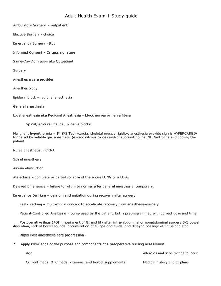
Adult Health Exam 1 Study guide Ambulatory Surgery - outpatient Elective Surgery - choice Emergency Surgery - 911 Informed Consent – Dr gets signature Same-Day Admission aka Outpatient Surgery Anesthesia care provider Anesthesiology Epidural block – regional anesthesia General anesthesia Local anesthesia aka Regional Anesthesia – block nerves or nerve fibers Spinal, epidural, caudal, & nerve blocks Malignant hyperthermia – 1st S/S Tachycardia, skeletal muscle rigidity, anesthesia provide sign is HYPERCARBIA triggered by volatile gas anesthetic (except nitrous oxide) and/or succinylcholine. NI Dantroline and cooling the patient. Nurse anesthetist - CRNA Spinal anesthesia Airway obstruction Atelectasis – complete or partial collapse of the entire LUNG or a LOBE Delayed Emergence – failure to return to normal after general anesthesia, temporary. Emergence Delirium – delirium and agitation during recovery after surgery Fast-Tracking – multi-modal concept to accelerate recovery from anesthesia/surgery Patient-Controlled Analgesia – pump used by the patient, but is preprogrammed with correct dose and time Postoperative ileus (POI) impairment of GI motility after intra-abdominal or nonabdominal surgery S/S bowel distention, lack of bowel sounds, accumulation of GI gas and fluids, and delayed passage of flatus and stool Rapid Post anesthesia care progression 2. Apply knowledge of the purpose and components of a preoperative nursing assessment Age Allergies and sensitivities to latex Current meds, OTC meds, vitamins, and herbal supplements Medical history and tx plans Surgical hx Previous anesthesia and responses to anesthesia Last oral intake Any medical implants or devices Piercings, dental implants, Nutritional deficiencies Family hx, social hx, smoking, drug and alcohol habits Hx of mental illness or abuse Support system and living conditions 3. Advance directives Identify the major nursing goals for each phase of the perioperative period. 3 PHASES of Perioperative period Preoperative phase – begins when the decision to have surgery is made, it ends when the client is transferred to the operating table Intraoperative phase – begins when the client is transferred to the OR table and ends when the client is admitted to PACU Postoperative phase – begins with the clients admission to the postanesthesia area and ends when the healing is complete 4. Identify the physical and psychosocial responses of clients to surgery. 5. Identify and analyze the components of preoperative client preparation. Lab Assess – type and crossmatch, CBC, renal and liver functions, liver enzymes, albumin, electrolytes, blood urea nitrogen, and creatinine. Urine for the presence of glucose, blood, protein, and specific gravity and the presence of keytones Radiological Assess – sometimes prior to surgery, MRI, CAT scan, sono or x-ray, imaging studies and an ECG Patient teaching – helps decrease anxiety = improved patient outcomes, PACU procedures, how the family will see the patient, and discharge information. Questions the family has Physical Prep – IV – 18 gauge cath (size for admin of all blood products), LABEL IV with gauge, time, date and nurses initials Bowel and B ladder – abdominal, intestinal, gyno, or rectal may be asked to do a bowel prep. Enema or laxatives, may also need a catheter Skin Prep – betadine or hexachloriphene soap at home or in the hospital room, shave with clippers only, double check that all body piercings are out at this time Medications – (see below) 6. Differentiate the purpose and types of common preoperative medications. Preoperative anxiety – Benzodiazepines – midazolam HCL, diazepam, or lorazepam for hx of N/V due to anesthesia - Antiemetics – metoclopramide HCL or ondansetron HCL 7. Identify nursing diagnoses and interventions that correspond to the intraoperative setting. 8. Identify complications that may occur during each phase of the perioperative period. Coughing, deep breathing and 9. Interpret the significance of data related to the perioperative client’s health status and operative risk. 10. Describe nursing interventions to prevent or treat postoperative complications 11. Describe the nursing role in the psychological and educational preparation of the surgical client. 12. Identify the special considerations of preoperative preparation for the older adult surgical client. CONTENT OUTLINE - Textbook I. II. III. IV. Preoperative phase Intraoperative phase Postoperative phase Pain Chapter 15 pp. 283 - 300 Chapter 16 pp. 301 - 322 Chapter 17 pp. 323 – 337 Chapter 11 pp. 175-214 LEARNING OPPORTUNITIES 1. Identify and describe the roles of the intraoperative health care team. 2. Develop a plan of care for the postoperative client including common potential postoperative complications. 3. Design a preoperative teaching plan for the client anticipating surgery. Cardiovascular Preload – volume of blood in ventricles at the end of diastole (end diastolic pressure) increased volume = increased stretch, which produces increased contraction (Starling’s Law) *****increases in MI, aortic stenosis, and hypervolemia*** Afterload - resistance to flow the ventricle must overcome to open the semilunar valves and eject its contents. This is related to BP, vessel lumen diameter, and/or vessel compliance. The valves can meet resistance on both the right and left through the pulmonary artery or aorta. Hypertension on the right or left is implicated in the negative effects of increased afterload. Affected by size of ventricle, wall tension, arterial blood pressure CO (HR X SV) blood ejected from the left side of the heart in 1 minute (normal 4~8 min.) HR ~controlled by ANS (unconscious) SV~amount of blood ejected from the ventricle with each beat, factors affecting are PRELOAD, CONTRACTILITY & AFTERLOAD. Ejection Fraction – how well the left ventricle pumps blood out of the heart Anemia – hemoglobin concentration is lower than normal , sign of underlying disorder (not enough RBC’s or not enough hb in cells. Classified by what is causing it – not making enough RBC’s hypoproliferative anemia, destruction of RBC’s (hemolytic), or loss of RBC’s (bleeding) Symptoms fatigue, dyspnea, dizziness, headache, feeling cold, weakness, breathlessness, lightheadedness, palpitations, angina pectoris Hypoproliferative Iron-deficiency Vitamin B12 deficiency (megaloblastic) Pernicious anemia Folate deficiency (megaloblastic) Decreased erythropoietin production (e.g. from renal dysfunction) Cancer / inflammation Hemolytic Altered erythropoiesis (sickle cell, thalessemia, other hemoglobinopathies) Hypersplenism (hemolysis) Blood loss Bleeding from GI tract, epistaxis, trauma, heavy menses Drug-induced anemia Autoimmune anemia Mechanical heart valve-related anemia Signs tachycardia or tachypnea if blood loss occurs rapidly Chronic – pale skin, mucous membranes or nail beds, fissures at the corner of the mouth. Aplastic anemia Pernicious anemia Anemia caused by deficient red cell production due to bone marrow disorders Idiopathic cases range from 40% to 70%, most common in adolescents and young adults; exposure to chemical and antineoplastic agents and ionizing radiation Chronic, an autoimmune macrocytic disease; parietal anemia marked cells of the by achlorhydria; stomach lining occurs most often fail to secrete in 40- to 80-year- enough intrinsic old northern factor to ensure Europeans of fair intestinal skin, but has been absorption of reported in other vitamin B12; due races and ethnic to atrophy of the glandular mucosa Bone marrow transplantation or immunosuppressive drugs Weakness, sore tongue, paresthesias (tingling and numbness) of extremities, and gastrointestinal symptoms such as diarrhea, nausea, vomiting, and pain; in severe anemia, there Vitamin B12 given parenterally or, in patients who respond, orally Folic acid deficiency groups; rare in of the fundus of may be signs of blacks and Asians the stomach and cardiac failure is associated with absence of HCl acid Anemia resulting Common in from a deficiency patients who are of folic acid; a experiencing cause of red cell nutritional enlargement deficiencies (e.g. (megaloblastic) alcoholics, patients with malabsorption) and during hemolysis or pregnancy. Iron-deficiency Anemia resulting anemia from a greater demand on stored iron than can be supplied. The red blood cell count may sometimes be normal, but there will be insufficient hemoglobin Caused by inadequate iron intake, malabsorption of iron, blood loss, pregnancy and lactation, intravascular hemolysis, or a combination of factors Foods rich in folic acid – green leafy vegetables, asparagus, broccoli, liver, organ meats, milk, eggs, yeast, wheat germ, kidney beans, beef, potatoes, dried peas and beans, wholegrain cereals, nuts bananas, cantaloupe, lemons, and strawberries Chronically Dietary intake of anemic patients iron is supplemented often complain of with oral ferrous fatigue and sulfate or ferrous dyspnea on gluconate (with exertion. Iron vitamin C to deficiency increase iron resulting from absorption) rapid bleeding may produce palpitations, orthostatic dizziness, or syncope Polycythemia – an excess of red blood cells; in a newborn it may reflect hemoconcentration due to hypovolemia or prolonged intrauterine hypoxia, or hypervolemia due to intrauterine twin-to-twin transfusion or placental transfusion resulting in delayed clamping of the umbilical cord (Taber’s) Information for table from Taber’s Polycythemia vera – a chronic, Symptoms – weakness, Treatment – permanent cure life-shortening fatigue, blood clotting, cannot be achieved today, but myeloproliferative disorder, vertigo, tinnitus, irritability, remissions of many years can be resulting from the reproduction flushing of the face, redness, produced. Phlebotomy, of a single cell clone; and pain or extremities occur radioactive phosphorus, characterized by an increase in red blood cell mass and hemoglobin concentration that occurs independently of erythropoietin stimulation commonly. Bone marrow shows increased cellularity. Peptic ulcers are often reported. cyclophosphamide, hydroxyurea, or melphalan is used. (also anagrelide (Agrylin) Atherosclerosis – obstruction of blood flow w/I coronary arteries, characterized by CAD, plaque w/I lumen of BV Capillary refill – press nail, return to color w/i 3 seconds = adequate perfusion Claudication – pain commonly in legs from too little blood flow usually from exercise Compensation – walls of heart enlarge trying to compensate for failure on one side of the heart or the other Friction Rub – scratching or grating sound heard during both Systole and Diastole, between the parietal and visceral pericardial layers, in the pericardial sac. (used in Dx for PERICARDITIS) Heart failure – thickening of the ventricle layers, Hemolytic Anemia – newborn, RH disease, red blood cells are destroyed faster than they are made Hypoxia – deficiency in the amount of oxygen reaching body tissues Intrinsic – substance produces & secreted by the parietal cells which enables the body to absorb B12, ties into pernicious anemia aka ADDISON’s ANEMIA Ischemia – heart muscle does not get enough oxygen and nutrients to meet demand, myocardial ischemia, PATHO of CAD Lymphedema – swelling of arms or legs, related to Cancer treatment pg 512 Hypertensive Crisis – BP higher than 180 and/or 120 Orthostatic Hypotension Diastolic Failure – major cause of morbidity and mortality, defined as symptoms of HF in a patient with preserved left ventricular function – stiff and doesn’t relax which leads to increased end diastolic pressure Systolic Failure Pulmonary Edema Paroxysmal nocturnal dyspnea Systemic Vascular Resistance Pernicious Anemia Phlebitis Polycythemia vera Primary Hypertension Secondary hypertension Sodium restriction Thrombophlebitis Varicosities Vasoconstriction Venous insufficiency Venous stasis Vitamin B12 Identify the significant subjective and objective assessment data related to the cardiovascular system that should be obtained from a client Describe the age-related changes of the cardiovascular system and differences in assessment findings Explain the rationale and nursing implications for selected diagnostic studies chest X-ray: Looking at cardiac silhouette for size and placement in chest, pericardial effusion and pulmonary congestion. Echocardiogram: ultrasound study with or w/o contrast. Evaluate valves, chamber size with contents, ventricular and septal movement and thickness, pericardial sac for fluid and ascending aorta for calcifications/aneurism. Can calculate E/F ejection fraction which gives a picture of left ventricular funct. Normal is 53-73%. It is possible to have “preserved EF” with heart disease. Means ventricle can’t relax btwn contractions. MRI: 3D view. Newer model pacemakers OK. Nuclear Studies: great way to see ventricular wall and septal mvmt. Also show coronary artery blood flow during exercise. Can be activity or chemically simulate effects of exercise. ECG: monitor electrical activity of heart. Deviations from normal – heart dysfunction. TEE: pt swallows' probe. In esophagus, gives great visualization of heart. No eating until gag returns. Cardiac cath: insertion femoral, radial , brachial/cephalic arteries. Injection of contrast. Post care: monitoring of distal pulses to insertion. Labs: Know normal, what information obtained from lab, any prep (ie fasting). Would also get electrolytes, renal/liver studies, why? BNP: elevated in heart failure. Measures hormone produced made by heart cells in ventricles from stretching. Troponin: released by cardiac muscle that are undergoing degrading. 80% MI have elevation, can be seen 2-3 hour, but definitive r/o is 12 hours. This is why admit chest pain for obs. Why clotting studies? PT, INR TEE & TTE – echocardiography, posterior of the heart (L atrium), clots in the atrium (RF for stroke) ECG – 10 electrodes CXR – chest x-ray – size, shape and portion of the heart Cardiac enlargement Pulmonary congestion Pneumonia Pneumothorax CHF Increased preload causes increased stretching of the muscle measured by – BNP Brain Naturetic Peptide >100 HF, blood test blood pressure and the mechanisms involved in its regulation Fig 33.1- p. 710 •can affect either CO or SVR, or both. •Regulation of BP is a complex process involving both short-term (seconds to hours) and longterm (days to weeks) mechanisms. •Short-term mechanisms, including the sympathetic nervous system (SNS) and vascular endothelium, are active within a few seconds. •Long-term mechanisms include renal and hormonal processes that regulate arteriolar resistance and blood volume. In a healthy person, these regulatory mechanisms function in response to the demands of the body. (Each mechanism is more fully described in the following slides.) Sympathetic nervous system •The nervous system, within seconds after a drop in BP, increases BP primarily by activating the SNS. •Increased SNS activity increases HR and cardiac contractility, produces widespread vasoconstriction in the peripheral arterioles, and promotes the release of renin from the kidneys. •The net effect of SNS activation is to increase BP by increasing both CO and SVR. Baroreceptors •Specialized nerve cells called baroreceptors (pressoreceptors) • are located in the carotid arteries and the arch of the aorta. • • important role in the maintenance of BP stability during normal activities. sensitive to stretching and, when stimulated by an increase in BP, send inhibitory impulses to the sympathetic vasomotor center. • Inhibition of the SNS results in decreased HR, decreased force of contraction, and vasodilation in peripheral arterioles. •When a fall in BP is sensed by the baroreceptors, the SNS is activated. The result is constriction of the peripheral arterioles, increased HR, and increased contractility of the heart. •In the presence of long-standing hypertension, the baroreceptors become adjusted to elevated BP levels and recognize this level as their new “normal.” Vascular Endothelium •The vascular endothelium is a single-cell layer that lines the blood vessels. It is essential to the regulation and maintenance of vasodilating and vasoconstricting substances. Nitric oxide and prostacyclin are both vasodilators. Endothelin (ET) is an extremely potent vasoconstrictor. A disruption or dysfunction of arterial tone (either through excessive constriction or dilation) is an early warning signal of CVD. Renal System •The kidneys contribute to BP regulation by controlling sodium excretion and extracellular fluid (ECF) volume. Sodium retention results in water retention, which causes an increase in ECF volume. This increases the venous return to the heart and stroke volume. Together, these increase CO and BP. •The renin-angiotensin-aldosterone system (RAAS) also plays an important role in BP regulation. The kidney secretes renin in response to SNS stimulation, decreased blood flow through the kidneys, or decreased serum sodium concentration. Renin is an enzyme that converts angiotensinogen to angiotensin I. Angiotensin I is then converted to angiotensin II by angiotensin-converting enzyme (ACE). A-II increases BP. •Prostaglandins (PGE2 and PGI2) secreted by the renal medulla have a vasodilator effect on the systemic circulation. This results in decreased SVR and lowering of BP. Endocrine System •Stimulation of the SNS results in release of epinephrine along with a small fraction of NE by the adrenal medulla. Epinephrine increases the CO by increasing the HR and myocardial contractility. Epinephrine activates β2-adrenergic receptors in peripheral arterioles of skeletal muscle, causing vasodilation. In peripheral arterioles with only α1-ardrenergic receptors (skin and kidneys), epinephrine causes vasoconstriction. •A-II stimulates the adrenal cortex to release aldosterone. Aldosterone stimulates the kidneys to retain sodium and water. This increases blood volume and CO. •An increased blood sodium and osmolarity level stimulates the release of ADH from the posterior pituitary gland. ADH increases the ECF volume by promoting the reabsorption of water in the distal and collecting tubules of the kidneys. The resulting increase in blood volume causes an increase in CO and BP. Discuss the pathophysiology, clinical manifestations, complications and nursing management for the client experiencing hypertension RF for Primary Hypertension o o o o o o o o o o o o o Age Alcohol Smoking Diabetes Elevated serum lipids Excess dietary sodium Gender Family history Ethnicity Sedentary lifestyle Socioeconomic status Stress Obesity BMI Categories: Underweight = <18.5 Normal weight = 18.5–24.9 Overweight = 25–29.9 Obese = BMI of 30 or higher Weight Reduction •Overweight persons have an increased incidence of hypertension and increased risk for CVD. •Weight reduction has a significant effect on lowering BP in many people, and the effect is seen with even moderate weight loss. •A weight loss of 22 lb (10 kg) may decrease SBP by approximately 5 to 20 mm Hg. •When a person decreases caloric intake, sodium and fat intake are usually also reduced. Although reducing the fat content of the diet has not been shown to produce sustained benefits in BP control, it may slow the progress of atherosclerosis and reduce overall CVD risk. •Weight reduction through a combination of calorie restriction and moderate physical activity is recommended for overweight patients with hypertension. DASH Eating Plan •The DASH eating plan emphasizes fruits, vegetables, fat-free or low-fat milk and milk products, whole grains, fish, poultry, beans, seeds, and nuts. •Compared with the typical American diet, the plan contains less red meat, salt, sweets, added sugars, and sugar-containing beverages. •The DASH eating plan significantly lowers BP and these decreases compare with those achieved with BP-lowering drug. •Additional benefits also include lowering of low-density lipoprotein (LDL) cholesterol. Dietary Sodium Reduction •This involves avoiding foods known to be high in sodium (e.g., canned soups, frozen dinners) and not adding salt in the preparation of foods or at meals. •Average sodium intake is about 4200 mg/day. •Patient and caregiver need to learn about sodium-restricted diets. Moderation of Alcohol Intake •Excessive alcohol intake is strongly associated with hypertension. Drinking three or more alcoholic drinks daily is also a risk factor for CVD and stroke. •Men should limit their intake of alcohol to no more than two drinks per day and women and lighterweight men to no more than one drink per day. One drink is defined as 12 oz of regular beer, 5 oz of wine (12% alcohol), or 1.5 oz of 80-proof distilled spirits. •Excessive alcohol intake that results in cirrhosis is the most frequent cause of secondary hypertension. Physical Activity •A physically active lifestyle is essential to promote and maintain good health. •The AHA and American College of Sports Medicine recommend that adults perform moderate-intensity aerobic physical activity for at least 30 minutes most days (i.e., more than 5) per week with a goal of at least 150 minutes per week. •Exercise goals can be accomplished by performing shorter periods of exercise that last at least 10 minutes or more. •Additionally, combinations of moderate and vigorous activity are acceptable (e.g., walking briskly for 30 minutes on 2 days of the week and jogging for 20 minutes on 2 other days). •For adults age 18 to 65, walking briskly at a pace that noticeably increases the pulse defines moderateintensity aerobic activity. Jogging at a pace that substantially increases the pulse and causes rapid breathing is an example of vigorous activity for this age group. •All adults should perform muscle-strengthening activities using the major muscles of the body at least twice a week. This helps to maintain or increase muscle strength and endurance. •Additionally, flexibility and balance exercises are recommended at least twice a week for older adults, especially for those at risk for falls. •Generally, physical activity is more likely to be done if it is safe and enjoyable, fits easily into one’s daily schedule, and is inexpensive. Avoidance of Tobacco Products •Nicotine contained in tobacco causes vasoconstriction and increases BP, especially in people with hypertension. •Smoking tobacco is also a major risk factor for CVD. •The cardiovascular benefits of stopping tobacco use are seen within 1 year in all age groups. Strongly encourage everyone, especially patients with hypertension, to avoid tobacco use. Advise those who continue to use tobacco products to monitor their BP during use. Management of Psychosocial Risk Factors •Psychosocial risk factors can contribute to the risk of developing CVD and to a poorer prognosis and clinical course in patients with CVD. •These risk factors include low socioeconomic status, social isolation and lack of support, stress at work and in family life, and negative emotions such as depression and hostility. Frequently, these risk factors are clustered together. For example, there tends to be higher rates of depression in individuals who experience job stress. •Psychosocial risk factors have direct effects on the cardiovascular system by activating the SNS and stress hormones. This can cause a wide variety of pathophysiologic responses, including hypertension •Psychosocial risk factors can contribute to CVD indirectly as well, simply by their impact on lifestyle behaviors and choices. Screening for psychosocial risk factors is important. Make appropriate referrals (e.g., counseling), when indicated. Suggest behavioral interventions such as relaxation training, stress management courses, support groups, and exercise training for individuals who are not in acute psychologic distress. The drugs currently available for treating hypertension have two main actions: (1) decrease the volume of circulating blood and (2) reduce SVR. The drugs used in the treatment of hypertension include: Diuretics promote sodium and water excretion, reduce plasma volume, and reduce the vascular response to catecholamines. Adrenergic-inhibiting agents act by diminishing the SNS effects that increase BP. Adrenergic inhibitors include drugs that act centrally on the vasomotor center and peripherally to inhibit norepinephrine release or to block the adrenergic receptors on blood vessels. Direct vasodilators decrease the BP by relaxing vascular smooth muscle and reducing SVR. Calcium channel blockers increase sodium excretion and cause arteriolar vasodilation by preventing the movement of extracellular calcium into cells. Angiotensin-converting enzyme (ACE) inhibitors prevent the conversion of angiotensin I to angiotensin II and reduce angiotensin II (A-II)–mediated vasoconstriction and sodium and water retention. A-II receptor blockers (ARBs) prevent angiotensin II from binding to its receptors in the walls of the blood vessels. •Once antihypertensive therapy is started, patients should return for follow-up and adjustment of drugs at monthly intervals until the goal BP is reached. More frequent visits are necessary for patients with stage 2 hypertension or with co-morbidities. After BP is at goal and stable, follow-up visits can usually be at 3- to 6-month intervals. •Side effects of antihypertensive drugs are common and may be so severe or undesirable that the patient does not adhere to the therapy. Patient and caregiver teaching related to drug therapy may help the patient comply with therapy. •Side effects may be an initial response to a drug and may decrease over time. Telling the patient about side effects that may decrease with time may help the person to continue taking the drug. The number or severity of side effects may relate to the dose. It may be necessary to change the drug or decrease the dosage. •A common side effect of several of the antihypertensive drugs is orthostatic hypotension. This condition results from an alteration of the autonomic nervous system’s mechanisms for regulating BP, which are needed for position changes. Consequently, the patient may feel dizzy and faint when assuming an upright position after sitting or lying down. •Sexual problems may occur with many of the antihypertensive drugs. This can be a major reason that patients do not comply with the treatment plan. Problems can range from reduced libido to erectile dysfunction. Rather than discussing a sexual problem with a HCP, the patient may decide just to stop taking the drug. •Diuretics cause dry mouth and frequent voiding. Sugarless gum or hard candy may help ease dry mouth. Taking diuretics earlier in the day may limit frequent voiding during the night and preserve sleep. Advise the patient to report all side effects to the health care provider who prescribes the drug ID risk factors for CAD and methods to alter select risks page 593 modifiable Smoking High total cholesterol, high LDL level, low HDL level, and high triglycerides HTN Diabetes Obesity, particularly central obesity Sedentary lifestyle/ physical inactivity Stress Excessive alcohol Nonmodifiable - gender, race, men >45, genetics/family HX, postmenopausal Clinical ,manifestations – ANGINA Stable Angina – chest pain or discomfort ass with physical activity Unstable Angina – chest pain at rest, precursor to MI, TX as emergency ID dietary management for the client with CAD American Heart Association (AHA) plan, DASH diet, Mediterranean diet, Ornish diet, and the US Dietary Guidelines for Americans. The heart-healthy benefits of the Medi-terranean diet continue to be the most widely examined. Encourage • Fresh, frozen, or canned fruits and vegetables (unsalted) • Olive, canola, or peanut oil when cooking • Raw or unsalted nuts and seeds, unsweetened nut butters • Fish, poultry, and small amounts of lean, red meat that are grilled, baked, or boiled (avoid fried) • Legumes and beans, such as great northern, pinto, black, garbanzo, and kidney beans • Low-fat dairy products, such as milk, cheese, yogurt, and cottage cheese • Whole grains in moderation, such as oatmeal, dried beans, barley, and brown rice Describe a plan of care for the client experiencing chronic cardiovascular dysfunction Compare and contrast the etiology, clinical manifestations, and management of selected chronic venous and arterial alterations Explain the nurse’s role in optimizing oxygenation in a client experiencing chronic cardiovascular disease Keep O2 greater than 93%, cardiac dysrhythmias, especially TACHYCARDIA & ANXIETY Discuss the psychosocial impact of chronic cardiovascular dysfunction on the client. Depression and anxiety in 20% of the cases I. Cardiovascular System Chapter 28 pp. 539-559 A. Structure and Functions of the Cardiovascular System (previously covered) B. Assessment of Cardiovascular System (previously covered) C. Diagnostic Studies of Cardiovascular System 1. Radiologic a. Chest X-ray b. Echocardiogram c. MRI d. Nuclear studies 2. Blood a. Serum lipid profile b. Cardiac Enzymes (Troponin I, CK-MB, myoglobin) c. Clotting studies e. CBC 3. Noninvasive monitoring a. Electrocardiogram (ECG) b. Holter monitor c. Stress testing d. Echocardiograms e. Angiography (Cardiac catheterization, etc.) II. Hypertension Chapter 31 pp. 620-629 III. Coronary artery disease Chapter 30 pp. 592-600 IV Chronic constrictive pericarditis VI. Congestive heart failure VII. Anemias A. Chapter 30 pp. 604-607 Additional resource article Chapter 30 pp. 611-618 Secondary to blood loss Chapter 34 pp. 614-615 B. C. Hemolytic D. Nutritional 1. Iron deficiency additional resources Chapter 34 pp. 711-714 2. Vitamin B12 deficiency Chapter 34 pp. 714-718 3. Folic acid deficiency Chapter 34 pp. 718-721 E. Aplastic anemia F. Polycythemia vera VIII. Chapter 34 pp. 726-729 Chapter 34 pp. 729-732 Vascular disease A. Arterial disorders 1. Peripheral vascular disease/ Peripheral Arterial Disease 2. carotid artery Disease Chapter 31 pp. 620-640 and additional resource articles B. Venous disorders/Peripheral Venous Disease Chapter 31 pp. 635-640 articles Veins 1. Thrombophlebitis 2. Venous insufficiency 3. Varicosities/Varicose Chapter 31 pp. 645-651 and additional resource LEARNING OPPORTUNITIES 1. Design a teaching plan for the client experiencing hypertension. 2. Design a discharge teaching plan for a patient with congestive heart failure. 3. Identify community services available to the client with coronary artery disease.
