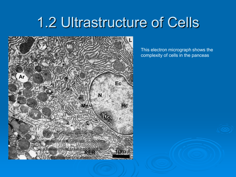
1.2 Ultrastructure of Cells This electron micrograph shows the complexity of cells in the panceas Two Fundamentally Different Cell Architectures A prokaryotic cell A eukaryotic cell 1.2.U1 Prokaryotes have a simple cell structure without compartmentalization Prokaryote means “before nucleus” http://www.wiley.com/college/boyer/0470003 790/animations/cell_structure/cell_structure. htm Prokaryotic Cell wall pili Plasma membrane Flagella Ribosomes (70 S) Nucleoid (region containing DNA) Prokaryotic Structures & Functions Cell Wall protects and maintains shape Composed of peptidoglycan (carbprotein) Some have additional layer to adhere to structures (teeth, skin) Plasma Membrane Controls movement of materials in and out Role in binary fission Cytoplasm is the complete interior No compartments DNA most visible structure Pili Prokaryotic Structures & Fucntions These hollow, hairlike structures made of protein allow bacteria to attach to other cells and specialized pili have a role in conjugation Flagella The purpose of flagella (sing., flagellum) is motility. Flagella are long appendages which rotate by means of a "motor" located just under the cytoplasmic membrane. Bacteria may have one, a few, or many flagella in different positions on the cell. http://highered.mcgrawhill.com/sites/007337525x/student_view0/exercises_3590/bacterial_locomotion.html Prokaryotic Structures & Functions Ribosomes site of protein synthesis small made up of two parts that come together, can be in large numbers 70S Note: S Svedberg unit, a measure of the rate of sedimentation in centrifugation rather than size and accounts for why fragment names do not add up (70S is made of 50S and 30S). Prokaryotic Structures & Functions Nucleoid region DNA in the bacterial cell is generally confined to this central region. Though it isn't bounded by a membrane, it is visibly distinct from the rest of the cell interior. Plasmid A plasmid is a DNA molecule that is separate from, and can replicate independently of, the chromosomal DNA. They are double stranded and, in many cases, circular. Plasmids usually occur naturally in bacteria, but are sometimes found in eukaryotic organisms 1.2.A2 Prokaryotes divide by binary fission http://www.classzone.com/books/hs/c a/sc/bio_07/animated_biology/bio_c h05_0149_ab_fission.html • asexual reproduction • semiconservative replication (unit 2.7 U1) •2 DNA loops attach to membrane •Membrane elongates and pinches off forming two cells •Daughter cells are clones (genetically identical) Drawing #1 Please complete in your sketchbook. You must be able to draw and label a diagram of the ultrastructure of a prokaryotic cell based on electron micrographs. The organelles previously discussed must be included in your drawing. Similarities Between Prokaryotes & Eukaryotes 1. DNA 1. Plasma membrane (a.k.a. “cell 3. Cytoplasm 4. Ribosomes membrane”) Differences Prokaryote Eukaryote DNA ring no protein 2. DNA free 3. No mitochondria 4. 70 S ribosomes 5. No internal compartmentalization 6. Less than 10 micrometers chromosomes nucleus mitochondria 80 S organelles greater than 10 micrometers 1. 1.2.U2 Eukaryotes have a compartmentalized structure Electron micrograph of a mammalian cell Why compartmentalize? Eukaryotic cells An idealized animal cell. An idealized plant cell Eukaryotic:Animal, algae, protozoa, fungi and Plant cells This shows the organelles of a liver cell Eukaryotic cells are usually 5 to 100 micrometers, nucleus is membrane bound, have membrane bound organelles 1 micrometer= 0.001mm 1nanometer= 0.001 µ m 1.2.A1 Structure and function of organelles within exocrine gland cells of the pancreas (animal cell) and within palisade mesophyll cells of the leaf (plant cell) Palisade mesophyll cells Pancreas cell The Nucleus • Generally spherical with double membrane • Pores in membrane • Contains genetic information in chromosomes/chromatin (DNA + histone proteins) Two meters of human DNA fits into a nucleus that’s 0.000005 meters across! The Mitochondrion Think of the mitochondrion as the powerhouse of the cell. Worn out mitochondria may be an important factor in aging • • • • • • • • Has a double membrane (smooth outer and folded inner) Folds are called cristae Shape varies Site of energy production (ATP) during cellular respiration they have their own DNA large surface for cellular metabolism produces and contains its own ribosomes (70S type) Can reproduce independently Free ribosomes Bound ribosomes are attached to another organelle called the endoplasmic reticulum and their protein products are shipped out of the cell The Endoplasmic Reticulum (ER) Endoplasmic Reticulum Smooth ER -no ribosomes -lipid synthesis - Fewer of these exist in pancreatic cells Rough ER • • • • • Consists of flattened membrane sacs called cisternae Often located near the nucleus 80 S ribosomes attached to outside Synthesizes proteins which are transported by vesicles to the golgi apparatus before secretion out of the cell Pancreatic exocrine cells would have an abundance of these do to the constant production of digestive enzymes Golgi apparatus The Golgi collects, packages, modifies and distribute materials the cell makes (especially protein) Golgi in the cytoplasm of a macrophage in lung tissue • Like the ER it is composed of flattened sacs called cisternae • However, it has no ribosomes and is often near the plasma membrane • Cisternae are shorter and more curved than those on ER Vesicles A single membrane with fluid inside Very small Used to transport materials outside of cell http://bcs.whfreeman.com/thelifewire/c ontent/chp04/0402002.html The Lysosome •Intracellular •arise from golgi •Spherical with a single membrane Functions: Contain digestive enzymes •Breakdown ingested food •Breakdown damaged or unwanted organelles •Cell suicide (a.k.a “suicide sac”) Stains dark in micrographs due to high concentration of enzymes Many diseases (e.g. Tay-Sachs) are caused by lysosome malfunction http://highered.mheducation.com/sites/0 072495855/student_view0/chapter2/ani mation__lysosomes.html The Lysosome This bacterium about to be eaten by an immune system cell will spend the last minutes of its existence within a lysosome. Vacuoles • Store food, waste, toxins, water • Increase SA to volume ratio • Allow plants to be rigid Cilia and Flagella Cilia Flagella tiny • hair like projections contains microtubules used to move the cell or move the fluids next to the cell • • Thin projection from cell surface Contains microtubules Used for movement Lung cilia Mature plant cells do not contain these structures but some plant gametes do Microtubules and Centrioles IN PLANTS NOT ANIMALS! The Chloroplast Think of the chloroplast as the solar panel of the plant cell. • • • • • • Starch granules maybe present if producing lots of food! Contain double membrane same size as bacteria Has own DNA has 70 S ribosomes can reproduce on own Inside are stacks of thylakoids (discs of flattened membrane) • Usually oval in shape • Site of photosynthesis (production of carbohydrates) Only plants and algae have chloroplasts Granumflattened sacs like solar panels (increase SA) Stroma-fluid, contains enzymes and chemicals Cell Wall • Extracellular component not an organelle • Secreted by all plant cells (also fungi and some protists) • In plants it contains mostly cellulose • Cellulose is permeable, strong and hard to digest (little maintenance required) IN PLANTS NOT ANIMALS! Plant Differences vs. Animal Cell wall Chloroplasts Large vacuoles Carbs stored as starch No centrosome Fixed angular shape http://www.youtube.com/watch?v=LP7xAr2 FDFU&feature=related http://www.youtube.com/watch?v=GigxU1 UXZXo&feature=fvwrel none none small or none glycogen centrosome flexible, rounded http://library.thi nkquest.org/C0 03763/flash/gai a1.htm Magnification- is how large the object will appear compared to its actual size Electron Micrographs A rat liver cell (with color enhancement to show organelles). Perioxsome Similar to lysosomes except peroxisomes bud off from the endoplasmic reticulum Have many roles and/or involvement in producing bile, fatty acid breakdown, cholesterol, myelin production, getting rid of H2O2 etc… Extracellular Matrix-animal cells composed of collagen and glycoproteins (sugar and protein) form fibres, anchor to the plasma membrane to strengthen it, cell to cell interaction possible altering of gene expression, leads to coordination of cell action may be involved in directing stem cells, and possibly cell migration and movement The Cytoskeleton The name is misleading. The cytoskeleton is the skeleton or scaffolding of the cell, but its also like the muscular system as well, able to change the shape of cells in a flash. An animal cell A Cytoskeleton Gallery The Cytoskeleton in Action A macrophage using the cytoskeleton to “reach out” for a hapless bacterium. The Cytoskeleton in Action Membrane ruffles on a crawling cell. Centrosome usually a pair of centrioles -Assemble microtubules for structure and movement -cell division - higher plants produce microtubules even though they do not have centrioles -
