Proper care
advertisement
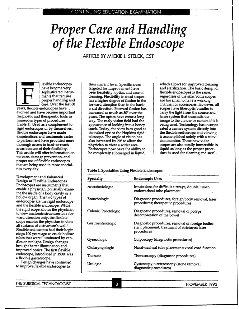
Proper Care and Hand ling o f the Flexible Endoscoae ARTICLE BY MlCKlE J. STELCK, CST lexible endoscopes have become very sophisticated instruments that require proper handling and care. Over the last 60 years, flexible endoscopes have . evolved and have become important diagnostic and therapeutic tools in numerous types of procedures (Table 1).Used as a complement to rigid endoscopes or by themselves, flexible endoscopes have made examinations and treatments easier to perform and have provided more thorough access to hard-to-reach areas because of their flexibility. This article will offer information on the care, damage prevention, and proper use of flexible endoscopes that are being used in more specialties every day. Development and Enhanced Design of Flexible Endoscopes Endoscopes are instruments that enable a physician to visually examine the inside of a body cavity or a hollow organ. The two types of endoscopes are the rigid endoscope and the flexible endoscope. While the rigid scope allows the physician to view anatomic structures in a forward direction only, the flexible scope enables the physician to view all surfaces of a structure's wa1l.l Flexible endoscopes had their beginnings 100 years ago as crude hollow tubes that were illuminated by candles or sunlight. Design changes brought better illumination and improved optics. The first flexible endoscope, introduced in 1930, was a flexible gastroscope. Design changes have continued to improve flexible endoscopes to their current level. Specific areas targeted for improvement have been flexibility, optics, and ease of cleaning. Flexibility in most scopes has a higher degree of flexion in the forward d i r e o n than in the backward direction. Forward flexion has increased as much as 30" over the years. The optics have come a long way. The early vision field had the appearance of looking into a honeycomb. Today, the view is as good as the naked eye or the Hopkins rigid telescope. The angle of vision has also increased by 20" to allow the physician to view a wider area. Endoscopes now have the ability to be completely submerged in liquid, which allows for improved cleaning and sterilization. The basic design of flexible endoscopes is the same, regardless of the size. Some scopes are too small to have a working channel for accessories. However, all scopes have fiberoptic bundles to carry the light from the source and lense system that transmits the image to the viewer or camera if it is being used. Technology has incorporated a camera system directly into the flexible endoscope and viewing is accomplished solely with a television monitor. These new video scopes are also totally immersible in liquid as long as the proper procedure is used for cleaning and steril- Table 1.Specialties Using Rexible Endoscopes Specialty Endoscopic Uses Anesthesiologic Intubations for difficult airways; double lumen endotracheal tube placement Bronchologic Diagnostic procedures; foreign body removal; laser procedures; therapeutic procedures Colonic, Proctologic Diagnostic procedures; removal of polyps; decompression of the bowel Gastroenterologic Diagnostic procedures; removal of foreign bodies; stent placement; treatment of strictures; laser procedures Gynecologic Colposcopy (diagnostic procedures) Otolaryngologic Nasal-tracheal tube placement; vocal cord function Thoracic Thoracoscopy (diagnosticprocedures) Urologic Cystoscopy; ureteroscopy (stone removal, diagnostic procedures) NOVEMBER 1993 Table 2. Accessories for Flexible Endoscopes Accessory Use Balloons Dilation; tamponade Baskets Foreign body removal Brushes Specimen biopsies Cameras (video and 35 mm) Photography of organs and lesions Electrosurgicalelectrodes and snares Coagulation Forceps Specimen biopsies; foreign body removal Laser fibers Incision, excision, vaporization, coagulation Needles Injection of medications; specimen biopsies ization. This new system allows for videotaping and still photography without changing cameras. Endoscope sizes range from 2.2 mm to 13.3 mm in diameter and come in various lengths based on the intended use. Endoscopes with a smaller diameter are the most fragde and should be carefully protected. Several companies specialize in the manufacturing of flexible endoscopes and their accessories (Table 2). In addition to the flexible endoscope, a compatible light source should be available. Light sources are available in various models, based on their intended use. Halogen lamps and xenon lamps are widely used. Halogen lamps provide sufficient light for most endoscopic procedures. However, if photography is incorporated, then a xenon lamp is necessary. If cost is a Figure 1.Xenon-type light source for flexible endoscopes that can be used for photography. THE SURGICAL TECHNOLOGIST primary consideration, the halogen lamp is less expensive (Figure 1). Video and still photography can expand an endoscopic procedure and serve as a tool for educating patients and medical staff. It can also serve as a source of documentation to track the progression of a patient's disease following prescribed treatment. Photographic equipment may include a 35-mm camera with an endoscopic attachment to allow for coupling to the flexible endoscope or video equipment. Videotaping equipment comes with several sizes of VCR tapes and different versions of camera equipment. Video cameras have become very sm$ and lightweight and do not add extra bulk when placed on the flexible endoscope (Figure 2). The importance of sterility during the procedure should be considered when selecting video equipment. Whether or not the camera can be disinfected or sterilized and presented onto a sterile field if necessary should be determined. Compatibility with flexible endoscopes and light sources is essential and must always be considered. Expense will also be a factor weighed when purchasing equipment. Cleaning, Disinfecting, and Sterilizing Endoscopes Cleaning. Cleaning is the manual act of removing debris from the out- side and the lumens of the endoscope with warm water and a neutral, nonresidual liquiddetergent solution. Cleaning should be done as soon as possible after the endoscope's use to prevent body fluids from drying on the scope, which makes it harder to clean. Thorough cleaning is necessary to reduce the bioburden and to allow the chemical agents used in disinfection and sterilization to reach all parts of the flexible endoscope to prevent corr~sion.~ Special cleaning brushes designed to go through the endoscope's lumens help to dislodge material that can not be removed by flushing with water and the soap solution alone. Ultrasonic cleaners should not be used because the vibrations from the cleaner could disrupt the optic and flexing mechanisms or damage the seals. The cleaning procedure for flexible endoscopes may vary from manufacturer to manufacturer. Lnstructions provided by the manufacturer should be followed by personnel responsible for cleaning and maintenance of the endoscopes. Cleaning instructions should be easily accessible to those who may be new to the handling of flexible endoscopes to ensure that all of the endoscopic equipment, including the lumens, is thoroughly and properly cleaned (Table 3). Procedures may be modified to fit the needs of the individual institution. Disinfecting. After manual cleaning, endoscopic equipment may be disinfected by one of several methods. There are three levels of disinfection, high, intermediate, and low, that may be used depending on the strength - of disinfection that is needed. L I Figure 2. Detachable video camera with a flexible endoscope adaptor. NOVEMBER 1993 Table 3. Example of Cleaning Procedure for Flexible Endoscopes 1. Suction povidone-iodine (Betadine)soap solution through the channel (approximately 100 cc). 2. Insert cleaning brush through the channel. 3. Suction more Betadine soap solution through the channel. 4. Suction distilled water through the channel until clear. 5. Remove any pieces that require separate cleaning. 6. Wash outside of flexible endoscope and all of its components and accessories in water and soap solution. 7. Rinse all parts thoroughly in water. 8. Proceed to disinfect or sterilize instrumentation. High-level disinfection is the act of exposing the instruments to a chemical disinfectant that kills most vegetative organism including the human immunodeficiency virus (HN) Instruments are exposed to activated 2% glutaraldehyde sohtion for a minimum of 20 minutes at 25'C.I The tuberculosis spore has a tough waxy coating on its outside so it is not killed by high-level disinfection. To ensure that the tuberculosis spore is killed, the instruments should be labeled tuberculocidal Figure 3. Large pan used to submerge instrumentation in glutaraldehyde solution. and exposure time should be increased to 45 minutes at 25OC. High-level disinfection is applied to any instrument that comes into direct contact with the patient. Intermediate disinfection kills resistant bacteria and viruses but it is not sporicidal. Agents used may be 70% and 95%alcohol or iodine compounds. Low-level disinfection includes housekeeping activities that use agents to kill the least resistant bacteria and viruses. Agents in this category include phenol or mercurial comp~unds.~ Disinfection takes less time to perform than sterilization. How- THE SURGICAL TECHNOLOGIST ever, unlike ste-tion, disinfection does not kill spores. Exposure to glutaraldehyde solution for a minimum of 20 minutes kills most microorganisms including HIV, (Figure 3) but the instruments are not sterile. Following immersion in the glutaraldehyde solution, instruments are thoroughly rinsed in distilled water to remove any excess solution, since it can be very imtating to mucous membranes. The endoscopic instruments are then completely dried before storage.' Sterilizing. Sterilization is the process of killing all microorganisms, including spores. Sterilization of flexible endoscopes and accessory instruments is recommended for highly infectious cases such as those involving tuberculosis or AIDS to reduce the risks of cross contamination. Sterilization should also be done for the more resistant viruses that cause serum hepatitis and infectious hepatitis. Sterilization can be accomplished by immersion in a 2% glutaraldehyde solution for 10 hours, but the harshness of thissolution and possible effect on the instruments may be a deterrent.' Cold solution sterilization does not require any special equipment and may be advantageous for this reason. Ethylene oxide gas (EO) steriiization is the most commonly used method of sterilization. Instruments undergoing sterilization can not be used for approximately 12 hours, including the aeration time to remove any residual ethylene oxide gas, which is required after EO sterilization. However, any type of flexible endoscope or accessory can be EO sterilized as long as the manufacturer's guidelines for time, temperature, and pressure are not exceeded. Instrumentation sent to be EO sterilized should be in a protective container that permits sterilization (Figure 4). To prevent damage to the equipment when transporting and sterilizing it, a fastening device is used to anchor everything securely. The flexible endoscope and accessories should be completely dried before they are sent for EO sterilization (Figure 5). Parts with lenses and some fiberoptic carriers can not be steam autoclaved.' A recent addition to the technology of sterilization methods involves the use of peracetic acid to sterilize any instrument that can be immersed in liquid. Specially designed machines allow both flexible and rigid endoscopes to be sterilized in approximately 30 minutes. The instruments are first cleaned as usual and then placed in the machine, at which time the cycle is started. The instruments are , exposed to a 35%concentrate of peracetic acid solution for 12 minutes and then rinsed four times in sterile water. Although this method has Figure 4. Protective container used to safely transport flexible endoscopes for EO sterilization. A, Open container with foam padding inside wire basket; B, Closed container for transporting endoscopes. NOVEMBER 1993 care should be taken to ensure that both the leak testing cap and the stem on the flexible endoscope are completely dry. Figure 5. Flexible endoscope with EO cap attached ready for EO sterilization. the advantage of providing sterility in a short period of time, not all instruments can fit into the machine because of their size or shape. EO sterilization is used for instruments that can not be totally immersed in liquid. As always, the manufacturer's guidelines should be followed. While the peracetic acid process allows instruments to be sterilized more quickly, this process should only be used when a shorter processing time is needed in order to use the instruments again sooner. It should not be used if the instruments will be stored on the shelf for a long time after sterilization. Leak Testing Leak testingafter every surgical procedure will ensure the integrity of the flexible endoscope's seals and demonstrate that no damage has occurred to either the outer coating or to the inner channel during use. Leak testing is accomplished by connecting the leak tester to the flexible endoscope and turning on the tester (Figure 6). The endoscope is submerged entirely in clear water and air is pumped into the endoscope to observe for air bubbles. If no air bubbles are seen, the leak test is negative and the cleaning and disinfection or sterilization procedures may be started. If a continuous stream of bubbles is noted, the leak test is positive. The endoscope is then cleaned and subjected to EO sterilization, after which it is sent for repair. In theory, leak testing should help prevent major internal moisture damage. However, there is a risk that moisture will be introduced during leak testing so THE SURGICAL TECHNOLOGIST Universal Precautions Specific procedures for following universal precautions for highly infectious diseases, such as HN, hepatitis, tuberculosis, and Creutzfeldt-Jakobdisease, vary from institution to institution. Hospitals should set policies detailing whether a cleaning, disinfecting, or sterilizing process is appropriate for specific instruments or equipment. The method of treatment should be determined based on an instrument's intended use. As a genera1 rule, any item that will touch sterile tissue must be ~terilized.~ While this may not always be possible, instrumentation sterility is prudent and hospital policies should be followed stringently. With the resurgence of drug-resistant tuberculosis in the United States, sterilization is preferred to disinfection and ideally should be used whenever possible in order to kill the spores and prevent cross contamination between patients. In addition to following procedures to prevent the transmission of diseases and infections between patients, health care workers involved in the processing of contaminated instruments must also take precautions to guarantee their own personal safety. Proper protective attire should always be worn. The Occupational Safety and Health Administration (OSHA)recommends the use of, "gloves, gowns, laboratory coats, face shields or masks, and eye protection, and mouthpieces, resuscitation bags, pocket masks, or other ventilation devices," when cleaning instruments man~ally.~ The Centers for Disease Control and Prevention's (CDC's) Universal Precautions include the following recommendation: "All health care workers should take precautions to prevent injuries...when cleaning used instruments." Furthermore, manual cleaning may cause splashes and the CDC recommends the use of protective clothing during procedures that are, "likely to generate splashes of Figure 6. Leak tester for flexible endoscopes. blood or other body fluids."' Storage of Endoscopic Equipment Storage of flexible endoscopes and instrumentation should provide protection while at the same time allowing for easy accessibility to the items. Vertical suspension of the flexibleendoscope prevents a memory curve from forming in the insertion portion of the endoscope (Figure 7). Drawers can also be used for storage but great care must be taken to prevent the endoscope from being crushed when closing the drawer. Before endoscopic instrumentation can be stored, all parts and accessories must be thoroughly dry,inside and out. Shipping cases provided by manufacturer's are not recommended because they are airtight; even a small amount of moisture can foster the growth of a rniamrganism. Closing shipping cases may also damage flexible endoscopes if the instruments are not carefully placed in the case. Storing flexible endoscopes in a cart with drawers with other needed supplies provides the mobility to travel to other departments for endoscopies. Keeping the endoscopy cart well-stocked for I I Figure 7. Flexible endoscopes stored vertically to prevent memory curve. NOVEMBER 1993 Figure 8. Cart with light source on top and drawers for endoscopes and supplies. emergency situations is an important consideration (Figure 8). When preparing to store the flexible endoscope, the optics and the flexing mechanisms should be checked to ensure there is no damage. A film over the lense or eyepiece is easily cleared with an alcohol swab. Sluggish flexion should be repaired as it will impair an examination. Damage Prevention Flexible endoscopes are expensive instnunents, both to purchase and to repair, and should be handled gently. New flexible endoscopes cost as much as $12,000 to $15,000. Repair costs vary from a few hundred dollars to a few thousand dollars if the endoscope can be effectively repaired. Damage is often so exten- Figure 9. Damaged endotracheal tube when a bite block was not secured. sive that repair is impossible and replacement is necessary. Damage may be caused by the physician, by other members of the operating room staff, or by the patient. Excessive bending or kinking of the fiberoptic bundle will result in breakage of the bundles, which will appear as black dots in the field of vision. The distal tip must be prevented from striking against objects that could crack the objective lense. A bite block of some kind should be used in bronchoscopic or gastroenterologicexaminations to prevent the patient from biting the endoscope (Figure 9). Accessory instruments should be gently inserted, not forced, though the channel when the distal portion of the flexible endoscope is flexed to prevent a puncture to the inner channel. Needles inserted through working channels of the flexible endoscope can very easily puncture the inner channel, so all staff should check to ensure that the needle is not exposed upon insertion and withdrawal (Figure 10). Extreme caution must be taken when using lasers with flexible endoscopes. Materials used in the construction of the flexible endoscope are extremely flammable. The laser fiber should be out of the flexible endoscope at least 2 an before firing. Injury or death could result during laser procedures involving the airway if the plastics of either the bronchoscope or the endotracheal tube ignite. Oxygen levels administered by anesthesia should be below 40% to help reduce the risk of a fire. Endotracheal tubes should be wrapped with a fire resistant, reflective material to further reduce the risk of a fire. Lubrication is very important when using flexible endoscopes. Adequate amounts of lubrication prevent the flexible endoscope from sticking or binding, which could cause the outer sheath to stretch or rip. Water-based lubricants such as surgical jelly should be used rather than petroleum-based lubricants since excessive use of petroleumbased products causes the outer plastics of the flexible endoscope to break down. Alcohol dries the mate- Figure 10. Small 22-gauge cytology specimen needle may cause extensive damage to an endoscope. rials and makes them brittle, so contad with alcohol on instruments should be avoided except on the objectibe lense and the eyepiece. Damage may also occur if the EO cap is not placed on the scope during sterilization or air shipment. Inner pressure build-up in the endoscope may cause a rupture of the outer sheath, leaving the inner flexing mechanism exposed. Although this type of damage is relatively inexpensive to repair, it will still cause the flexible endoscope to be out of use while it is being repaired. Flexible endoscopes should be treated as the expensive and delicate instruments that they are. Having a flexible endoscope out for repair may be more than an inconvenience for the medical team-it may affect a patient's diagnosis or treatment. A References 1. Atkinson LJ. Berry and Kohn's Operating Room Technique.7th ed. St Lo+, Mo: Mosby-Year Book,Inc;1992. 2.1993 AORN Standards and Recommended Practices. Denver, Colo: AORN,Inc.; ' 1993. 3. Fairchild SS. Penenoperative Nursing Pin- (Continued on page 22) MD,at the Mnyo Clinic. . NOVEMBER 1993 CE programs, except those organized directly by the AST national organization, are generally privately sponsored; have no official relationship to AST; and to the extent that AST is involved in the CE process, AST is in no way guaranteeing the accuracy of the CE program contents or the experience, knowledge, or qualifications of the CE instructor. AST does not, in any way, represent that, upon completion of any CE course, the person completing such course will have the experience, knowledge, or competency to perform any of the tasks or job descriptions covered by such course. November Baton Rouge, LA: November 6. Chapter 25. Session title: High Tech Surgery in the 90s. Contact: Laura Kendrick, 7744 LaSalle Ave, #40, Baton Rouge, LA 70806; 504-924-3124. Denver, CO: November 6-7. Colorado Surgical Assisting, Inc. Session title: Wound Closure/Suturing and Tying Techniques HandsOn. Contact: Ron Coulter, 3033 S Parker Rd, Market Tower 1, Suite 504, Aurora, CO 80014; 303-7459509. Greenville, NC: November 13. Chapter 205. Session title: TBA. Contact: Laura Setliff, Rt 14, Box 77, Greenville, NC 27834; 919-7580824. Portland, OR: November 20-21. Colorado Surgical Assisting, Inc. Session title: Wound Closure/ Suturing and Tying Techniques Hands-on. Contact: Ron Coulter, 3033 S Parker Rd, Market Tower 1, Suite 504, Aurora, CO 80014; 303-745-1261. Abilene, TX: November 20-21. Chapter 248. Session title: TBA. Contact: Naomi Kline, 1818 University Blvd, Abilene, TX 79603; 915-675-0304. HE SURGICAL TECHNOLOGIST The programs listed here are seeking or have received conditional approval by the AST Continuing Education Committee. Conditional approval is awarded when the program dates have been confirmed and the program agenda and speaker fact sheets have been received by the Continuing Education Committee representative. Final approval of CE credit is contingent upon the submission and review of the appropriate postprogram documentation. When advertising a program, the program sponsor must use the proper terminology. The program may be advertised as "conditional approval pending by the AST Continuing Education Committee for XX CE credits" if the program agenda and speaker fact sheets have not been approved. If this documentation has been approved, the program may be advertised as "conditionally approved by the AST Continuing Education Committee for XX CE credits." 1994 WORKSHOPS April February St Louis, MO: April 2-3. Chapter 52. Session title: Scope-A-Rama. Contact: Ron Novak, 4769 Ambs Rd, St Louis, MO 63128; 314-4871494. Hartford, CT: April 16. Region 1 Meeting. Chapter 56. Session title: TBA. Contact: Lillie Harris, 2824 Ellington Rd, South Windsor, CT 06074; 203-644-8086. A Tulsa, OK: February 5-6. Chapter 53. Session title: Pediatric Overview. Contact: Nina Michael, Rt 2, Box 437H, Coweta, OK 74429; 918-486-3302. Dallas, TX: February 19. Region 6 Meeting. Chapter 254. Session title: We Cycle Organs. Contact: Carrie Lee, 5401 Knollridge, Garland, TX 75043; 214-226-8436. Las Vegas, NV: February 26-27. Region 8 Midyear Meeting. Session title: TBA. Contact: James Kephart, 408 Peregrine Ave, Henderson, NV 89015; 702-565-9068. March Savannah, GA: March 5. Region 3 Meeting. Chapter 212. Session title: TBA. Contact: Margaret Cashion, 3984 Lacewood Dr, Memphis, TN 38115; 901-3670878. Louisville, KY: March 5. Chapter 5. Session title: TBA. Contact: Beverly Crady, 1520 Sanders Ln, Louisville, KY 40214; 502-3665965. Ft Worth, TX: March 5-6. Chapter 32. Session title: Best Little Workshop in Texas. Contact: Violet Berna, 635 Oak St, Burleson, TX 76028; 817-295-2534. Buffalo, NY: March 19. Chapter 62. Session title: Orthopedic Update. Contact: Maria Szefler, 162 Castle Wood, Cheektowaga, NY 14227; 716-668-4761. Spring Regional Meetings Region 6 Meeting Date: February 19 Region 8 Meeting Date: February 26-27 Region 3 Meeting Date: March 5 Region.1 Meeting Date: April 16 ciples and Practice. Boston, Mass: Jones and Bartlett Publishers; 1993. 4. Perkins JJ. Principles and Methods of Sterilization in Health Sciences. 2nd ed. Springfield, Ill: Charles C. Thomas Publishing; 1982. 5. Gamer JS, Favero MS. CDC guideline for handwashing and hospital environmental control. Today's OR Nurse. April 1986. 6. Reichert M. Laparoscopic instruments: Patient care, cost issues. AORN Journal. March 1993. 7. Centers for Disease Control and Prevention. Recommendations for prevention of HIV transmission in health-care settings. MMWR. 1987;36.
