Medical Applications of Virtual Reality
advertisement
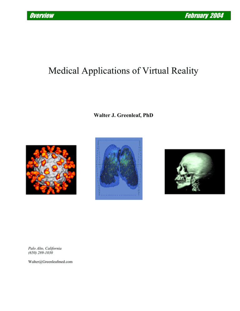
Overview February 2004 Medical Applications of Virtual Reality Walter J. Greenleaf, PhD Palo Alto, California (650) 289-1030 Walter@Greenleafmed.com "The major pivotal point was in 1989 when the first virtual laparoscopic gallbladder operation was performed. In addition to minimally invasive surgery, virtual reality in the future will offer, among other benefits, remote surgery, greatly improved medical and surgical training, visualization of massive medical databases, and innovative rehabilitation techniques.” COL Richard Satava, MD 1 1 COL Richard Satava, M.D. F.A.C.S., Clinical Associate Professor of Surgery (USUHS) is a practicing clinical and research general surgeon. He was assigned to General Surgery at Walter Reed Army Medical Center, as Special Assistant in Advanced Technologies at the US Army Medical Research and Materiel Command, and to research at the Advanced Research Projects Agency (ARPA). He has also been with the Yale School of Medicine and the HITLab. INTRODUCTION TO VIRTUAL REALITY TECHNOLOGY Virtual Reality (VR) is the term commonly used to describe a novel human-computer interface that enables users to interact with computers in a radically different way. VR consists of a computer-generated, multi-dimensional environment and interface tools that allow users to: 1) immerse themselves in the environment, 2) navigate within the environment, and 3) interact with objects and other inhabitants in the environment. The experience of entering this environment—this computer-generated virtual world—is compelling. To enter, the user usually dons a helmet containing a head-mounted display (HMD) that incorporates a sensor to track the wearer’s movement and location. The user may also wear sensorclad clothing that, likewise tracks movement and location. The sensors communicate position and location data to a computer, which updates the image of the virtual world accordingly. By employing this garb, the user "breaks through" the computer screen and becomes completely immersed in this multi-dimensional world. Thus immersed, one can walk through a virtual house, drive a virtual car, or run a marathon in a park still under design. Recent advances in computer processor speed and graphics make it possible for even desk-top computers to create highly realistic environments. The practical applications are far reaching. Today, using VR, architects design office buildings, NASA controls robots at remote locations, and physicians plan and practice difficult operations. Virtual reality is quickly finding wide acceptance in the medical community as researchers and clinicians alike become aware of its potential benefits. Several pioneer research groups have already demonstrated improved clinical performance using VR imaging, planning and control techniques. Overview of Virtual Reality Technology The term “Virtual Reality” describes the experience of interacting with data from within the computer-generated data set. The computer-generated data set may be completely synthetic or remotely sensed, such as X-ray, MRI, PET, etc. images. Interaction with the data is natural and intuitive and occurs from a first-person perspective. From a system perspective, VR technology can be segmented as shown in Figure 1. The computer-generated environment, or virtual world content, consists of a 3D graphic model, typically implemented as a spatially-organized, object-oriented database; each object in the database represents an object in the virtual world. A separate modeling program is used to create the individual objects for the virtual world. For greater realism, texture maps are used to create visual surface detail. The data set is manipulated using a real-time dynamics generator that allows objects to be moved within the world according to natural laws such as gravity and inertia, or to other variables such as spring-rate and flexibility that are specified for each particular experience by application-specific programming. The dynamics generator also tracks the position and orientation of the user's head and hand using input peripherals such as a head tracker and DataGlove. Powerful renderers are applied to present 3D images and 3D spatialized sound in realtime to the observer. Modeling Program Content • 3D Graphics Model • Texture Maps Translators • CAD Database Input * VRML Input * Med-Web Data Input Application-Specific Programming Renderers • 3D Image • Spatialized Sound Dynamics Generator Input Peripherals • DataGlove • Head Tracker • Microphone Output Peripherals • HMD Video • HMD Audio • Motion Platform User Actions • Turn • Grab • Speak Figure 1 - A complete VR system. The common method of working with a computer (the mouse/keyboard/monitor paradigm), based, as it is, on a two-dimensional desk-top metaphor, is inappropriate for the multi-dimensional virtual world. Therefore, one long-term challenge for Virtual Reality developers has been to replace the conventional computer interface with one that is more natural, intuitive and allows the computer— not the user— to carry a greater proportion of the interface burden. Not surprisingly, this search for a more practical multi-dimensional metaphor for interacting with computers and computer-generated 2 artificial environments has spawned the development of a new generation of computer interface hardware. Ideally, the interface hardware should consist of two components, sensors for controlling the virtual world and effectors for providing feedback to the user. To date, three new computerinterface mechanisms, not all of which manifest the ideal sensor/effector duality, have evolved: Instrumented Clothing, the Head Mounted Display (HMD), and 3D Sound Systems. Instrumented Clothing The DataGlove™ and DataSuit™ use dramatic new methods to measure human motion dynamically in real time. The clothing is instrumented with sensors that track the full range of motion of specific activities of the person wearing the Glove or Suit, for example as the wearer bends, moves, grasps or waves. Tactile Feedback Device Flexion Sensors Fiber Optic Cables Cable Guides Absolute Position and Orientation Sensor Abduction Sensors Figure 2 - the DataGlove, a VR control device. The DataGlove is a thin lycra glove with bend-sensors running along its dorsal surface. When the joints of the hand bend, the sensors bend and the angular movement is recorded by the sensors. These recordings are digitized and forwarded to the computer, which calculates the angle at 3 which each joint is bent. On screen, an image of the hand moves in real time, reflecting the movements of the hand in the DataGlove and immediately replicating even the most subtle actions. The DataGlove is often used in conjunction with an absolute position and orientation sensor that allows the computer to determine the three-space coordinates, as well as the orientation of the hand and fingers. A similar sensor can be used with the DataSuit and is nearly always used with an HMD. The DataSuit is a customized body suit fitted with the same sophisticated bend-sensors found in the DataGlove. While the DataGlove is currently in production as both a VR interface and as a data-collection instrument, the DataSuit is available only as a custom device. As noted, DataGlove and DataSuit are utilized as general purpose computer interface devices for Virtual Reality. There are several potential applications of this new technology for clinical and therapeutic medicine. Head Mounted Display (HMD) The best-known sensor/effector system in Virtual Reality is a head-mounted display (HMD). It supports first-person immersion by generating an image for each eye, which, in some HMDs may provide stereoscopic vision. 3D Spatialized Sound The impression of immersion within a virtual environment is greatly enhanced by inclusion of 3D spatialized sound Stereo-pan effects alone are inadequate since they tend to sound as if they are originating inside the head. Research into 3D audio has shown the importance of modeling the head and pinea and using this model as part of the 3D sound generation. A Head Related Transfer Function (HRTF) can be used to a generate the proper acoustics. Other VR Interface Technology A sense of motion can be generated in a VR system by a motion platform. These have been used in flight simulators to provide cues that the mind integrates with visual and spatialized sound cues to generate perception of velocity and acceleration. Haptics is the science of touch. Haptic interfaces generate the percepts of touch and resistance in VR. Most systems to date have focused on providing force feedback to enable users to sense the inertial qualities of virtual objects, and/or kinesthetic feedback to specify the location of virtual object in the world. A few prototype systems exist that generate tactile stimulation, which allows users to feel the surface qualities of virtual objects. Many of the haptic systems developed thus far consist of exo-skeletons that provide position sensing as well as active force application. Some preliminary work has been conducted on generating the sense of temperature in VR. Small electrical heat pumps have been developed that produce sensations of heat and cold as part of the simulated environment. 4 VR APPLICATION EXAMPLES Virtual reality had been researched for years in government laboratories and universities, but because of the enormous computing power demands and associated high costs, applications had been slow to migrate from the research world to other areas. Continual improvements in the price/performance ratio of graphic computer systems, however, have made VR technology more affordable and, thus, used more commonly in a wider range of application areas Applications today are diverse and represent dramatic improvements over conventional visualization and planning techniques: Public Entertainment: VR made its first major inroads in the area of public entertainment, with ventures ranging from shopping mall game simulators to low-cost virtual reality games for the home. Major growth continues in home VR systems, partially as a result of 3D games on the Internet. Computer-Aided Design: Using VR to create "virtual prototypes" in software allows engineers to test potential products in the design phase, even collaboratively over computer networks, without investing time or money for conventional hard models. All of the major automobile manufacturers and many aircraft manufacturers rely heavily on virtual prototyping. Military: With VR, the military’s solitary cab-based systems have evolved to extensive networked simulations involving a variety of equipment and situations. All levels of the military now have the ability to practice as teams in a verity of complex simulation scenarios, practicing search and rescue missions, for example, using acquired details from target-areas modeled into virtual worlds. Architecture/Construction: VR allows architects and engineers and their clients to "walk through" structural blueprints. Designs may be understood more clearly by clients who often have difficulty comprehending them even with conventional cardboard models. Data Visualization: By allowing navigation through an abstract "world" of data, VR helps users rapidly visualize relationships within complex, multi-dimensional data structures. This is particularly important in financial-market data, where VR supports faster decisionmaking. VR is commonly associated with exotic “fully immersive applications” because of the over dramatized media coverage on helmets, body suits, entertainment simulators and the like. Equally important are the “Window into World” applications where the user or operator is allowed to interact effectively with “virtual” data, either locally or remotely. 5 Current Status of Virtual Reality Technology The commercial market for VR, while taking advantage of advances in VR technology at large, is nonetheless contending with the lack of integrated systems and the frequent turnover of equipment suppliers. Over the last few years, VR users in academia and industry have developed different strategies for circumventing these problems. In academic settings, researchers buy peripherals and software from separate companies and configure their own systems to maintain the greatest application versatility. In industry, however, expensive, state-of-the-art VR systems are vertically integrated to address problems peculiar to the industry. Each solution is either too costly or too risky for most medical organizations; what is required is a VR system tailored to the needs of the medical community. Unfortunately, few companies offer integrated systems that are applicable to VR medical market. This situation is likely to change in the next few years, as VR-integration companies develop to fill this void. At the same time, the nature of the commercial VR medical market is changing as the price of high-performance graphics systems continues to decline. High-resolution graphics monitors are becoming more cost-effective for even markets that rely solely on desktop computers. Technical advances are also occurring in networking; visual photo-realism; tracker latency through predictive algorithms; and variable-resolution image generators. Improved database access methods are underway. Hardware advances, such as eye gear that provides an increased field of view with highresolution, untethered VR systems, and inexpensive intuitive input devices, e.g. DataGloves, have lagged behind advanced in computational, communications, and display capabilities. 6 OVERVIEW OF VR IN MEDICINE The use of VR in medical applications provides for better image manipulation, improved image understanding, improved quantitative comparisons, and better surgical planning. Healthcare's potential use of interactive 3D technologies is broad. To date, most of the media's attention has centered on two application areas: surgical training and planning, and computer-aided surgery systems. However, the possible uses are much broader. The following categories represent current and emerging applications of VR in medicine: Application Medical / Dental Surgical Training Pre-Surgical Planning Computer-Aided Surgery Systems Interactive 3D Diagnostic Imaging Description Training and rehearsing a surgical procedure using surgical instruments linked to a realistic simulation – may or may not include haptic feedback. Using 3D radiological images and computer workstation tools to design and plan an operative procedure. Using 3D images overlaid in real-time on the operating field to facilitate surgery. Tools for data analysis and quantitative comparisonscapturing and manipulating medical imaging data in a 3D format. Collaborative environments. Radiation Treatment Planning and Control Design of radiation treatment procedure to match patients’ anatomy precisely. 3D design and control systems. Medical Education Case histories, 3D anatomy lessons and virtual cadavers, procedure training, ER ward simulation, palpation training, etc. 3D Visualization for Telemedicine Telesurgery Radiological image tele-consultation and second opinions, shared data for tumor review boards, remote patient examination, and specialty consults. Computer-assisted surgery at a distance. Predictive algorithms, 3D surgical planning. 7 Application Description Rehabilitation and Sports Medicine Simulated environments for evaluation and rehabilitation - occupational therapy, physical therapy, ergonomics, orthopedics, and sports medicine Disability Solutions Neurological Evaluation Psychiatric and Behavioral Healthcare Augmented Reality environments for treatment of autism and other cognitive impairments. Environmental control systems. Standardized simulated environments for evaluation of cognitive processing, stroke deficits, memory disorders, movement disorders, and higher-functions. Evaluation and treatment of cognitive and behavioral disorders: phobias, anxiety, social affect disorders, ADHD, post-traumatic stress disorder, addiction treatment. Historical Economic Barriers Over the last decade, developers for VR in medicine have been faced with numerous obstacles. While government research and development money has been relatively forthcoming, venture funding has been difficult to come by. Historically, corporate investment has been limited to a few significant backers such as Johnson & Johnson, and Abbott Laboratories. It is estimated that less than $20 million is currently being spent annually on VR technologies for use in healthcare worldwide. In the US, government funding is primarily coming from the military at about $6 million 2 per year . There have been two types of economic barriers for commercial market development: 1) initial upfront costs of the technology, and 2) insurance reimbursement. Cost Barrier • 2 The first historic roadblock had been the cost of the technology. In order to produce a sense of realism for medical purposes, developers have been faced with providing organ fidelity, interactivity, physical and physiological properties of organs, and sensory input. Together, an integrated package required extreme amounts of computing power. Only expensive systems such as SGl's RealityEngine had the capabilities to even begin to integrate all those requirements. The costs of such computing power for full-scale medical simulations thwarted many interested healthcare professionals. See [1] 8 Insurance Reimbursement Barrier • The second roadblock stemmed from medical economics. A medical procedure must be shown to directly affect the patient before it can become billable to the patient. In addition, in today’s cost-conscious environment, the procedure must be shown to be more cost-effective than existing options. This is not always easy to demonstrate, given hurdles in conducting cost-effectiveness studies and in securing reimbursement. Recent Changes Over the last five years, the economic barriers to VR's use in medicine have been lessened. Basic computing power has dropped in price and VR developers are already working on products based on lower-cost desktop systems as well as Web-based solutions. Furthermore, an alternative solution is emerging that is based on part-task training or 'pieces' of a total learning experience. Products that 'break apart' a total learning experience and provide training for subsets of a medical procedure typically require less computing power and less cost than a full-blown VR simulation. One of the first companies to utilize the “break apart” approach was Virtual Presence, a Londonbased company that has developed, MIST, a training package that teaches the standard psycho-motor skills necessary for laparoscopic surgery. (Note that US-based Mentice, Inc. purchased the medical division of Virtual Presence in 2000.) Surgeons can practice movements such as tissue cut and lift, or arterial/duct clipping on MIST with the attached "surgical scissors" The individual user's movements are analyzed against a large database that assesses specific physical skills and provides spreadsheet feedback. By separating the psychomotor skills and using simple geometric objects onscreen, MIST avoids any requirement for the interactivity or organ fidelity/receptivity of full-scale 3 simulation graphics. . The Internet itself is embracing VR; technologies like Java 3D and VRML (Virtual Reality Modeling Language) are replacing 2D home pages with a true sense of place. At some point in the future there will be 3D browsers and full interactivity will take place over the Web. Current Dynamics With the availability of inexpensive computational power and the need for cost-effective solutions in healthcare, medical technology products are being commercialized at an increasingly rapid pace. VR is already incorporated into several emerging products in the areas of medical education, radiology, surgical planning and procedures, physical rehabilitation, disability solutions, and mental health. For example, VR is helping surgeons learn invasive techniques before operating, and allowing physicians to conduct real-time remote diagnosis and treatment. In addition, the contribution of robotics has accelerated the replacement of many open surgical treatments with more efficient minimally invasive surgical techniques using 3D visualization techniques. Other applications of VR include the modeling of molecular structures in three dimensions as well as aiding in genetic mapping and drug synthesis. 3 Proceedings of the Medicine Meets Virtual Reality Conference, 2004. 9 APPLICATIONS OF VR IN MEDICINE Interactive 3D Diagnostic Imaging Tchnology for diagnostic imaging, compared to the other medical specialties, is large and well-developed, with thousands of imaging systems deployed. The latest generation of these systems has the capability to collect large amounts of highresolution image data, but lacks the sophisticated software to easily navigate through this data. Due to the large capital outlay for these systems, there is a clear economic imperative for system owners to generate additional revenue to justify the equipment expenditure. Adding Interactive 3D visualization software to the installed hardware base provides value by reducing personnel time and costs, as well as improving clinical efficacy. The imaging modalities that represent computer-aided applications of VR in radiology include CT, MRI, X-ray imaging, NM, ultrasound, and computed radiography. Existing picture, archiving and communications systems (PACS) transfer and display radiological images acquired from all of these image modalities. The most common modalities in the PACS environment are CT, followed by CR, MRI, and mammography. Despite important difference in their core technologies, all major medical imaging modalities showed some degree of convergence in development trends during the past several years. These trends include: • Transition to streamlined, all digital data processing • Use of real-time image acquisition and display • Enhancement of 3D reconstruction and tissue differentiation capabilities • Establishment of standardized data storage and communications (networking) protocols • Emergence of broad-based interpretive diagnostic information systems with synergetic data sharing and fusion capabilities In addition to these trends, there are specific technologies that are enhancing the usefulness of diagnostic imaging. These include: • Multi-slice CT for faster and higher resolution anatomical imaging, • Image fusion, combining functional (PET) and anatomical (CT, X-ray) visualization, Molecular imaging, using high-resolution probes that can characterize levels of specific molecular targets in the tissue, for early cancer detection and other applications 10 Surgical / Medical and Dental Procedure Training “Called to the emergency, a physician faced a tough intubation. As other doctors and nurses swarmed around, and as the patient's blood oxygen level dropped, the doctor picked up the endoscope and performed a procedure that he had practiced on Immersion Medical's flexible bronchoscopy simulator, and quickly nailed it. ‘After doing it on the simulator, a lot of the stress is 4 gone. You know what to expect. It's not all foreign to you’, he said.” This is an application of VR that has launched numerous companies with increasing acceptance among medical professionals and institutions. There is a growing requirement in training to practice techniques and operations in a way that does not put patients at risk. Also, it is no wonder that as surgical technology advances, traditional training methods are becoming inefficient and unsuitable. VR offers great potential as a technology for computer-based training and simulation. The realistic visual environment that VR offers, together with its intuitive forms of interaction, makes it attractive for a variety of training applications. VR allows surgeons / medical students to practice hundreds of procedures prior to patient contact, and the practice carries no risk to live patients. Since doctors with less experience are, in general, more likely to produce errors, VR simulations could theoretically reduce physician error and save money as a result of fewer complications. VR also has the potential to reduce the high costs of training, as there is no need for lab animals and there is less demand on physicians’ time. Initial VR surgical simulation development efforts have concentrated on minimally invasive surgery (MIS) partly because the paradigm for MIS already involves a physician looking at a monitor. A MIS simulation involves putting the instruments through openings and displaying a computergenerated model overlaid upon visuals of surgical representations. Other types of surgical simulations and simulations of medical and dental procedures have also been developed, for example a training simulator for epidural administration. Current surgical training tools offer force feedback, simulating touch, and make organs not only look like real organs, but act like real organs– undulating and changing light reflection when touched, and compressing when squeezed or cut. VR applications also allow training for uncommon emergency scenarios that practitioners would otherwise encounter for the first time without the experience necessary to guide them. Learning assessment can also be strengthened by allowing users to compare their performance on VR simulators with that of their peers. The largest market for VR training simulators includes medical schools, dental schools, nursing schools, and colleges where medical technicians are trained Pre-Surgical Planning “The Nepalese twins, Jamuna and Ganga Shrestha, were connected at the tops of their heads and shared the same brain cavity. The successful separation surgery was complicated by the fact that 4 www.hitl.washington.edu/information/press_releases/nbbj.html 11 their brains, partially fused, shared many overlapping blood vessels. For the operation, preplanning was often conducted collaboratively on two different continents over the course of six months, with two key surgeons simultaneously observing the same data, in this case virtual versions of the babies' bodies…the surgery team leader, Singapore-based Dr. Keith Goh, was able to discuss complex questions with Dr. Benjamin Carson, a surgeon based on the other side of the world at the 5 Johns Hopkins University in Baltimore.“ Surgical planning systems are similar to training systems, with one major exception - a surgical planning device takes actual physical data from an individual patient and combines it with computer-generated data; incorporating real-time interaction with computer graphics that mimic a patient’s anatomy. This is then used to make a simulation that will help plan, and rehearse a surgical procedure - both for training and advanced planning of an operation. The representation of information in three dimensions as opposed to two provides medical students and practitioners with the tools for more accurate diagnosis and planning of surgical procedures. VR simulators afford repetitive training that allows students to practice complex procedures over and over again before an operation. There are numerous companies developing surgical planning software and hardware. In fact, several companies have developed successful commercialized systems that are setting the standard for this field. Driven by the increased use of MIS, surgical planning systems will continue to be an important tool for surgeons moving into the future. Computer-aided Surgery Systems / Visualization for Minimally Invasive Surgery Surgery as we know it is undergoing a fundamental change that will render it unrecognizable in the near future based on use of computer-assisted technologies and applications of VR. As surgery becomes more based on MIS, the demand for new tools and methods will continue to increase to help reduce injury and improve outcomes. Numerous companies have responded to this challenge by developing computer-aided surgery systems that are making MIS procedures a standard in surgical practice. Image-guided surgery is currently being used in neurosurgery, minimally invasive prostate therapy, breast biopsy, orthopedic surgery, and cardiac surgery, among other areas. In fact, image-guided surgery has been employed in neurosurgery since the mid-1990s. This technique relies on a powerful computer system that assists the surgeon in precisely localizing a lesion, in planning each step of the procedure via a threedimensional model on the computer screen, and in calculating the ideal access to a tumor before the operation. The tumor and its surroundings can be viewed from different angles and in relation to landmark structures, such as the optic nerve or the brain stem. During the operating procedure, the movement of the instruments in use inside the brain can be 5 http://www.hoise.com/vmw/01/articles/vmw/LV-VM-05-01-20.html 12 tracked on the monitor with a precision of 1-2 millimeters, through which damage to healthy tissue and to critical areas can be avoided as much as possible. Image-guided surgery techniques have been gaining increased acceptance because this approach reduces the size of the access corridor and affected zones, thereby shortening in-patient and recovery times. Key to this revolution are new computational and image processing technologies. Real-time feedback systems, the integration of optics, spectroscopy, imaging methods, and telemetry are helping expand the information available to the surgeon. This is already making an impact in the OR with methods such as interventional MRI and real-time tissue analysis. In addition, the use of disposable micro-endoscopic / microscopic vision technologies have greatly enhanced visualization capabilities into the direct surgical field; and frameless surgical navigation technology currently is making an impact as well. Further, remote monitoring and surgical control will facilitate the involvement of experts at a distance, giving all patients access to the best care. Doctors can also be aided by sophisticated computational techniques from such diverse fields as signal processing and artificial intelligence. Pattern recognition and feature extraction algorithms as well as data fusion techniques and diagnostic expert systems will soon be widely available to assist surgeons with diagnoses. As these approaches mature, they will act as capability multipliers, facilitating doctors' diagnoses in both the dimensions of speed and accuracy. Anatomically Keyed Displays with Real-time Data Fusion These systems give the physician "quasi-X-ray eyes" with which to integrate real-time data from an actual patient into a medical procedure. For example, images presented via a "heads up" display could in principle provide a surgeon with an MRI defined map of the surgical field, scaled to actual size and location. A surgeon using a surgical microscope would be able to mount a VR display device on the microscope that will overlay a three dimensional image of the patient's tumor within the defined targeting area. Micromanipulation An area of evolving interest is VR applications in microtechnology and, eventually, nanotechnology. Academicians in the Europe, US, UK and Japan, have been exploring the use of VR to help scientists visualize the microscopic surfaces of very small materials. VR is already aiding genetic mapping, and drug synthesis. It is possible that at some point a robosurgeon, perhaps using a headset or a stereo projection system, and a haptic feedback system, will be able to carry out an operation using microsubmersible, such as those that are being experimented with in Japan for placing retinal implants. MicroTEC, a company in Germany, is using VR to develop microsubmersibles, and other systems for use in the future in advanced surgery. The actual MicroTEC submersible, which is 6 powered by a tiny induction motor, is about 3mm long and 450 microns in diameters. 6 http://www.microtec-d.com/html/a_homepage/e_start.html 13 Non-invasive Virtual Diagnostic Radiology First appearing in the early 1990s, non-invasive virtual diagnostic radiology combined detailed imaging technology with advanced graphics software to make exquisite three-dimensional, highly accurate images of internal structures. These interactive virtual applications use three-dimensional models constructed from MRI, CT or PET cross-sectional images to provide three-dimensional viewing of anatomical structures. Many of the systems developed support exploration and analysis in real-time. These technologies are primarily designed to be used for non-invasive diagnostic tasks. For example, virtual colonoscopy has excellent potential for examining the entire colon for early cancer and the polyps that precede colon cancer. Virtual colonoscopy detects colorectal cancer, the second leading cause of death from cancer, killing 60,000 people annually. If cancerous growths are detected early, patients have a 90 percent chance of surviving, but relatively few people undergo traditional colonoscopy due to the discomfort. Another opportunity is in the area of CT contrast injection, such as for newer cardiovascular diagnostic techniques like CT angiography. Moving into the future, computer-aided imaging techniques are expected to lead to greater advancements in the understanding, diagnosis and surgical treatment of cardiovascular diseases; heart disease and stroke, among other areas in medicine. While currently clinicians are using virtual endoscopy to image the colon and airways - anything that contains air or fluid is a candidate for virtual endoscopy. In addition to diagnosing ailments, doctors are using virtual endoscopy to plan out many types of surgery. It is estimated that thousands of patients across the country have undergone some type of virtual imaging, illustrating the most salient feature of the new technology - it is more comfortable than endoscopy for many patients, and they are more likely to choose it over standard imaging, even if it means that they have to pay for it out of pocket until reimbursement is established. Radiation Treatment Planning Another interesting application of VR is in radiosurgery – the non-invasive radiation treatment of conditions, such as intracranial brain lesions, using focally directed beams of X-rays from a medical linear accelerator. It constitutes an important approach in cases when other treatments fail or tumors are not operable. Currently, in clinical practice, treatment planning systems do not fully take into account an individual patient’s anatomy. As a result, radiation treatment is not always as effective as it could be, and often complications due to damage of surrounding tissue leaves patients with 7 disabilities or side effects that affect quality of life. Using CT data, research groups are working to develop VR-based radiosurgery computer simulation systems with the goal of making sure that tumors receive a lethal dose, but the surrounding tissue remains undamaged. Clinical studies are underway in Europe and other parts of the world. As this application of VR becomes more available, it is expected that clinicians will be hard-pressed to approach radiation planning any other way. Medical Education VR is increasingly being used in medical training and education. In fact, CyberEdge believes that it 8 is one of the vertical markets that will be the most active in the future. Applications focus on the use of three-dimensional inter-active graphics to give the students and clinicians a much better 7 http://www.radionics.com/ 8 Market for Visual Simulation/Virtual Reality Systems, CyberEdge, September 2001, 14 feeling for, and understanding of, anatomy than can be gained by looking at two-dimensional pictures or by reading text. For example, computer-generated images can be used to model the visual field loss caused by glaucoma. By being able to experience the visual impairments of the condition, medical professionals are able to recognize the kinds of problems patients may be experiencing and increase the sensitivity of friends and family to the patients' difficulties. Such systems may even help to convince noncompliant patients of the serious nature of their disease and increase adherence with medical protocols. Anatomical images are essential in training as well as in the surgeon’s toolkit, but they are often not available where and when they are needed. Advancements in digital image storage, retrieval, and manipulation may finally have solved this dilemma. The National Library of Medicine's Visible Human project, an online digital database of body images, has become a resource from which VR systems continue to be developed and commercialized. This digital biomedical library represents the entire anatomy of a man and a woman in volumetric electronic data, obtained by scanning millimetric cross-sectional slices of frozen cadavers. Traditionally, textbook images or cadavers were used for training purposes, the former limiting one's perspective of anatomical structures to the two-dimensional plane, and the latter limited in supply and generally allowing one-time use only. Today, VR-based medical education tools are becoming the training method of choice in medical schools. Unlike textbook examples, VR simulations allow users to view the anatomy from a wide range of angles and "fly through" organs to examine bodies from the inside. Telemedicine Telemedicine is quite broad and refers generally to the use of information technologies such as satellite transmission, video conferencing or electronic data transfer for healthcare education, consultation, and delivery. One area of telemedicine that overlaps with VR is the use of 'tele-present' medical experts, who interact remotely with a patient by making use of VR technology. Other applications of VR in telemedicine include remote medical or surgical consultation where two or more clinicians interact with the same three-dimensional image(s). The key reasons for the growth of telemedicine are essentially economic, particularly for applications that enable health-care providers to increase their market size and enable health-care payers to lower costs. However, other factors contributing to market growth include technology development, which has lowered equipment and telecommunications costs and increased performance to levels where telemedicine is finally becoming cost-effective. There are many aspects to telemedicine where VR applications apply. Teleradiology, which is the transmission of radiologic images for remote analysis and interpretation, is the largest and most mature segment of the telemedicine market. Telemammography, telefluoroscopy, and remote ultrasound imaging represent emerging opportunities that will benefit from increased availability of high bandwidth connections. Other emerging applications include telepathology, telepsychiatry, teledermatology, and tele-surgery. Although the long-term outlook for telemedicine is bright, there are obstacles impeding the shortterm growth prospects of the market. Briefly, the obstacles facing telemedicine include access to high bandwidth connections, confidentiality of patient information, licensing of providers across 15 borders, reimbursement, malpractice issues, antitrust limitations, fraud and abuse laws, and telecommunications contracting. Significant progress has been made on all fronts in the last five years, and companies working in this areas are banking on continued progress as telemedicine takes its invitable place as part of standard medical practice. Telesurgery Using computer-assisted technologies and VR, robotic surgery is now a reality. Hundreds of surgeons around the world are using surgical robots to fix hearts (valve repairs as well as bypasses), 9 remove gallbladders, repair fallopian tubes and remove prostates, among other procedures. Telesurgery will never eliminate the need for a trained surgeon in the same room as the patient, but it could allow doctors to draw on the expertise of surgeons far away. Using fiber optic cables to transmit signals across the ocean and high-speed data decompression systems to interpret the signals at each end, researchers have cut the time lag down to about three-twentieths of a second, an almost imperceptible delay. Many researchers are confident there will be other, equally extraordinary developments in robotic surgery in the next 10 to 20 years. The key to many of them is “data fusion,” which will merge the robotic surgery systems with diagnostic technologies, like MRI and CT scans. It is expected that robosurgeons will eventually operate while looking at real-time diagnostic images of the patient’s anatomy, and that the instruments will also have haptic feedback, a computerized sense of touch, so the robosurgeons will be able to “feel” the tissue they are operating on. Data fusion will also enable robosurgeons to load a patient’s diagnostic imagery into the robot and practice the operation in advance, in a virtual dry run. It is believed that data fusion will eventually lead to robotic systems that can do pre-op diagnosis and pre-op planning. Rehabilitation and Sports Medicine VR in this area has been applied to diagnostic and therapeutic use in sports’ medicine, physiotherapy, orthopedics, neurology, and certain degenerative conditions, including Parkinson's disease and multiple sclerosis. The use of VR for rehabilitation purposes offers the advantages of testing patients in a controlled environment. The testing is dynamic, allowing for scenarios that can be difficult to present by other means. Environments and programs can be adjusted depending on the patient's degree of impairment and treatment goals. The result is that the time required for rehabilitation can be shortened by as much as 50% for certain neuromuscular and skeletal disorders.10 Some VRbased rehabilitation systems use VR components such as the DataGlove and DataSuit. Both offer a basis for collecting data dynamically in three-dimensional space, and enabling human motion capability measurements to be used as the basis for decisions about treatment. There is an increase in the number of companies entering this field. Some of the large corporations providing rehabilitation services, including the military, are very interested in developing VR-based rehabilitation systems to expand their geographical reach and to increase the productivity of their 9 http://www.mos.org/cst/article/1623/ 10 http://www.devicelink.com/mddi/archive/00/05/004.html 16 staff. Also, large sports training chains are also developing systems to differentiate themselves from competitors. Examples of products in development include a system that employs a force-feedback, such as a glove, to simulate virtual deformable objects. Before rehabilitation, the patient places his or her hand in a sensing glove that measures the force exerted by the fingers. Information from the sensing glove is received by an interface and transmitted to a computer, which uses the information to diagnose the patient's manual capability and generates rehabilitation-control signals for a force-feedback glove. The patient places a hand in this glove and attempts to bring the fingers together, as though grasping the virtual object. The force-feedback glove resists the squeezing movement in a manner that simulates the feel of the object. The force exerted by the patient's fingers is fed back to the computer control system, where it can be recorded or used to modify future rehabilitation-control signals. The basic concept can also be applied to the arms, legs, neck, knees, elbows, and other articulated 11 joints . Disability Solutions Here, VR technologies are used as adaptive devices to enable individuals with physical disabilities to accomplish tasks and have experiences otherwise not available to them. Headmounted displays, position/orientation sensing, tactile feedback, eye tracking, three-dimensional sound systems, data input devices, image generation and optics are all under study for possible use as adaptive technologies. This field had early leadership and co-ordination from Dr. Harry Murphy, Founder and Director for the California State University Center on Disabilities. Consequently, numbers of prototype VR projects are underway for the disabled community. Patient Evaluation , Neurological Testing, Psychiatric Care, and Behavioral Intervention This medical market involves the use of VR technology to accurately measure human movement, cognition, and behavior. Applications in ergonomics and human factors require the ability to accurately and objectively measure movement of the human body, for example wrist movement of an office worker at risk for repetitive strain injury. Furthermore, the ability to simulate a believable and reproducible environment allows for objective testing in neurology, psychiatry, and all other areas of behavioral medicine. There are numerous clinical groups using VR technology as part of the treatment of phobias, chronic pain, PTST, and other cognitive impairments. 11 http://www.columbia.edu/cu/21stC/issue-1.4/doctor.html 17 SUMMARY Virtual Reality tools and techniques are being rapidly developed in the scientific, engineering and medical areas. This technology will directly affect medical practice. Computer simulation will allow physicians to practice surgical procedures in a virtual environment in which there is no risk to patients, and where mistakes can be recognized and rectified immediately by the computer. Procedures can be reviewed from new, insightful perspectives that are not possible in the real world. Specific application areas that have move beyond prototype into the area of applied clinical care include: • Surgical Training • Pre-Surgical Planning • Computer-Aided Surgery Systems • Interactive 3D Diagnostic Imaging • Radiation Treatment, Planning and Control • Medical Education • 3D Visualization for Telemedicine • Telesurgery • Rehabilitation and Sports Medicine • Disability Solutions • Neurological Evaluation • Psychiatric and Behavioral Healthcare The innovators in medical VR will be called upon to refine technical efficiency and increase physical and psychological comfort and capability, while keeping an eye to reducing costs for health care. The mandate is complex, but like VR technology itself, the possibilities are very exciting. While the possibilities -- and the need -- for medical VR are immense, approaches and solutions using new VR-based applications require diligent, cooperative efforts among technology developers, medical practitioners and medical consumers to establish where future requirements and demand 18 will lie. 19
