Quick reference chart for clinical breast examination
advertisement

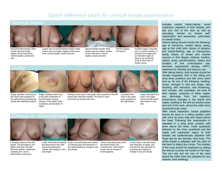
Quick reference chart for clinical breast examination Clinical breast examination (CBE) Normal breast 1 Normal female breasts: Note similar size and shape, nipples at the same level, normal nipples, areola and skin 2 3 Lateral view of normal female breasts: Note similar size and shape, nipples at the same level, normal nipples, areola and skin 4 Normal female breasts: Note similar size and shape, nipples at the same level, normal nipples, areola and skin 5 Normal nippple and areola 6 Inverted nipple, horizontal slit is a normal variation. It should not be diagnosed as retracted nipple as there is no underlying lump or other sign of breast cancer Lump 7 Single, painless, hard lump in the lower outer quadrant of the right breast just below the areola with restricted mobility 8 Single, painless, hard lump in the lower quadrants of the left breast causing fullness of the areola. Note crustations and deviation of nipple 9 10 Painless, hard lump in the upper outer quadrant of the left breast with restricted mobility. The lump is more prominent on raising both arms 11 Lobulated hard lump in the upper outer quadrant of the right breast 12 Large, lobulated hard lump in the upper half of the left breast with fixation to the skin Change in volume/shape 13 Increased volume of the left breast. The left nipple is at a higher level than the right. Note the reddish, inflammed overlying skin, due to inflammatory carcinoma 14 Large, hard lump occupying the lateral half of the right breast increasing the volume with change in size and shape 15 Lump in the left breast with fixation of overlying skin and retraction of the nipple leading to change in size and shape 16 Lump in the lateral part of the left breast fixed to the overlying skin. Such lateral lumps may be missed in mammography 17 12 A hard mass in the left breast with retraction of nipple, skin involvement and retraction of overlying skin leading to change in size and shape includes careful history-taking, visual inspection, palpation of both breasts, arm pits and root of the neck as well as educating women on breast selfexamination and awareness, particularly on breast lumps. History-taking should include the following: age at menarche, marital status, parity, age at first child birth, history of lactation and breast-feeding, age at menopause, family history of breast and ovarian cancers in first degree relatives (mother, sisters, aunts, grandmothers), history and duration of oral contraceptive use, hormone replacement therapy (HRT), treatment for infertility and tobacco use. After taking history, both breasts should be visually inspected, both in the sitting and lying down positions and with arms down and up, for any of the following: swelling, lumps, changes in size and shape, skin dimpling, skin retraction, skin thickening, skin nodules, skin ulceration, the level of both nipples, retraction of either nipple, any discharge from the nipple(s), eczematous changes in the areola and nipple, swelling in the arm pit (axillae area) and root of the neck, above the collar bone (supraclavicular area). After visual inspection, breast palpation should be done in a sitting position both with arms by one’s side and raised above the head. Following this, examination is repeated in a lying down position with arms above the head. Outer quadrants followed by the inner quadrants and the nipple and subareolar region of both breasts are systematically palpated with finger tips for any discrete, hard lump. The whole breast is palpated with the palm of the hand to detect any lumps. The mobility of the lump should be assessed by asking the woman to press her hands on her hips. The arm pits and the root of the neck above the collar bone are palpated for any nodular, hard swellings. Quick reference chart for clinical breast examination Changes in nipple 18 Blood-stained discharge from the nipple 19 Deviated nipple with underlying hard lump under the areola of the right breast. The nipple is pulled towards the lump 20 Partially retracted nipple in the left breast due to underlying cancerous lump in the upper inner quadrant. Note the left nipple is at a higher level than the right 21 22 Complete retraction of Crusting and itching of the nipple due to the nipple due to underlying hard lump infiltration by cancer. Palpation and mammogram revealed hard lump in the outer quadrant 26 24 Complete destruction and puckering of the nipple and areola due to underlying cancer 25 Eczema with complete destruction of the nipple due to a large, cancerous lump distorting the size and shape of the right breast Any one or more of the following findings require urgent referral to a doctor: Changes in skin Retraction of skin in the lower outer quadrant of the left breast due to underlying cancerous lump 23 Eczema-like rash with crusting, bleeding and itching of the nipple due to Paget’s disease 27 Lump in the lower half of the left breast involving overlying skin and part of the areola and nipple. The skin is fixed to the lump 28 Ulcerated, fungating cancerous lump in the lower outer quadrant of the right breast 29 Note the striking change in size and shape of the right breast, with features of eczema, reddish inflammed skin, ‘peau d’orange’ (orange peel) appearance and a hard lump Inflammatory cancer 30 Hard lump in inframammary area with involvement of overlying skin. This lump could be missed in the standing position • Discrete hard lump in the breast • Signs which are highly suggestive of cancer such as: - recent nipple retraction or distortion - skin dimpling or retraction - change in size and shape of the breast - skin nodule - ulceration - eczema of the nipple - blood stained nipple discharge - a swelling in the armpit If no abnormalities are discovered, women are educated on breast lumps and changes in the breast skin, areola and nipple and are advised to self examine their breasts on a monthly basis. They should consult a doctor or a trained health care worker promptly, if they ever suspect a lump or any of the above findings. Findings in figures 7, 8, 9, 10, 19, 20, 22, 23, 26 resulted in diagnosis of early stage breast cancers. 31 Diffuse hard mass occupying almost the entire right breast with reddish, inflammed overlying skin and ‘peau d’orange’ (orange peel) appearance with puckering of the nipple and areola 32 Red inflammed swollen left breast due to underlying inflammatory cancer 33 Thickening of the areola and ‘peau d’orange’ (orange peel) appearance of the skin due to underlying inflammatory carcinoma in the left breast World Health Organization - International Agency for Research on Cancer (IARC), Indian Cancer Society, Mumbai, India International Network for Cancer Treatment and Research (INCTR) International union Against Cancer (UICC) Source: A. Kurkure, E. Lucas , R. Sankaranarayanan. Breast digital atlas http://screening.iarc.fr


