Up-regulated expression of long non
advertisement
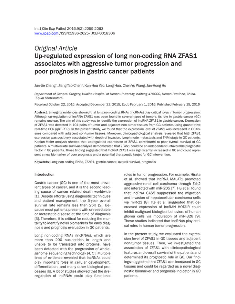
Int J Clin Exp Pathol 2016;9(2):2059-2063 www.ijcep.com /ISSN:1936-2625/IJCEP0018306 Original Article Up-regulated expression of long non-coding RNA ZFAS1 associates with aggressive tumor progression and poor prognosis in gastric cancer patients Jun-Jie Zhang*, Jiang-Tao Chen*, Kun-Hou Yao, Long Hua, Chen-Yu Wang, Jun-Hong Hu Department of General Surgery, Huaihe Hospital of Henan University, Kaifeng 475000, Henan Province, China. * Equal contributors. Received October 22, 2015; Accepted December 22, 2015; Epub February 1, 2016; Published February 15, 2016 Abstract: Emerging evidences showed that long non-coding RNAs (lncRNAs) play critical roles in tumor progression. Although up-regulation of lncRNA ZFAS1 was been found in several types of tumors, its role in gastric cancer (GC) remains unclear. The aim of this study was to identify the expression of lncRNA ZFAS1 in gastric cancer. Expression of ZFAS1 was detected in 104 pairs of tumor and adjacent non-tumor tissues from GC patients using quantitative real-time PCR (qRT-PCR). In the present study, we found that the expression level of ZFAS1 was increased in GC tissues compared with adjacent non-tumor tissues. Moreover, clinicopathological analysis revealed that high ZFAS1 expression was positively associated with depth of invasion, lymph node metastasis and TNM stage in GC patients. Kaplan-Meier analysis showed that up-regulated expression of ZFAS1 contributed to poor overall survival of GC patients. A multivariate survival analysis demonstrated that ZFAS1 could be an independent unfavorable prognostic factor in GC patients. Those finding suggested that lncRNA ZFAS1 was significantly increased in GC and could represent a new biomarker of poor prognosis and a potential therapeutic target for GC intervention. Keywords: Long non-coding RNAs, ZFAS1, gastric cancer, overall survival, prognosis Introduction Gastric cancer (GC) is one of the most prevalent types of cancer, and it is the second leading cause of cancer related death worldwide [1]. Despite efforts using diagnostic techniques and patient management, the 5-year overall survival rate remains less than 25% [2]. Because most patients present with unresectable or metastatic disease at the time of diagnosis [3]. Therefore, it is critical for reducing the mortality to identify novel biomarkers for early diagnosis and prognosis evaluation in GC patients. Long non-coding RNAs (lncRNAs), which are more than 200 nucleotides in length and unable to be translated into proteins, have been detected with the progression of wholegenome sequencing technology [4, 5]. Multiple lines of evidence revealed that lncRNAs could play important roles in cellular development, differentiation, and many other biological processes [6]. A lot of studies showed that the dysregulation of lncRNAs could play functional roles in tumor progression. For example, Hirata et al. showed that lncRNA MALAT1 promoted aggressive renal cell carcinoma through Ezh2 and interacted with miR-205 [7]. Hu et al. found that lncRNA GAS5 suppressed the migration and invasion of hepatocellular carcinoma cells via miR-21 [8]. Ke et al. suggested that decreased expression of lncRAN HOTAIR could inhibit malignant biological behaviors of human glioma cells via modulation of miR-326 [9]. These studies indicated that lncRNAs play critical roles in human tumor progression. In the present study, we evaluated the expression level of ZFAS1 in GC tissues and adjacent non-tumor tissues. Then, we investigated the association of ZFAS1 with clinicopathological features and overall survival of the patients and determined its prognostic role in GC. Our findings suggested that ZFAS1 was increased in GC tissues and could be regarded as a novel diagnostic biomarker and prognosis indicator in GC patients. LncRNA ZFAS1 expression in gastric cancer pital of Henan University between 2008 and 2010, and were diagnosed with gastric cancer based on histopathological evaluation. No patients received neoadjuvant chemotherapy or radiotherapy before surgery. All specimens were immediately frozen in liquid nitrogen and stored at -80°C until RNA extraction. The study was approved by the Research Ethics Committee of Huaihe Hospital of Henan University. Informed consents were obtained from all patients. Figure 1. LncRNA ZFAS1 expression in 104 pairs of GC tissues and adjacent non-tumor tissues detected by qRT-PCR analysis. ZFAS1 expression was significantly increased in GC tissues when compared with adjacent non-tumor tissues. *P<0.05. Table 1. Association of lncRNA ZFAS1 expression with clinicopathological features in GC patients Clinicopathological features Total Age (years) <60 ≥60 Gender Male Female Tumor size (cm) <5 ≥5 Differentiation Well Moderate + Poor Depth of invasion T1 + T2 T3 + T4 Lymph node metastasis No Yes TNM stage I + II III + IV 43 61 64 40 55 49 36 68 43 61 71 33 47 57 LncRNA ZFAS1 P expression value Low High 0.550 23 20 29 32 0.687 33 31 19 21 0.169 31 24 21 28 0.216 15 21 37 31 0.001 30 13 22 39 0.000 44 27 8 25 0.000 38 9 14 43 Materials and methods Quantitative real-time PCR Total RNA was extracted from GC tissues and adjacent non-tumor tissues with RNAiso Plus (Takara). The isolated total RNA was reverse transcribed using the PrimeScript RT Master Mix (Takara) according to manufacturer instructions. The sequence-specific forward and reverse primers sequences for ZFAS1 were 5’-TCTGACCAACGGCTCTTAGAC-3’ and 5’-GTGCCATAGTTGACCAGAGTC-3’ respectively. Forward and reverse primers sequences for GAPDH were 5’-AGAAGGCTGGGGCTCATTTG-3’ and 5’-AGGGGCCATCCACAGTCTTC-3’ respectively. qPCR was performed using SYBR Premix Ex TaqTM II (Takara) on a Light Cycler (Roch). Relative quantification of lncRNA ZFAS1 expression was calculated by using the 2-ΔΔCt method. Each experiment was performed in triplicate. Statistics analyses All computations were carried out using the software of SPSS version 18.0 for windows. Data were expressed as means ± standard deviation (SD). Statistical significance was tested by a Student’s t-test or a Chi-square test as appropriate. Survival analysis was performed using the Kaplan-Meier method, and the logrank test was used to compare the differences between patient groups. The Cox proportional hazards model for multivariate survival analysis was used to assess predictors related to survival. Differences were considered statistically significant when P was less than 0.05. Results Patients and specimens Expression of lncRNA ZFAS1 is up-regulated in GC tissues A total of 104 GC tissues and their adjacent non-tumor tissues were obtained from the Department of General Surgery, Huaihe Hos- In order to explore the role of ZFAS1 in GC progression, we performed qRT-PCR to detect the expression level of ZFAS1 in 104 pairs of GC 2060 Int J Clin Exp Pathol 2016;9(2):2059-2063 LncRNA ZFAS1 expression in gastric cancer Association of lncRNA ZFAS1 expression with GC patients’ survival In order to identify the prognostic value of ZFAS1 expression for GC, we explored the association between the levels of ZFAS1 expression and overall survival through Kaplan-Meier analysis and logrank test. Our data showed that the overall survival of GC patients with high ZFAS1 expression was significantly poorer compared to those patients with low ZFAS1 expression (Figure 2, P<0.05). Univariate analysis showed that depth of invasion, lymph node metastasis, TNM stage, Figure 2. Kaplan-Meier curves for GC patients according to the expression and ZFAS1 expression were of lncRNA ZFAS1. The group with high ZFAS1 expression exhibited a poorer significantly correlated with overall survival compared with the group with low ZFAS1 expression (P<0.05, overall survival of GC patients log-rank test). (Table 2, P<0.05). To evaluate the prognostic value of ZFAS1 tissues and adjacent non-tumor tissues. qRTexpression, variables with a value of P<0.05 PCR showed that the expression of ZFAS1 was were selected for multivariate analysis. Our significantly up-regulated in GC tissues comresults revealed that depth of invasion, lymph pared to adjacent non-tumor tissues (Figure 1, node metastasis, TNM stage, and ZFAS1 P<0.05). These findings indicated that abnorexpression were independent prognostic facmal ZFAS1 expression may be related to gastric tors for overall survival of GC patients (Table 2, cancer progression. P<0.05). Taken together, these data suggested that high ZFAS1 expression level was an in Association between clinicopathological feadependent risk factor for GC patients. tures and lncRNA ZFAS1 expression in GC patients Discussion We further analyzed the association between the expression of ZFAS1 and clinicopathological features of GC patients. 104 GC patients were classified into the low expression group (n=52) and the high expression group (n=52) according to the median ratio of relative ZFAS1 expression in tumor tissues. The association between clinicopathological features and ZFAS1 expression levels in GC patients was summarized in Table 1. Our data showed that high expression of ZFAS1 was positively correlated with deeper depth of invasion, lymph node metastasis, and advanced TNM stage in GC patients (P<0.05). However, ZFAS1 expression level was not associated with other clinicopathological features such as age, gender, tumor size, and differentiation in GC patients (P>0.05). 2061 GC is a highly heterogeneous disease. Mainstream tumorigenic processes involved in GC are characterized by phenotypic multistep progression cascades [10]. The reliable identification of GC progression-specific targets has huge implications for its prevention and treatment [11, 12]. However, identification of the molecular mechanisms underlying tumorigenesis still remains a challenge. LncRNA dysregulation contributes to a range of biological functions and provides a cellular growth advantage, resulting in progressive and uncontrolled tumor growth [13]. Effective control of both cell proliferation and invasion is critical to the prevention of oncogenesis and successful cancer therapy [14]. Therefore, Int J Clin Exp Pathol 2016;9(2):2059-2063 LncRNA ZFAS1 expression in gastric cancer Table 2. Univariate and multivariate analysis of overall survival in GC patients Clinicopathological features Age (years) ≥60 vs <60 Gender Male vs Female Tumor size ≥5 cm vs <5 cm Differentiation Moderate + Poor vs Well Depth of invasion T3 + T4 vs T1 + T2 Lymph node metastasis Yes vs No TNM stage III + IV vs I + II LncRNA ZFAS1 High vs Low Univariate analysis Hazard Ratio 95% CI 0.873 0.514-1.824 P 1.134 0.638-2.149 0.316 2.214 0.776-3.817 0.178 2.843 0.713-4.219 0.203 2.158 0.823-4.236 0.019 1.938 0.747-3.926 0.013 3.721 1.186-7.022 0.017 3.347 1.071-6.758 0.007 3.381 1.253-6.915 0.014 3.074 1.185-5.831 0.004 2.953 1.318-7.043 0.002 2.573 1.251-6.836 0.003 identification of GC associated lncRNAs and investigation of their clinical significance may provide a missing piece of the well known oncogenic and tumor suppressor network puzzle. Recently, lots of studies showed that lncRNAs were dysregulated in multiple cancers including GC. For example, Zhou et al. suggested that down-regulation of lncRNA LET correlated with clinical progression and unfavorable prognosis in GC [15]. Fei et al. reported that decreased lncRNA LINC00982 could be identified as a poor prognostic biomarker in GC and regulate cell proliferation [16]. Li et al. found that upregulated expression of lncRNA BANCR was associated with clinical progression and poor prognosis in GC [17]. Kong et al. revealed that increased expression of lncRNA PVT1 indicated a poor prognosis of GC and promoted cell proliferation through epigenetically regulating p15 and p16 [18]. However, little is known about the clinical significance of ZFAS1 in GC patients. In the present study, we explored the expression of lncRNA ZFAS1 in GC, our results showed that the expression levels of ZFAS1 in GC tissues were significantly higher than those in adjacent non-tumor tissues. High ZFAS1 expression was associated with deeper depth of invasion, lymph node metastasis and advanced TNM stage in GC patients. Then, Kaplan-Meier analysis showed that patients with high ZFAS1 expression were associated with shorter over2062 P 0.274 Multivariate analysis Hazard Ratio 95% CI all survival. Finally, Univariate and multivariate analyses revealed that increased expression of ZFAS1 was an independent prognostic biomarker for shorter overall survival of GC patients. Previous studies showed that ZFAS1 was dysregulated in types of cancers and play important roles in tumor progression. For example, Askarian-Amiri et al. showed that ZFAS1 was highly expressed in the mammary gland and down-regulated in breast tumors. Furthermore, they demonstrated that decreased expression of ZFAS1 in mammary epithelial cells resulted in a significant increased in proliferation and metabolic activity, suggesting that ZFAS1 act as a putative tumor suppressor gene in breast cancer [19]. However, Li et al. found that ZFAS1 was increased and associated with intrahepatic and extrahepatic metastasis and poor prognosis of hepatocellular carcinoma. Furthermore, they showed that ZFAS1 could function as an oncogene in hepatocellular carcinoma progression by binding miR-150 and abrogating its tumor suppressive function in this setting [20]. Thorenoor et al. showed that ZFAS1 was increased in colorectal cancer (CRC) and promoted the proliferation of CRC cells in vitro [21]. Our finding expanded the tumor oncogenic role of ZFAS1 in GC progression. In conclusion, this is the first study demonstrated that the increased expression of lncRNA Int J Clin Exp Pathol 2016;9(2):2059-2063 LncRNA ZFAS1 expression in gastric cancer ZFAS1 was associated with advanced clinical features and poor prognosis of GC patients, indicating that increased expression of ZFAS1 could act as an unfavorable prognostic biomarker in GC patients. However, further studies are needed to investigate the precise molecular mechanism of ZFAS1 in the development and progression of GC. Acknowledgements The present study was supported by the Scientific and Technological Research Projects of the Henan Provincial Education Department (grant no. 14A320064). Disclosure of conflict of interest None. Address correspondence to: Dr. Jun-Hong Hu, Department of General Surgery, Huaihe Hospital of Henan University, 8 Baogonghu North Road, Kaifeng 475000, Henan Province, China. E-mail: hujunhong73@163.com [9] [10] [11] [12] [13] [14] [15] [16] References [1] [2] [3] [4] [5] [6] [7] [8] Jemal A, Bray F, Center MM, Ferlay J, Ward E and Forman D. Global cancer statistics. CA Cancer J Clin 2011; 61: 69-90. Brenner H, Rothenbacher D and Arndt V. Epidemiology of stomach cancer. Methods Mol Biol 2009; 472: 467-477. Japanese Gastric Cancer Association. Japanese gastric cancer treatment guidelines 2010 (ver. 3). Gastric Cancer 2011; 2: 113-123. Mercer TR, Dinger ME and Mattick JS. Long non-coding RNAs: insights into functions. Nat Rev Genet 2009; 10: 155-159. Spizzo R, Almeida MI, Colombatti A and Calin GA. Long non-coding RNAs and cancer: a new frontier of translational research? Oncogene 2012; 31: 4577-4587. Gibb EA, Brown CJ and Lam WL. The functional role of long non-coding RNA in human carcinomas. Mol Cancer 2011; 10: 38. Hirata H, Hinoda Y, Shahryari V, Deng G, Nakajima K, Tabatabai ZL, Ishii N and Dahiya R. Long Noncoding RNA MALAT1 Promotes Aggressive Renal Cell Carcinoma through Ezh2 and Interacts with miR-205. Cancer Res 2015; 75: 1322-1331. Hu L, Ye H, Huang G, Luo F, Liu Y, Yang X, Shen J, Liu Q and Zhang J. Long noncoding RNA GAS5 suppresses the migration and invasion of hepatocellular carcinoma cells via miR-21. Tumour Biol 2015; [Epub ahead of print]. 2063 [17] [18] [19] [20] [21] Ke J, Yao YL, Zheng J, Wang P, Liu YH, Ma J, Li Z, Liu XB, Li ZQ, Wang ZH and Xue YX. Knockdown of long non-coding RNA HOTAIR inhibits malignant biological behaviors of human glioma cells via modulation of miR-326. Oncotarget 2015; 6: 21934-21949. Guggenheim DE and Shah MA. Gastric cancer epidemiology and risk factors. J Surg Oncol 2013; 107: 230-236. Al-Batran SE, Ducreux M and Ohtsu A. mTOR as a therapeutic target in patients with gastric cancer. Int J Cancer 2012; 130: 491-496. Kato M and Asaka M. Recent development of gastric cancer prevention. Jpn J Clin Oncol 2012; 42: 987-994. Khorkova O, Hsiao J and Wahlestedt C. Basic biology and therapeutic implications of lncRNA. Adv Drug Deliv Rev 2015; 87: 15-24. Oliveira-Mateos C and Guil S. LncRNAs and the Control of Oncogenic Signaling. Current Pathobiology Reports 2015; 3: 203-207. Zhou B, Jing XY, Wu JQ, Xi HF and Lu GJ. Downregulation of long non-coding RNA LET is associated with poor prognosis in gastric cancer. Int J Clin Exp Pathol 2014; 7: 8893. Fei ZH, Yu XJ, Zhou M, Su HF, Zheng Z and Xie CY. Upregulated expression of long non-coding RNA LINC00982 regulates cell proliferation and its clinical relevance in patients with gastric cancer. Tumour Biol 2015; [Epub ahead of print]. Li L, Zhang L, Zhang Y and Zhou F. Increased expression of LncRNA BANCR is associated with clinical progression and poor prognosis in gastric cancer. Biomed Pharmacother 2015; 72: 109-112. Kong R, Zhang EB, Yin DD, You Lh, Xu TP, Chen WM, Xia R, Wan L, Sun M and Wang ZX. Long noncoding RNA PVT1 indicates a poor prognosis of gastric cancer and promotes cell proliferation through epigenetically regulating p15 and p16. Mol Cancer 2015; 14: 82. Askarian-Amiri ME, Crawford J, French JD, Smart CE, Smith MA, Clark MB, Ru K, Mercer TR, Thompson ER and Lakhani SR. SNORDhost RNA Zfas1 is a regulator of mammary development and a potential marker for breast cancer. RNA 2011; 17: 878-891. Li T, Xie J, Shen C, Cheng D, Shi Y, Wu Z, Deng X, Chen H, Shen B, Peng C, Li H, Zhan Q and Zhu Z. Amplification of Long Noncoding RNA ZFAS1 Promotes Metastasis in Hepatocellular Carcinoma. Cancer Res 2015; 75: 3181-3191. Thorenoor N, Vychytilová P, Mlčochová J, Sonja H, Markus K, Šána J and Slabý O. ZFAS1, long non-coding RNA with significance in colorectal cancer. 2015. Int J Clin Exp Pathol 2016;9(2):2059-2063
