Measurement of Centi-Gray X-Ray Dose Levels With Radiochromic
advertisement

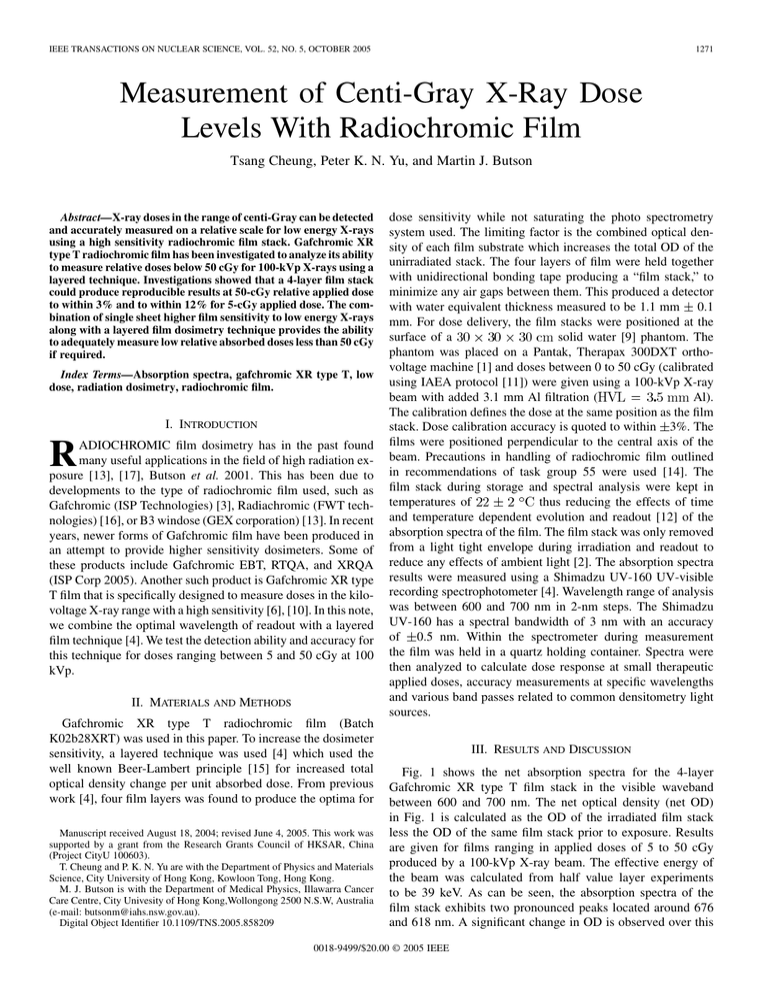
IEEE TRANSACTIONS ON NUCLEAR SCIENCE, VOL. 52, NO. 5, OCTOBER 2005 1271 Measurement of Centi-Gray X-Ray Dose Levels With Radiochromic Film Tsang Cheung, Peter K. N. Yu, and Martin J. Butson Abstract—X-ray doses in the range of centi-Gray can be detected and accurately measured on a relative scale for low energy X-rays using a high sensitivity radiochromic film stack. Gafchromic XR type T radiochromic film has been investigated to analyze its ability to measure relative doses below 50 cGy for 100-kVp X-rays using a layered technique. Investigations showed that a 4-layer film stack could produce reproducible results at 50-cGy relative applied dose to within 3% and to within 12% for 5-cGy applied dose. The combination of single sheet higher film sensitivity to low energy X-rays along with a layered film dosimetry technique provides the ability to adequately measure low relative absorbed doses less than 50 cGy if required. Index Terms—Absorption spectra, gafchromic XR type T, low dose, radiation dosimetry, radiochromic film. I. INTRODUCTION R ADIOCHROMIC film dosimetry has in the past found many useful applications in the field of high radiation exposure [13], [17], Butson et al. 2001. This has been due to developments to the type of radiochromic film used, such as Gafchromic (ISP Technologies) [3], Radiachromic (FWT technologies) [16], or B3 windose (GEX corporation) [13]. In recent years, newer forms of Gafchromic film have been produced in an attempt to provide higher sensitivity dosimeters. Some of these products include Gafchromic EBT, RTQA, and XRQA (ISP Corp 2005). Another such product is Gafchromic XR type T film that is specifically designed to measure doses in the kilovoltage X-ray range with a high sensitivity [6], [10]. In this note, we combine the optimal wavelength of readout with a layered film technique [4]. We test the detection ability and accuracy for this technique for doses ranging between 5 and 50 cGy at 100 kVp. II. MATERIALS AND METHODS Gafchromic XR type T radiochromic film (Batch K02b28XRT) was used in this paper. To increase the dosimeter sensitivity, a layered technique was used [4] which used the well known Beer-Lambert principle [15] for increased total optical density change per unit absorbed dose. From previous work [4], four film layers was found to produce the optima for Manuscript received August 18, 2004; revised June 4, 2005. This work was supported by a grant from the Research Grants Council of HKSAR, China (Project CityU 100603). T. Cheung and P. K. N. Yu are with the Department of Physics and Materials Science, City University of Hong Kong, Kowloon Tong, Hong Kong. M. J. Butson is with the Department of Medical Physics, Illawarra Cancer Care Centre, City Univesity of Hong Kong,Wollongong 2500 N.S.W, Australia (e-mail: butsonm@iahs.nsw.gov.au). Digital Object Identifier 10.1109/TNS.2005.858209 dose sensitivity while not saturating the photo spectrometry system used. The limiting factor is the combined optical density of each film substrate which increases the total OD of the unirradiated stack. The four layers of film were held together with unidirectional bonding tape producing a “film stack,” to minimize any air gaps between them. This produced a detector with water equivalent thickness measured to be 1.1 mm 0.1 mm. For dose delivery, the film stacks were positioned at the solid water [9] phantom. The surface of a phantom was placed on a Pantak, Therapax 300DXT orthovoltage machine [1] and doses between 0 to 50 cGy (calibrated using IAEA protocol [11]) were given using a 100-kVp X-ray Al). beam with added 3.1 mm Al filtration ( The calibration defines the dose at the same position as the film stack. Dose calibration accuracy is quoted to within 3%. The films were positioned perpendicular to the central axis of the beam. Precautions in handling of radiochromic film outlined in recommendations of task group 55 were used [14]. The film stack during storage and spectral analysis were kept in thus reducing the effects of time temperatures of and temperature dependent evolution and readout [12] of the absorption spectra of the film. The film stack was only removed from a light tight envelope during irradiation and readout to reduce any effects of ambient light [2]. The absorption spectra results were measured using a Shimadzu UV-160 UV-visible recording spectrophotometer [4]. Wavelength range of analysis was between 600 and 700 nm in 2-nm steps. The Shimadzu UV-160 has a spectral bandwidth of 3 nm with an accuracy of 0.5 nm. Within the spectrometer during measurement the film was held in a quartz holding container. Spectra were then analyzed to calculate dose response at small therapeutic applied doses, accuracy measurements at specific wavelengths and various band passes related to common densitometry light sources. III. RESULTS AND DISCUSSION Fig. 1 shows the net absorption spectra for the 4-layer Gafchromic XR type T film stack in the visible waveband between 600 and 700 nm. The net optical density (net OD) in Fig. 1 is calculated as the OD of the irradiated film stack less the OD of the same film stack prior to exposure. Results are given for films ranging in applied doses of 5 to 50 cGy produced by a 100-kVp X-ray beam. The effective energy of the beam was calculated from half value layer experiments to be 39 keV. As can be seen, the absorption spectra of the film stack exhibits two pronounced peaks located around 676 and 618 nm. A significant change in OD is observed over this 0018-9499/$20.00 © 2005 IEEE 1272 IEEE TRANSACTIONS ON NUCLEAR SCIENCE, VOL. 52, NO. 5, OCTOBER 2005 Fig. 1. Net optical density visible absorption spectra for the 4-layer Gafchromic XR type T radiochromic film stack dosimeter when exposed to 100-kVp X-rays. Doses up to 50 cGy are shown when analyzed in the wavelength range of 600 to 700 nm. TABLE I MEAN PERCENTAGE ERROR IN MEASUREMENT USING THE 4 -LAYER XR TYPE T RADIOCHROMIC FILM STACK A linear line of best fit approximates dose response of the film stack detector at doses less than 50 cGy with standard deviation of 3% at 676 nm and 6.5% at 600–700 nm average. When results are analyzed at specific wavelengths a relative dose response curve can be produced as shown in Fig. 2. Results are for the relative dose response of the stacked film detector at specific wavelengths, which typify the most common densitometers used for Gafchromic film analysis. Results show that at the highest sensitivity wavelength, a 0.0156-OD units per cGy relationship applies. This equates to a 1.56-OD units per Gray applied dose for the film stack dosimeter. Higher sensitivities and thus lower percentage errors are seen at the main absorption peak. When compared to other detectors, the XR type T stack produces a higher level of uncertainty at low dose levels. For example at 5-cGy applied doses a MOSFET detector system produces an approximately 5% uncertainty and LiF (Mg,Ti) Thermoluminescent dosimeters produce an approximate 7% uncertainty (12% for film stack). These values decrease to 2.5% and 5%, respectively at 10-cGy applied dose (9% for film stack) ([7] submitted). Although the film stack does not provide as high a level of accuracy at low doses as compared to other detectors, the film stack has increased the sensitivity of radiochromic film to place it in a useable range for clinical use. It is acknowledged that an absolute dose response is unable to be performed easily with radiochromic film due to inherent variations in chemical composition and other parameters which effect absolute dose measurement. The film however can provide a relatively accurate relative dose analysis when the radiation source is already calibrated with another primary dosimeter such as a ionization chamber. The radiochromic film stack has the added ability to measure a 2-D dose map instead of a point dose. IV. CONCLUSION Fig. 2. OD to dose sensitivity comparison for Gafchromic XR type T radiochromic film when analyzed at various wavelengths and band passes. The highest sensitivity is seen at the absorption peak (676 nm) producing a coloration of 0.0156 OD per cGy or 1.56 OD per Gray. dose range, e.g., at 676 nm a net OD of 0.78 is found for 50-cGy applied dose. As the response of XR type T is energy dependent [8], each X-ray beam used will require absolute dose measurement with an ionization chamber prior to relative dose measurements. The energy dependant nature of the film may also affect results measured at depth or off axis due to spectral changes with beam penetration. This may need to be taken into account for profile or depth dose assessment. Table I shows the reproducibility and error associated with measurements of doses at these levels using the radiochromic film. Results show that at 676 nm wavelength the average percentage error in relative dose measurement using a film stack detector was 12% for 5 -cGy applied dose ranging down to 1.5% at 50-cGy applied dose and are derived from the linear fits displayed in Fig. 2. Using a 600–700 nm average (which relates to a common broadband filtered red light source) these values become 22% and 2.5%, respectively. A 4-layered Gafchromic XR type T radiochromic film detector produces a high sensitivity radiation detector with the ability to adequately measure relative doses in the range of centi Grays at 100-kVp X-ray energy. Results showed that a 5-cGy relative applied dose can be measured with an uncertainty of within 12% at 100 -kVp X-ray energy. The uncertainty decreases to within 2% at 50-cGy relative applied dose. The added advantage of a film dosimeter is the ability to measure 2-D relative dose results much easier and quicker than conventional dosimeters such as ionization chambers. REFERENCES [1] M. Butson, J. Mathur, and P. Metcalfe, “Dose characteristics of a therapax 300 kVp orthovoltage machine,” Austral. Phys. Eng. Sci. Med., vol. 18, no. 4, pp. 133–138, 1995. [2] M. Butson, P. Yu, and P. Metcalfe, “Effects of readout light sources and ambient light on radiochromic film,” Phys. Med. Biol., vol. 43, pp. 2407–2412, 1998. [3] M. J. Butson, K. N. Yu, T. Cheung, and P. E. Metcalfe, “Radiochromic film for medical radiation dosimetry,” Mater. Sci. Eng. R: Rep., vol. 41, pp. 61–120, 2003. [4] M. J. Butson, T. Cheung, and P. K. Yu, “Corresponding dose response of radiographic film with layered gafchromic film,” Phys. Med. Biol., vol. 47, no. 22, pp. N285–N289, Nov. 21, 2002. [5] M. Butson, P. K. Yu, T. Cheung, M. G. Carolan, K. Y. Quach, A. Arnold, and P. E. Metcalfe, “Dosimetry of blood irradiation with radiochromic film,” Transfus Med., vol. 9, no. 3, pp. 205–208, Sep. 1999. CHEUNG et al.: CENTI-GRAY X-RAY DOSE LEVELS [6] M. Butson, T. Cheung, and P. K. N. Yu, “Visible absorption properties of radiation exposed XR type T radiochromic film,” Phys. Med. Biol., Aug. 2004. [7] T. Cheung, K. N. Yu, and M. J. Butson, “Low dose measurement with a MOSFET in high energy radiotherapy applications,” Radiation Measurements, 2004. [8] T. Cheung, M. J. Butson, and P. K. Yu, “Experimental energy response verification of XR type T radiochromic film,” Phys Med Biol., vol. 49, no. 21, pp. N371–N376, Nov. 7, 2004. [9] C. Constanitinou, F. Atix, and B. Paliwal, “A solid water phantom material for radiotherapy x-ray and gamma ray beam ray calculations,” Med. Phys., vol. 9, pp. 436–441, 1982. [10] E. R. Giles and P. H. Murphy, “Measuring skin dose with radiochromic dosimetry film in the cardiac catheterization laboratory,” Health Phys., vol. 82, no. 6, pp. 875–880, Jun. 2002. [11] International Atomic Energy Agency (IAEA), Absorbed dose determination in photon and electron beams, Vienna, Austria, 1987. [12] P. H. Meigooni, P. H. Sanders, and P. H. Ibbot, “Dosimetric characteristics of an improved radiochromic film,” Med. Phys., vol. 23, pp. 1883–1888, 1996. 1273 [13] R. B. Miler, G. Loda, R. C. Miller, R. Smith, D. Shimer, C. Seidt, M. MacArt, H. Mohr, G. Robison, P. Creely, J. Bautista, T. Oliva, L. M. Young, and D. DuBois, “A high-power electron linear accelerator for food irradiation applications,” Nucl. Instr. Meth. Phys. Res. Section B: Beam Interactions With Materials and Atoms, vol. 211, pp. 562–570, 2003. [14] A. Niroomand-Rad, C. Blackwel, B. Coursey, K. Gall, J. Galvin, W. McLaughlin, A. Meigooni, R. Nath, J. Rodgers, and C. Soares, “Radiochromic film dosimetry: recommendation of AAPM radiation therapy task group 55,” Med. Phys., vol. 25, no. 11, pp. 2093–2115, 1998. [15] V. Pit, Dictionary of Physics. Middlesex, U.K.: Penguin, 1984. [16] F. C. Young, J. R. Boler, S. J. Stephanakis, T. G. Jones, and J. M. Neri, “Radiachromic film as a detector for intense MeV proton beams,” Rev. Scient. Instr., vol. 70, no. 1, pp. 1201–1204, Jan. 1999. [17] F. Ziaie, H. Afarideh, S. M. Hadji-Saeid, and S. A. Durrani, “Investigation of beam uniformity in industrial electron accelerator,” Rad. Measure., pp. 609–613, Jun. 2001.

