Gel Electrophoresis - University of Colorado Boulder
advertisement

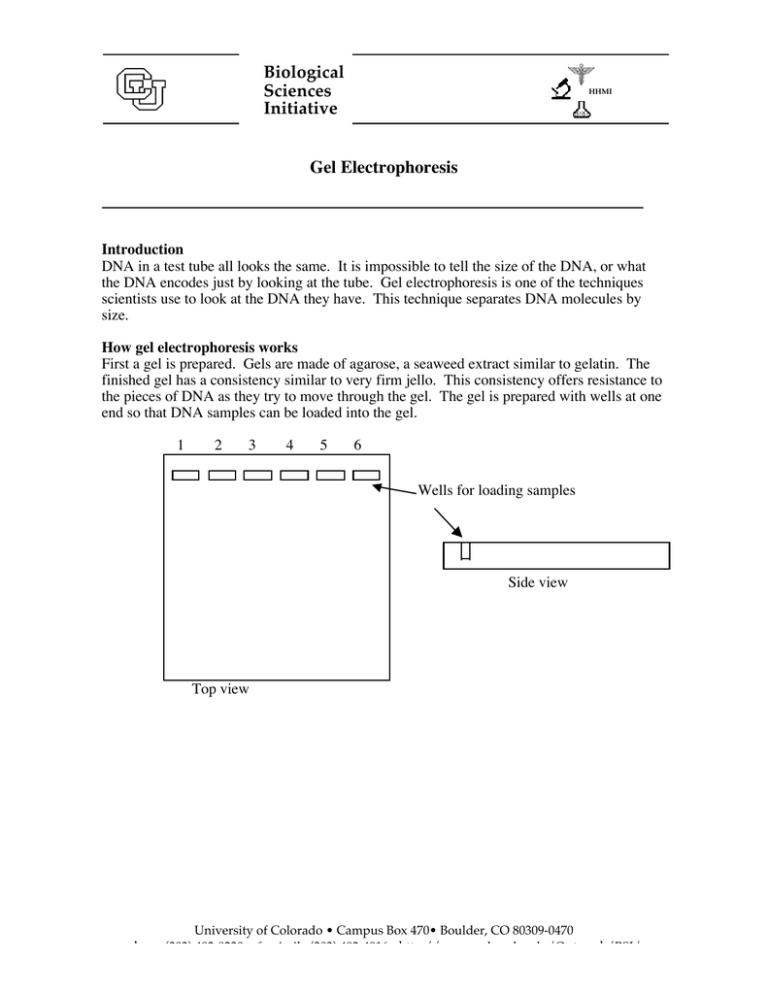
Biological Sciences Initiative HHMI Gel Electrophoresis Introduction DNA in a test tube all looks the same. It is impossible to tell the size of the DNA, or what the DNA encodes just by looking at the tube. Gel electrophoresis is one of the techniques scientists use to look at the DNA they have. This technique separates DNA molecules by size. How gel electrophoresis works First a gel is prepared. Gels are made of agarose, a seaweed extract similar to gelatin. The finished gel has a consistency similar to very firm jello. This consistency offers resistance to the pieces of DNA as they try to move through the gel. The gel is prepared with wells at one end so that DNA samples can be loaded into the gel. 1 2 3 4 5 6 Wells for loading samples Side view Top view University of Colorado • Campus Box 470• Boulder, CO 80309-0470 phone (303) 492-8230 • facsimile (303) 492-4916• http://www.colorado.edu/Outreach/BSI/ Once the DNA samples are loaded onto the gel, an electric current is applied to the gel. DNA is negatively charged due to all the phosphate groups in the backbone of DNA. Thus, DNA will move towards the positive electrode. - electrode direction of migration - electrode + electrode Top view + electrode Top View Side View + electrode As the pieces of DNA move through the gel, they will meet with resistance. Larger pieces of DNA will have more difficulty moving through the gel than smaller fragments. Thus, larger fragments will move slower than smaller fragments. This allows separation of all different sizes of DNA fragments. 1 1 2 2 3 4 5 6 1 2 3 4 10 minutes 20 minutes 10 min 20 min 3 4 5 5 6 6 Here you can see the migration of different fragments of DNA over time. Lane 2 contains a small fragment only, lane 3 a large fragment only, and lane 4 both a small and a large fragment. Larger fragment Smaller fragment 30 minutes 30 min Uses of Gel Electrophoresis Gel electrophoresis is used to provide genetic information in a wide range of data fields. Human DNA can be analyzed to provide evidence in criminal cases, to diagnose genetic diseases, and to solve paternity cases. Samples can be obtained from any DNA-containing tissue or body fluid, including cheek cells, blood, skin, hair, and semen. In many analyses, polymerase chain reaction (PCR) is used to amplify specific regions of DNA that are known to vary among individuals. A person’s “DNA fingerprint” or “DNA profile” is constructed by using gel electrophoresis to separate the DNA fragments from several of these highly variable regions. These DNA profiling techniques are also used by scientists in other fields of biology. Conservation biologists use DNA profiling to determine genetic similarity and kinship among populations or individuals. This information is particularly important to captive breeding programs, such as those at zoos, so that the deleterious effects of inbreeding can be minimized. Biologists who study animal behavior use DNA profiling to determine kinship among members of a group and its effects on relationships. The genetic variation revealed by DNA profiling is used by taxonomists to distinguish species. Evolutionary biologists use DNA profiles to compare the similarities and differences among species, constructing hypothetical family trees. Proteins can also be run on gels. Most commonly proteins are run on gels made of polyacrylamide in the presence of SDS. In SDS PAGE (SDS polyacrylamide gel elecrtrophoresis) all proteins are coated with SDS and are thus negatively charged. This technique separates proteins by size. More rarely proteins are separated by charge in gels without SDS. Scientific dyes can also be separated by gel electrophoresis. Like DNA most scientific stains are negatively charged. Scientific dyes are often used in classroom setting to simulate DNA since the chemicals required to visualize DNA in a gel cause cancer.
