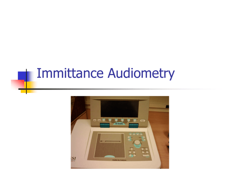
Immittance Audiometry
advertisement

Immittance Audiometry Terminology Immittance: Immittance Immittance is a generic term that encompasses impedance, admittance, and their components Impedance (Z - in acoustic ohms) in the middle ear system is defined as the total opposition of this system to the flow of the acoustic energy. Admittance (Y - in acoustic mmhos) is the reciprocal of impedance and is the amount of acoustic energy that flows into the middle ear system. Currently available immittance instruments typically measure admittance. Simple Harmonic Motion M Variables That Determine Admittance Compliance (the inverse of the stiffness) stiffness – C/S Mass – M: the admittance offered by mass elements in : the admittance offered by stiffness elements in the middle ear system which is called compliant susceptance and is denoted by BS (also stiffness reactance, negative reactance, or -Xs in impedance terms) the middle ear system which is called mass susceptance and is denoted by Bm (also mass reactance, positive reactance, or Xm in impedance terms) Friction or Resistance – R:determines the absorption or dissipation of acoustic energy. In admittance terms, this element is called conductance and is denoted by G (also resistance, or R in impedance system). Admittance Layout Total susceptance (or total reactance in impedance terms) which store acoustic energy is the algebraic sum of the mass and compliance elements as plotted along the Y-axis Xm R the compliant susceptance (Bs) is on the RESISTANCE positive axis that begins at zero and extends upward indefinitely, whereas the ∅ -Xs mass susceptance (Bm) is on negative |Z| axis that begins at zero and extends downward indefinitely. If the total susceptance is positive, a system is Impedance - Z stiffness controlled; if this value is negative, the system is mass controlled Conductance (G) is plotted on the X-axis. The value of conductance is always positive. |Y| Bs ∅ -Bm CONDUCTANCE G Admittance- Y Admittance is a Complex Number The admittance of the system (|Y|) is a two dimensional quantity and is a vector sum of conductance (G) and the total susceptance (Bt). Mathematically, admittance can be expressed in rectangular notation or in polar notation. In rectangular notation, admittance is expressed as the sum of its conductance (G) and susceptance (Bt) elements. Y = G + jBt In polar notation admittance is expressed by its magnitude and phase angle. |Y| ∠ Øy Complex Acoustic Admittance Y Btotal = Bs+ Bm Mathematical Correlation Between Polar & Rectangular Notation Admittance_Y |Y| < Øy (Polar notation) G + jB t (Rectangular notation) G= |Y| Cos Øy B= |Y| Sin Øy Ytm = Gtm 2 + Btm 2 Tan Øy= B/G Øy= arctan (B/G) Relationship Between Admittance Components & Frequency Acoustic conductance (the frictional component) is independent of frequency compliance and mass susceptance are frequency dependent Mass susceptance is directly proportional to frequency compliance susceptance is inversely proportional to frequency Therefore, as frequency increases, the total susceptance progresses from positive values (stiffness controlled) toward zero (resonance) to negative value (mass controlled). Resonance of the middle ear system is achieved when the compliant and mass susceptance are equal, i.e., total susceptance is equal to 0 mmhos. Relationship Between Admittance Components & Frequency B< G: 1B1G or 3B1G 226Hz 1 791Hz 904Hz 0 1017Hz -1 1130Hz -2 -90° 0 1 2 3 4 5 CONDUCTANCE (G) mmho 0° Bt=0 STIFFNESS 565Hz Resonance MASS SUSCEPTANCE (B) in mmho 2 45° B=G: 1B1G B>G : 1B1G 90° Admittance Meter Standard Low Frequency Tympanometry Tympanometry is the measurement of the acoustic immittance of the ear as a function of ear canal air pressure (ANSI, S3.391987). For clinical purposes, the admittance of the middle ear is measured using tympanometry to gain information regarding middle ear function. Standard clinical tympanometry is performed using a low probe tone frequency, usually 220 or 226 Hz, and measures the admittance magnitude |Y| as a function of ear canal air pressure. The result is a graphic display called a tympanogram. At the low probe tone frequency used in standard tympanometry, the normal middle ear system is stiffness dominated and susceptance (the stiffness element) contributes more to overall admittance than conductance (the frictional element) Standard Low Frequency Tympanometry Traditional parameters obtained from low frequency tympanometry: Static admittance (SA) Tympanometric Shapes Tympanometric peak pressure (TPP) Ear Canal Volume (ECV) Tympanometric width (TW) Tympanogram 226 Hz T ym panogram 1.50 1.25 1.00 0.75 Peak Y tm 0.50 0.25 T PP 0.0 -500 -400 -300 -200 -100 0 100 Air Pressure (daPa) Probe Ear:Right -296Ya=0.4m mho T PP=-18daPa Peak Ytm=0.6mmho 200 300 400 500 Plane of The measurement Because the probe tip of the admittance measurement system is remote from the surface of the tympanic membrane, admittance measured at the probe tip jointly reflects the admittance of the external auditory canal and the admittance of the middle ear (plane of the measurement) The dimensions of the external auditory canal vary depending on the depth of insertion of the probe tip as well as individual differences in ear canal size. This produces substantial variation in the admittance due to the external ear Therefore, to derive a measure of middle ear admittance alone, it is necessary to subtract the admittance due to the external ear canal from the overall admittance measure. Static Admittance (SA) Measuring admittance under changes in air pressure provides a way to derive an estimate of the admittance due to ear canal volume This is accomplished through placing the eardrum under sufficient tension by a high positive or negative pressure to drive the impedance of the middle ear toward infinity The admittance measured at the probe tip under these extreme pressures provides a reasonable estimate of the ear canal admittance alone This estimate (e.g., at -296Ya in previous figure) is then subtracted from the peak value (tympanometric peak pressure –TPP) which jointly reflects the admittance of the external auditory canal and the middle ear to arrive at a value that reflects only the admittance of the tympanic membrane and middle ear According to ANSI, (1987) the resulting value (Peak Ytm in the previous figure) is properly referred to as the peak-compensated static acoustic admittance Variables Affecting SA The choice of pressure value for compensation of ear canal admittance: The compensated static admittance is typically higher when extreme negative (rather than extreme positive) pressure is used to estimate ear canal admittance (Margolis & Smith, 1977; Rabinowitz, 1981; Shanks & Lilly, 1981). ). This asymmetry occurs because friction contributes less to admittance at extreme negative pressures than at extreme positive pressure (Margolis & Smith, 1977). The rate of ear canal pressure: Higher SA values for faster pump speeds (Van Camp, 1974)and more frequenct notching of high frequency tympanograms recorded at faster pump speeds (Van Camp et al., 1976) The direction of ear can pressure change: SA is greater for negative to positive (-/+)than for positive to negative (+/-) pressure change (Wilson et al., 1984). The incidence of notched tympanograms also is higher for -/+ (Margolis et al, 1978) Variables Affecting SA Ear canal correction: Because admittance is a vector quantity, it cannot be added or subtracted unless the phase angle of the two admittance vectors is identical. Subtracting admittance vector data at standard low probe tone frequency results in negligible error since the phase difference between the admittance vector of middle ear and the ear canal is small. At higher probe tone frequencies a marked error occurs because a significant phase shift for the admittance vector takes place. Therefore, at higher probe tone frequencies it is necessary to compensate for the effect of ear canal from admittance rectangular components (susceptance and conductance), and then convert the data back to admittance vector (Margolis & Hunter, 1999; Shanks, Wilson, & Cabron, 1993). Admittance Vectors (Phasor) additions Variables Affecting SA Probe tone frequency: As probe tone frequency increases the SA also changes. At low probe tone frequency, regardless of the pathology tested, the middle ear system is stiffness dominated. One effect of the middle ear disease is to shift the resoonant frequency of the normal middle ear system. The greatest effect of the disease on on SA occurs near resonant frequency (Liden et al, 1974; Shahnaz & Polka, 1997). The superiority of higher probe tone frequency over 226 Hz has been shown both in low impedance pathologies (Van Camp et al., 1980) and high impedance pathologies (Shahnaz & Polka, 1997). Guidelines for Measuring SA Ear canal volume should be estimated with the ear canal pressurized to the value that results in the minimum admittance value (MIN), however, if test re test reliability is an issue the + 200 daPa should be used. SA should be calculated at the ear canal pressure corresponding to the peak value for single peaked tympanograms (MAX). For notched admittance tympanograms, the static value should be calculated at the minimum in the notch. When susceptance (B) and or conductance (G) tympanograms are notched, Static susceptance should be calculated at the ear canal pressure corresponding to the minimum in the susceptance notch. Guidelines for Measuring SA Either direction of ear canal pressure change can be used for tympanograms obtained with a low frequency probe (226 Hz). The decreasing (+/-) pressure direction, however, should be used with high frequency probe (e.g., 678 Hz) to minimize the occurrence of multipeaked tympanograms. In normal ears. Both admittance components ( B & G) should be recorded simultaneously. Calculating SA from Notched Tympanogram Right 900 Hz Tympanogram 6.00 5.00 4.00 3.00 2.00 1.00 0.0 -500 Ga: Ba: -400 -300 -200 -100 0 100 Air Pressure (daPa) 200 300 400 500 SA Norms for Adults @ 226 Hz Normal (adults) N=68 Y+ B+ Y- B- Mean 0.65 0.59 0.74 0.69 SD 0.31 0.27 0.31 0.27 90% Range 0.32 | 1.28 0.3 | 1.11 0.39 | 1.26 0.39 | 1.15 95% CI 0.57 | 0.72 0.53 | 0.65 0.66 | 0.81 0.62 | 0.75 Descriptive statistics on static immittance (mmhos) for admittance (Y) and susceptance (B) using positive (+) and negative (-) compensation @ 226 Hz. The results are shown for the normal. Re.: Shahnaz & Polka (1997) and Shahnaz (Ph.D. dissertation) Suggested Diagnostic Criteria for SA @ 226 Hz Group Adult (Shahnaz & Polka, 1997) Fail 90% Normal Range mmho 0.30 -130 < 0.30 (+ 250 daPa compensation) (≥ 18 y) Adult (Margolis & Goycoolea, 1993) Children (Hunter, 1993) (3-10 years) (≥ 18 y) 0.30 – 1.70 0.25 – 1.05 < 0.30 < 0.40 < (+ 200 daPa compensation (Negative compensation) 0.20 < (+ 200 daPa compensation) 0.30 (Negative compensation) Dreena’s Thesis SA + compensation - compensation Mean SD 90% Range Mean SD 90% Range (mmho) (mmho) (mmho) (mmho) (mmho) (mmho) M 0.70 0.34 0.24-1.46 0.76 0.36 0.32-1.59 F 0.74 0.52 0.34-2.49 0.81 0.54 0.40-2.61 C 0.72 0.43 0.34-1.55 0.79 0.46 0.39-1.69 M 0.58 0.34 0.22-1.47 0.63 0.33 0.24-1.51 F 0.43 0.28 0.14-1.22 0.47 0.28 0.17-1.17 C 0.50 0.32 0.19-1.23 0.55 0.31 0.20-1.19 M 0.58 0.29 0.30-1.10 -- -- -- F 0.52 0.28 0.20-1.30 -- -- -- C 0.55 0.28 0.20-1.10 -- -- -- M 0.87 0.46 0.30-1.80 -- -- -- F 0.58 0.27 0.30-1.12 -- -- -- C 0.72 0.40 0.30-1.19 -- -- -- M 0.77 0.37 -- -- -- -- F 0.65 0.21 -- -- -- -- C 0.72 0.32 0.27-1.38 -- -- -- Wiley et al. (1996) 0.66 -- 0.20-1.50 -- -- -- Holte (1996) 0.84 0.53 0.30-1.80 0.86 0.55 0.30-1.90 Shahnaz & Polka (1997) -- -- -- 0.85 0.47 0.40-1.60 Shahnaz & Polka (2002) 0.65 0.31 0.30-1.70 0.74 0.31 0.39-1.26 Margolis & Goycoolea (1993) 0.79 0.37 0.30-1.70 0.88 0.37 0.40-1.70 Shanks et al. (1993) 0.40 -- -- -- -- -- Investigator Current Study (Caucasian) Current Study (Chinese) Wan & Wong (2002) Roup et al. (1998) Margolis & Heller (1987) Tympanometric Width (TW) Tympanometric width (also referred to as tympanometric gradient) refers to the width of tympanogram (in daPa) measured at one half the compensated static admittance as illustrated This measure provides an index of the shape of the tympanogram in the vicinity of the peak It quantifies the relative sharpness (steepness) or roundness of the peak A large tympanometric width is measured when the tympanogram is rounded and a small tympanometric width results when the tympanogram has a sharp peak TW Measurement 226 H z T ym p ano g ram 1.50 1.25 1.00 Ytm/2 0.75 Y tm 0.50 TW 0.25 0.0 -500 -400 -300 -200 -100 0 100 Air P ressu re (d aP a) P ro b e E ar:R ig h t -296Y a= 0.4m m h o T P P = -18d aP a P eak Y tm = 0.5m m h o T . W id th = 94.6d aP a 200 300 400 500 TW Norms 90% Normal Range daPa Fail Adult (Shahnaz & Polka, 1997) 30 – 125 > 125 Adult (Margolis & heller, 1987) 51-114 > 115 80-159 > 160 Group (≥ 18 y) (≥ 18 y) Children (Hunter, 1993) (3-10 years) SA + compensation - compensation Mean SD 90% Range Mean SD 90% Range (mmho) (mmho) (mmho) (mmho) (mmho) (mmho) M 0.70 0.34 0.24-1.46 0.76 0.36 0.32-1.59 F 0.74 0.52 0.34-2.49 0.81 0.54 0.40-2.61 C 0.72 0.43 0.34-1.55 0.79 0.46 0.39-1.69 M 0.58 0.34 0.22-1.47 0.63 0.33 0.24-1.51 F 0.43 0.28 0.14-1.22 0.47 0.28 0.17-1.17 C 0.50 0.32 0.19-1.23 0.55 0.31 0.20-1.19 M 0.58 0.29 0.30-1.10 -- -- -- F 0.52 0.28 0.20-1.30 -- -- -- C 0.55 0.28 0.20-1.10 -- -- -- M 0.87 0.46 0.30-1.80 -- -- -- F 0.58 0.27 0.30-1.12 -- -- -- C 0.72 0.40 0.30-1.19 -- -- -- M 0.77 0.37 -- -- -- -- F 0.65 0.21 -- -- -- -- C 0.72 0.32 0.27-1.38 -- -- -- Wiley et al. (1996) 0.66 -- 0.20-1.50 -- -- -- Holte (1996) 0.84 0.53 0.30-1.80 0.86 0.55 0.30-1.90 Shahnaz & Polka (1997) -- -- -- 0.85 0.47 0.40-1.60 Shahnaz & Polka (2002) 0.65 0.31 0.30-1.70 0.74 0.31 0.39-1.26 Margolis & Goycoolea (1993) 0.79 0.37 0.30-1.70 0.88 0.37 0.40-1.70 Shanks et al. (1993) 0.40 -- -- -- -- -- Investigator Current Study (Caucasian) Current Study (Chinese) Wan & Wong (2002) Roup et al. (1998) Margolis & Heller (1987) Sensitivity & Specificity of Tympanometry & Otoscopy Varia bles Criter Sens ion (%) OT Spec (%) PPV (%) NPV (%) 86 71 78 79 AR Absent 86 65 76 77 Ytm ≤ 0.2 46 92 88 58 TW > 275 81 82 85 78 Nozza et al., 1994; N = 249; diagnosis of MEE; Gold Standard = Myringotomy Multifrequency Tympanometry The selection of 220 or 226 Hz probe tone frequency in standard tympanometry was partly for ease of calibration and not because it necessarily provided the most clinically useful information (see Terkildsen & Thomson, 1959) Now it is possible to record tympanograms at multiple probe tone frequencies and at multiple components (B & G) In normal ears, a low probe tone frequency tympanogram has a single peak. In contrast, tympanograms recorded at higher frequencies often have multiple peaks Recording Methods Sweep Frequency (SF): pressure is held constant while frequency is swept across multiple frequencies Sweep Pressure (SP): frequency is held constant while the pressure is swept across a given range Sweep Frequency (SF) 6.00 Admittance (mmho) 5.00 1000 Hz 4.00 3.00 500 Hz 2.00 1.00 0.0 -500 250 Hz -400 -300 -200 -100 0 100 Air Pressure (daPa) 200 300 400 500 Multifrequency Tympanometry Parameters Tympanometric configuration Vanhuyse Pattern Resonant frequency (RF) Frequency corresponding to admittance phase angle of 45 degree (F45°) Multifrequency Tympanometry Vanhuyse, Creten, & Van Camp (1975) developed a model which predicts the shape of susceptance (B) and conductance (G) tympanograms at 678 Hz in normal ears and in various pathologies The Vanhuyse model categorizes the tympanograms based on the number of peaks or extrema on the susceptance (B) tympanogram and the conductance (G) tympanogram and predicts four tympanometric patterns at 678 Hz Vanhuyse Model Vanhuyse, Creten, & Van Camp (1975) developed a model which predicts the shape of susceptance (B) and conductance (G) tympanograms at 678 Hz in normal ears and in various pathologies The Vanhuyse model categorizes the tympanograms based on the number of peaks or extrema on the susceptance (B) tympanogram and the conductance (G) tympanogram and predicts four tympanometric patterns at 678 Hz Vanhuyse Pattern & Frequency Vanhuyse Pattern Interpretation Except in neonates (< 4 months of age), notched tympanograms should always be considered abnormal at standard low probe tone frequency. With high probe frequency, a notched tympanogram should be considered normal if the following conditions are met: The number of peaks (both maxima and minima) must not exceed five for B and 3 for G tympanograms. The distance (in daPa) between the outermost G maxima must not exceed the distance between the B maxima The distance between the outermost maxima must not exceed 75 dPa for tympanograms with three pekas (3B3G0 and must not exceed 100 daPa for tympanograms with five peaks (e.g., 5B3G) Guidelines for Measuring SA Either direction of ear canal pressure change can be used for tympanograms obtained with a low frequency probe (226 Hz). The decreasing (+/-) pressure direction, however, should be used with high frequency probe (e.g., 678 Hz) to minimize the occurrence of multipeaked tympanograms. In normal ears. Both admittance components ( B & G) should be recorded simultaneously. Calculating SA from Notched Tympanogram Right 900 Hz Tympanogram 6.00 5.00 4.00 3.00 2.00 1.00 0.0 -500 Ga: Ba: -400 -300 -200 -100 0 100 Air Pressure (daPa) 200 300 400 500 Resonant Frequency (RF) Is the frequency at which the total susceptance is zero. The resonant frequency of the middle ear system may be shifted higher or lower compared to healthy ears by various pathologies Resonant is directly proportional to the stiffness of the middle ear system, e.g., Otosclerosis increases the resonant frequency of the middle ear Resonant is inversely proportional to the mass of the middle ear system RF Estimation-Virtual or GSI Right 900 Hz Tympanogram 6.00 5.00 4.00 3.00 Positive Tail 2.00 1.00 0.0 -500 Ga: Ba: -400 -300 -200 -100 0 100 Air Pressure (daPa) 200 300 400 500 RF Estimation- GSI F45° This parameter may also be shifted higher or lower by various middle ear pathologies. Preliminary findings suggest that the frequency corresponding to a 45° phase angle may be a better index than resonant frequency with respect to distinguishing healthy ears from otosclerotic ears (Shanks, Wilson, & Palmer, 1987; Shahnaz, Polka, 1997). F45° Estimation Right 710 Hz Tympanogram Admittance - mmho 6.00 5.00 4.00 3.00 2.00 1.00 0.0 -500 G B -400 -300 -200 -100 0 100 Air Pressure (daPa) 200 300 400 500 RF norm SF + compensation - compensation Mean (Hz) SD (Hz) 90% Range (Hz) Mean (Hz) SD (Hz) 90% Range (Hz) M 1084 168 805-1400 1377 298 905-1800 F 1105 158 900-1400 1444 283 1120-2000 C 1094 161 900-1400 1411 289 1000-1990 M 997 157 715-1250 1168 225 805-1600 F 973 138 572-1120 1098 212 577-1400 C 985 146 714-1250 1133 219 805-1590 Margolis & Goycoolea (1993) 1135 306 800-2000 1315 377 710-2000 Hanks & Rose (1993) 1003 216 650-1400 -- -- -- Shanks et al. (1993) 817* -- 565-1130 1100 * -- 678-1243 Valvik et al. (1994) 1049 261 650-1500 -- -- -- Holte (1996) 905 184 630-1250 1001 257 710-1400 Hanks & Mortenson (1997) 908 188 650-1300 1318 308 900-1750 Shahnaz & Polka (1997) 894 166 630-1120 1043 290 710-1400 M 826 146 -- 993 259 -- F 898 189 -- 1076 297 -- C 866 175 -- 1039 283 -- 955 206 612-1347 1124 309 710-1600 Investigator Current Study (Chinese) Current Study (Caucasian) Wiley et al. (1999) Shahnaz (2000) Adult > 18 yrs RF Norm SP + compensation - compensation Mean (Hz) SD (Hz) 90% Range (Hz) Mean (Hz) SD (Hz) 90% Range (Hz) M 990 148 810-1250 1141 243 810-1780 F 1032 133 805-1250 1214 202 715-1600 C 1011 141 800-1250 1177 223 800-1600 M 905 132 710-1244 1036 195 800-1393 F 881 134 634-1120 1036 225 715-1590 C 893 132 710-1120 1035 208 800-1400 Margolis & Goycoolea (1993) 990 290 630-1400 1132 337 710-2000 Hanks & Rose (1993) -- -- -- -- -- -- Shanks et al. (1993) -- -- -- -- -- -- Valvik et al. (1994) -- -- -- -- -- -- Holte (1996) -- -- -- -- -- -- Hanks & Mortenson (1997) -- -- -- -- -- -- 615 148 400-870 508 127 355-686 M -- -- -- -- -- -- F -- -- -- -- -- -- C -- -- -- -- -- -- 841 168 560-1120 974 253 630-1250 Investigator Current Study (Chinese) Current Study (Caucasian) Shahnaz & Polka (1997) Wiley et al. (1999) Shahnaz (2000) Box-and-whisker plot showing a significant race effect for resonant frequency with race (collapsed genders) as a between-subject factor and estimate (SF+, SF-, SP+, SP-) as a within-subject factor. RF Norms – Children F45° Norms Age Group Adults > 18 yrs Shahnaz & Polka (1997) SF (Hz) SP (Hz) Mean = 615 90 % range: 400-870 < 400 Hz & > 870 Hz Mean = 508 90 % range: 355-686 < 400 Hz & > 870 Hz Box-and-whisker plot showing F45° by gender and race (between-subject factors) and estimate (within-subject factor). Genders are not collapsed as significant gender differences were found in the Chinese adults. Compensated B & G as a Function O\of Frequency The Choice of Probe Tine Frequency For Measuring SA Shahnaz & Polka (paper submitted for publication) 5 S I/Y + S I/Y + S I/B + S I/B + Immittance Magnitude - mmho 4 3 - H e a lth y E a rs O to s c le ro tic E a rs H e a lth y E a rs O to s c le ro tic E a rs 2 1 0 -1 -2 -3 0 200 400 600 800 P ro b e T o n e F re q u e n c y - H z 1000 1200 Low vs. High Probe Tone Frequency Y+ @ 630 Hz Y+ @ 226 Hz 100 Sensitivity 80 60 40 20 0 0 20 40 60 False Positive 80 100 SA Norms at Multiple Frequencies Freq. Hz Y+ mmho Mean SD 226 0.65 0.31 355 1.60 1.15 450 2.10 1.21 560 2.75 1.40 630 3.07 1.54 710 3.34 1.50 800 3.57 1.44 900 3.81 1.45 1000 3.97 1.52 B+ mmho 90% Range 0.321.28 Mean SD 0.59 0.27 0.623.50 0.774.88 0.955.33 1.145.64 1.335.83 1.536.04 1.826.76 1.756.78 1.23 0.67 1.33 0.66 1.10 0.75 0.65 1.03 0.14 1.33 -0.34 1.72 -1.30 1.65 -1.95 1.83 Ymmho 90% Range 0.301.11 Mean SD 0.74 0.31 0.542.90 0.492.32 -0.222.17 -1.691.88 -2.941.49 -3.841.30 -4.400.73 -5.710.21 1.70 1.13 2.21 1.18 2.78 1.34 3.03 1.46 3.20 1.38 3.00 2.61 3.40 1.35 3.47 1.24 Bmmho 90% Range 0.391.26 Mean SD 0.69 0.27 0.703.55 0.914.92 1.055.00 1.255.20 1.515.59 1.195.75 1.706.16 1.555.87 1.41 0.68 1.62 0.69 1.56 0.62 1.19 0.80 0.81 1.05 0.63 1.25 -0.30 1.30 -0.85 1.38 90% Range 0.391.15 0.653.02 0.792.62 0.732.60 -0.332.25 -1.601.84 -1.172.10 -3.311.20 -4.080.85 Y+ Frequency Y- Mean SD 90% range Mean SD (mmho) (mmho) (mmho) (mmho) (mmho) 90% range (mmho) 226 Hz 315 Hz 400 Hz 500 Hz 630 Hz 710 Hz 800 Hz 900 Hz 1000 Hz 1120 Hz 1250 Hz M 0.70 0.34 0.24-1.46 0.76 0.36 0.32-1.59 F 0.74 0.52 0.34-2.49 0.81 0.54 0.40-2.61 M 1.09 0.54 0.44-3.60 1.14 0.58 0.44-2.45 F 1.11 0.78 0.35-3.60 1.18 0.80 0.44-3.76 M 1.40 0.62 0.54-2.74 1.49 0.66 0.64-2.94 F 1.41 1.03 0.32-4.60 1.46 1.05 0.32-4.75 M 1.81 0.82 0.72-3.35 1.95 0.88 0.71-3.58 F 1.75 1.16 0.47-5.04 1.82 1.17 0.43-5.13 M 2.37 1.12 0.96-4.36 2.49 1.18 0.86-4.44 F 2.13 1.10 0.42-5.05 2.14 1.08 0.61-5.09 M 2.63 1.23 1.00-4.83 2.75 1.26 1.02-4.76 F 2.23 1.14 0.61-5.12 2.23 1.15 0.52-5.05 M 2.66 1.16 1.07-4.80 2.76 1.20 0.99-4.99 F 2.50 1.14 1.04-4.74 2.42 1.10 1.01-4.52 M 2.56 0.93 1.22-4.15 2.53 0.93 1.12-4.02 F 2.58 1.08 1.02-4.25 2.48 1.03 0.98-4.02 M 2.55 1.06 1.15-3.94 2.48 0.76 1.20-3.77 F 2.62 1.06 1.07-4.55 2.49 0.96 1.04-4.15 M 2.41 0.76 1.23-3.61 2.60 1.11 1.02-4.81 F 2.31 0.65 1.27-3.29 2.41 0.98 0.99-4.48 M 2.24 0.70 1.02-3.49 2.15 0.57 1.17-4.47 F 2.56 1.16 0.91-5.52 2.30 0.93 1.00-4.47 Caucasian Y+ Frequency Y- Mean SD 90% range Mean SD (mmho) (mmho) (mmho) (mmho) (mmho) 90% range (mmho) 226 Hz 315 Hz 400 Hz 500 Hz 630 Hz 710 Hz 800 Hz 900 Hz 1000 Hz 1120 Hz 1250 Hz M 0.58 0.34 0.22-1.47 0.63 0.33 0.24-1.51 F 0.43 0.28 0.14-1.22 0.47 0.28 0.17-1.17 M 0.92 0.42 0.43-1.88 0.96 0.42 0.43-1.96 F 0.67 0.42 0.25-1.92 0.75 0.43 0.29-1.80 M 1.12 0.59 0.49-2.28 1.19 0.56 0.56-2.27 F 0.75 0.41 0.30-1.93 0.83 0.45 0.38-2.23 M 1.52 0.87 0.44-3.20 1.62 0.85 0.65-3.20 F 0.99 0.54 0.41-2.53 1.11 0.60 0.46-3.00 M 1.85 1.23 0.41-4.66 1.94 1.16 0.76-4.55 F 1.40 0.90 0.57-4.42 1.54 0.95 0.63-4.85 M 2.05 1.47 0.40-5.54 3.12 1.41 0.77-5.34 F 1.58 1.07 0.71-5.36 1.71 1.09 0.82-5.58 M 2.25 1.61 0.45-6.19 2.31 1.52 0.78-6.15 F 1.68 1.02 0.76-5.17 1.83 1.01 0.96-5.23 M 2.57 1.58 0.54-6.47 2.29 1.23 0.81-4.55 F 1.81 0.91 0.77-4.50 1.93 0.86 1.00-4.55 M 2.31 1.27 0.76-5.60 2.30 1.17 0.96-5.33 F 1.90 0.84 0.94-4.19 2.01 0.78 1.18-4.07 M 2.24 1.13 0.80-5.03 2.21 1.04 0.93-4.84 F 1.91 0.78 1.07-4.00 1.95 0.69 1.13-3.74 M 2.22 1.11 0.62-5.08 2.13 0.98 0.84-4.81 F 1.91 0.78 1.07-3.98 1.91 0.64 1.19-3.50 Chinese Positive compensation Negative compensation Case 1: OM (Fowler & Shanks, 2002) Case 2: OM (Fowler & Shanks, 2002) Case 3: TM Pathology vs. Disarticulation (Fowler & Shanks, 2002) Case 4: Otosclerosis (Fowler & Shanks, 2002) Case 5: Middle Ear Problems (Fowler & Shanks, 2002) Tympanometry in Infants The clinical value of standard and multifrequency tympanometry in infants under 4 months of age is controversial This is mainly due to the presence of mesenchyme (unresorbed fetal tissue), amniotic fluid, and other cellular debris (Eavey, 1993) Method (Pilot study, Shahnaz 2001; Polka, Shahnaz, Zeitoni, 2001) Thirty ears of sixteen 3-weeks old infants were tested using Virtual 310 middle ear analyzer with EHF option Sweep pressure recording was used to record tympanograms at nine probe tone frequencies (226 – 1000 Hz) in roughly 100 Hz intervals All infants, except one, passed Algo-II automatic ABR protocol for both ears at the time of birth and at 3-weeks of age Results Percent of the total While eighteen ears had multiple peak or irregular patterns on Y tympanogram at standard low probe tone frequency (226 Hz), 22 ears had a single peak and essentially normal shape tympanogram on G component at 800 Hz and Y @ either 800 or 1000 Hz. One infant who failed Algo-II protocol in both ears at the time of birth and at 3-weeks of age, had an irregular Y tympanogram at 226 Hz and single peak G tympanogram at 800 Hz. This infant was later diagnosed to have a moderate to severe bilateral sensorineural 90 80 70 60 50 40 30 20 10 0 Y B G 226 Hz 630 HZ 800 1000 Hz HZ 100 Percent of the total =/> 3 peaks/Flat 80 60 Y-Newborn Y-Adult 40 20 0 226 Hz 630 HZ 800 Hz 1000 HZ Results & Conclusions The findings of the current study suggest the importance of multifrequency, multicomponent tympanometry in newborns and young infants Further studies are needed to seek out the optimum probe tone frequency and admittance component in measuring the middle ear status of newborns and young infants with documented conductive component. Case II 800 Hz Tympanogram 6.00 M mmho a 5.00 g 4.00 n i 3.00 t 2.00 u d 1.00 e 0.0 -500 800 Hz Tympanogram -300 -100 0 100 300 Air Pressure (daPa) Probe Ear:Right Probe Ear:Left Pme=-18daPa 239Ya=1.5mmho Peak Ytm=1.6mmho T. Width=-112.8daPa f= 800Hz 1000 Hz Tympanogram 6.00 M mmho a 5.00 g 4.00 n i 3.00 t 2.00 u d 1.00 e 0.0 -500 -300 -100 0 100 300 Air Pressure (daPa) Probe Ear:Right Probe Ear:Left Pme=-23daPa 244Ya=1.8mmho Peak Ytm=2.2mmho T. Width=-150.4daPa f= 1000Hz 500 6.00 A mmho d 5.00 m 4.00 i t 3.00 t 2.00 a n 1.00 c 0.0 e -500 -300 Ga: Case I -100 0 100 300 Air Pressure (daPa) 500 Ba: freq = 800 Hz Probe Ear:Right 1000 Hz Tympanogram 500 6.00 A mmho d 5.00 m 4.00 i t 3.00 t 2.00 a n 1.00 c 0.0 e -500 Ga: -300 -100 0 100 300 Air Pressure (daPa) Ba: freq = 1000 Hz Probe Ear:Right 500 Tympanometry in Infants Wideband Reflectance Definition: is a ratio of energy reflected from a surface to the energy that strikes the surface (incident energy) All the figures are from P. Feeney, 2001 Wideband Reflectance If all the energy is reflected from the eardrum the energy reflectance (ER) would be 1. Wideband Reflectance If all the energy is absorbed by the middle ear, the ER would be 0. Wideband Reflectance ER is derived from wideband impedance measures using a chirp or click as probe stimulus (0.25-8 KHz) Measures obtained in seconds using computer averaging Energy Reflectance Graph In normal adults ears, more than 90% of low frequency energy is reflected The location of the notch corresponds to the range that energy is most effectively transmitted into the middle ear Reflectance Norm Reflectance & Age Application of The Reflectance Reflectance tympanometry (Margolis et al, 1999) Prediction of CHL (Piskorski et al., 1999) Neonatal hearing screening programs (Keefe et al, 2000)


