A muscle fiber contraction uses ATP in a cycle that repeats during
advertisement

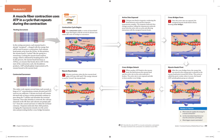
Module 9.7 Watch A muscle fiber contraction uses ATP in a cycle that repeats during the contraction Mitosis The contraction cycle is a series of interrelated steps that begins with the arrival of calcium ions within the zone of overlap in a sarcomere. 1 ADP + P In the resting sarcomere, each myosin head is already “energized”—charged with the energy that will be used to power a contraction. Each myosin head points away from the M line. In this position, the myosin head is “cocked” like the spring in a mousetrap. Cocking the myosin head requires energy, which is obtained by breaking down ATP. In this process, the myosin head functions as ATPase, an enzyme that breaks down ATP. At the start of the contraction cycle, the breakdown products, ADP and phosphate (represented as P), remain bound to the myosin head. Ca2+ Once the active sites are exposed, the energized myosin heads bind to them, forming cross-bridges. ADP + P Tropomyosin ADP + P Ca2+ Ca2+ Troponin Actin 3 Cytosol ADP + P Ca2+ Myosin reactivates when the free myosin head splits ATP into ADP and P. The energy released is used to “recock” the myosin head. Cross-Bridges Detach Myosin Heads Pivot When another ATP binds to the myosin head, the link between the myosin head and the active site on the actin molecule is broken. The active site is now exposed and able to form another cross-bridge. After the cross-bridges form, the energy that was stored in the resting state is released as the myosin heads pivot toward the M line. This action is called the power stroke; when it occurs, the bound ADP and phosphate group are released. Ca2+ Ca2+ Ca2+ Ca2+ ADP P + 4 ADP + P ATP Ca2+ ADP + P ADP P + 5 6 Ca2+ Ca2+ Active site ADP + P The entire cycle repeats several times each second, as long as Ca2+ concentrations remain elevated and ATP reserves are sufficient. Calcium ion levels will remain elevated only as long as action potentials continue to pass along the T tubules and stimulate the terminal cisternae. Once that stimulus is removed, the calcium channels in the SR close and calcium ion pumps pull Ca2+ from the cytosol and store it within the terminal cisternae. Troponin molecules then shift position, swinging the tropomyosin strands over the active sites and preventing further cross-bridge formation. Calcium ions bind to troponin, weakening the bond between actin and the troponin– tropomyosin complex. The troponin molecule then changes position, rolling the tropomyosin molecule away from the active sites on actin and allowing interaction with the energized myosin heads. Myosin head Myosin Reactivates Contracted Sarcomere Cross-Bridges Form 2 Contraction Cycle Begins Resting Sarcomere Active Sites Exposed Ca2+ ADP + P ATP Module 9.7 Review a. What molecule supplies the energy for a muscle contraction? b. List the interrelated steps that occur once the contraction cycle begins. c. What triggers myosin reactivation? 318 • Chapter 9: Skeletal Muscle Tissue M09_MART0000_00_SE_CH09.indd 318-319 9.7 Describe the role of ATP in a muscle contraction, and explain the steps involved in the contraction of a skeletal muscle fiber. Section 1: Functional Anatomy of Skeletal Muscle Tissue • 319 7/19/13 3:28 PM


