Document 17853471
advertisement
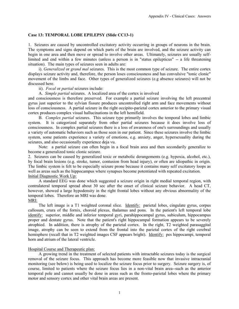
Appendix IV - Clinical Cases: Answers Case 13: TEMPORAL LOBE EPILEPSY (Slide CC13-1) 1. Seizures are caused by uncontrolled excitatory activity occurring in groups of neurons in the brain. The symptoms and signs depend on which parts of the brain are involved, and the seizure activity can begin in one area and then move or spread to involve other areas. Ultimately, seizures are usually selflimited and end within a few minutes (unless a person is in "status epilepticus" -- a life threatening situation). The main types of seizures seen in adults are: i). Generalized or grand mal seizures. This is the most common type of seizure. The entire cortex displays seizure activity and, therefore, the person loses consciousness and has convulsive "tonic clonic" movement of the limbs and face. Other types of generalized seizures (e.g absence seizures) will not be discussed here. ii). Focal or partial seizures include: A. Simple partial seizures. A localized area of the cortex is involved and consciousness is therefore preserved. For example a partial seizure involving the left precentral gyrus just superior to the sylvian fissure produces uncontrolled right arm and face movements without loss of consciousness. A partial seizure in the right occipito-parietal cortex anterior to the primary visual cortex produces complex visual hallucinations in the left hemifield. B. Complex partial seizures. This seizure type primarily involves the temporal lobes and limbic system. It is categorized separately from other partial seizures because it does involve loss of consciousness. In complex partial seizures there is a loss of awareness of one's surroundings and usually a variety of automatic behaviors such as those seen in our patient. Since these seizures involve the limbic system, some patients experience a variety of emotions, e.g. anxiety, anger, hypersexuality during the seizures, and also occasionally experience deja vu. Note: a partial seizure can often begin in a focal brain area and then secondarily generalize to become a generalized tonic clonic seizure. 2. Seizures can be caused by generalized toxic or metabolic derangements (e.g. hypoxia, alcohol, etc.), by focal brain lesions (e.g. stroke, tumor, contusion from head injury), or often are idiopathic in origin. The limbic system is felt to be especially seizure prone because it contains many self excitatory loops as well as areas such as the hippocampus where synapses become potentiated with repeated excitation. Initial Diagnostic Work Up: A standard EEG was done which suggested a seizure origin in right medial temporal region, with contralateral temporal spread about 30 sec after the onset of clinical seizure behavior. A head CT, however, showed a large hypodensity in the right frontal lobes without any obvious abnormality of the temporal lobes. Therefore an MRI was done. MRI: The left image is a T1 weighted coronal slice. Identify: parietal lobes, cingulate gyrus, corpus callosum, crura of the fornix, choroid plexus, thalamus and pons. In the patient's left temporal lobe identify: superior, middle and inferior temporal gyri, parahippocampal gyrus, subiculum, hippocampus proper and dentate gyrus. Note that the patient's right hippocampal formation appears to be severely atrophied. In addition, there is atrophy of the parietal cortex. In the right, T2 weighted parasaggital image, atrophy can be seen to extend from the frontal into the parietal cortex of the right cerebral hemisphere (recall that in T2 weighted images CSF appears bright). Identify: pes hippocampi, temporal horn and atrium of the lateral ventricle. Hospital Course and Therapeutic plan: A growing trend in the treatment of selected patients with intractable seizures today is the surgical removal of the seizure focus. This approach has become more feasible now that invasive intracranial monitoring (see below) is being used to localize the seizure focus prior to surgery. Seizure surgery is, of course, limited to patients where the seizure focus lies in a non-vital brain area--such as the anterior temporal pole and cannot usually be done in areas such as the fronto-parietal lobes where the primary motor and sensory cortex and other vital brain areas are present. 1 Appendix IV - Clinical Cases: Answers Based on the above information it was not possible to determine if our patients seizures were originating from the abnormal right fronto-parietal or temporal lobe. A PET scan was done in an attempt to improve localization of the functionally abnormal brain tissue, however, this again was not helpful since it showed an extensive area of hypometabolism involving both the right fronto-parietal and temporal regions. Since preliminary standard EEG recordings with surface electrodes suggested that the seizure focus may lie in the temporal lobe it was decided, together with the patient, to do invasive intracranial monitoring to determine if the seizure focus is amenable to surgery. Using three-dimensional stereotactic localization based on an MRI scan (see slide), depth electrodes were placed in the right hippocampal formation and amygdala, and surface grid electrodes were placed over the lateral and medial surfaces of the right fronto-parietal lobes. The intracranial EEG was monitored in the hospital continuously for five days with simultaneous video recordings of the patient's activities during which time she had two seizures demonstrating a clear temporal lobe origin on the EEG. Prior to surgery, the patient also underwent a "WADA" test in order to predict potential deficits which could occur due to resection of the brain regions surrounding the seizure focus. In this test, sodium amytal is injected into the internal carotid artery on each side while the awake patient undergoes extensive neurological examination. Based on the WADA test, the patient's language functions were found to be localized to the left cerebral hemisphere, and memory functions where found to be distributed bilaterally. The patient has, therefore, been scheduled for surgery to resect the anterior 5 cm of the right temporal lobe (what visual field deficit would you predict from more posterior resection of the temporal lobe?). Studies have demonstrated a success rate of about 70% in controlling complex partial seizures with this type of surgery. 2

