Document 14675115
advertisement
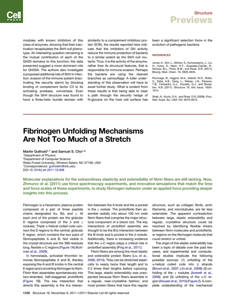
Structure Previews modules with known inhibitors of this class of enzymes, showing that their inactivation recapitulates the StrH null phenotype. An interesting question remaining is the mutual contribution of each of the GH20 domains to this function; the data presented suggest a more dominant role for GH20A. The authors also investigate a proposed additional role of StrH in infection: evasion of the immune system (inactivating the security alarm) by blocking binding of complement factor C3 to its activating protease, convertase. Even though the StrH structure was found to have a three-helix bundle domain with similarity to a complement inhibitory protein SCIN, the results reported here indicate that the inhibitors of GH activity reduce the immune protection of bacteria to a similar extent as the StrH null mutants. Thus, it is the activity of the enzyme, rather than its structural features, that is responsible for immune evasion. Perhaps the bacteria are using the cleaved branches as camouflage. A fuller understanding of this observation will have to await further study. What is evident from these results is that being able to clear a path through the security hedge of N-glycans on the host cell surface has been a significant selection force in the evolution of pathogenic bacteria. REFERENCES Jones, K., Sim, L., Mohan, S., Kumarasamy, J., Liu, H., Avery, S., Naim, H.Y., Quezada-Calvillo, R., Nichols, B.L., Pinto, B.M., and Rose, D.R. (2011). Bioorg. Med. Chem. 19, 3929–3934. Pluvinage, B., Higgins, M.A., Abbott, W.D., Robb, C., Dalia, A.B., Deng, L., Weiser, J.N., Parsons, T.B., Fairbanks, A.J., Vocadlo, D.J., and Boraston, A.B. (2011). Structure 19, this issue, 1603– 1614. Shah, N., Kuntz, D.A., and Rose, D.R. (2008). Proc. Natl. Acad. Sci. USA 105, 9570–9575. Fibrinogen Unfolding Mechanisms Are Not Too Much of a Stretch Martin Guthold1,* and Samuel S. Cho1,2 1Department of Physics of Computer Science Wake Forest University, Winston-Salem, NC 27109, USA *Correspondence: gutholdm@wfu.edu DOI 10.1016/j.str.2011.10.008 2Department Molecular explanations for the extraordinary elasticity and extensibility of fibrin fibers are still lacking. Now, Zhmurov et al. (2011) use force spectroscopy experiments, and innovative simulations that match the time and force scales of these experiments, to study fibrinogen behavior under an applied force providing deeper insights into this process. Fibrinogen is a hexameric plasma protein composed of a pair of three peptide chains designated Aa, Bb, and g. At each end of the protein are the globular D regions comprised of the b and g nodules. Triple a-helical coiled coils connect the D regions to the central, globular E region, which contains the two pairs of fibrinopeptides A and B. Not visible in the crystal structure are the 389 residues long, flexible a-C regions (Figure 1A) (Kollman et al., 2009). In hemostasis, activated thrombin removes fibrinopeptides A and B, thereby exposing the A and B knobs in the central E region and converting fibrinogen to fibrin. Fibrin then assembles spontaneously into two-stranded, half-staggered protofibrils (Figure 1B). The key interaction that directs this assembly is the A:a interac- tion between the A knob and the a pocket in the g nodule. The protofibrils then assemble radially into about 100 nm wide fibrin fibers that comprise the major structural component of a blood clot. The key interactions of protofibril assembly are thought to be the B:b interaction between the B knob and b pocket in the b nodule. Additionally, there is increasing evidence that the a-C region plays a critical role in protofibril assembly (Ping et al., 2011). Fibrin fibers are among the most elastic and extensible protein fibers (Liu et al., 2006, 2010). They can be stretched elastically to nearly twice their length and to 2.5 times their lengths before rupturing. This large, elastic extensibility was unexpected because fibrin fibers assemble in a regular, near-crystalline fashion, and most protein fibers that have this regular 1536 Structure 19, November 9, 2011 ª2011 Elsevier Ltd All rights reserved structure, such as collagen fibrils, actin filaments, and microtubules, are far less extensible. The apparent contradiction between large, elastic extensibility and regular, crystalline structure could be resolved by identifying flexible linkers between fibrin molecules and protofibrils, or regions on the fibrinogen molecule that could stretch or unfold. The origin of this elastic extensibility has been a topic of debate over the past few years, and experimental and computational studies implicate the following possible sources: (1) unfolding of the a-helical coiled coils into b strands (Brown et al., 2007; Lim et al., 2008); (2) unfolding of the g nodules (Averett et al., 2008); and (3) unfolding of the a-C region (Houser et al., 2010) (Figure 2). A complete understanding of the mechanical Structure Previews A Figure 1. Fibrin Assembly Molecular structures of human (A) fibrinogen and (B) fibrin fibers. The molecular structures were based on the X-ray structures of the human fibrinogen (PDB: 1GHG; Kollman et al., 2009) and D dimer (PDB: 1FZE). The unstructured a-C regions are shown as schematics. The fibrin structure was constructed by performing a best fit of each end of fibrinogen to each D region of the D dimer. also occurs on a similar timescale when a modest force is applied to the system in experiments. For atomistic simulations to observe unfolding events under mechanical force, the only recourse is to apply unnaturally high forces to observe unfolding (Lim et al., 2008). Coarsegrained simulations increase the timeB scales of simulations, while still accurately capturing the folding mechanisms of biologically relevant biomolecules (Cho et al., 2009; Hills and Brooks, 2009). In these classes of simulations, groups of atoms, such as residues, are represented by a bead, thereby simplifying the calculations. Unfortunately, the computational demands of even coarse-grained simulations require very high forces to observe unfolding events. properties of fibrin will improve our un- istic detail, even though biologically releAnother approach to optimize MD derstanding and treatment of thrombotic vant biomolecules are much larger and simulations is to perform them on events, such as heart attacks, ischemic can fold much slower. Indeed, a single graphics processing unit (GPU) hardware, strokes, and pulmonary embolisms. protein chain is, on average, 400 amino which readily lends itself to a high degree To address these outstanding issues, acids long and takes considerably longer of parallelization and the very fast floatingZhmurov et al. (2011) [in this issue of than 1 ms to fold. Mechanical unfolding point calculations. GPUs are specialized Structure] employed atomic video cards that were origiforce microscopy (AFM)nally designed to perform based force spectroscopy floating-point calculations and molecular dynamics that are characteristic of (MD) simulations to identity operations for rendering to the underlying molecular a graphical display. In this mechanism of fibrin’s extenparallel architecture many sibility and elasticity. A main processors independently reason why their study is of execute the same set of particular note is because instructions on large sets of simulating relevant biomoledata. GPUs are well suited cules on timescales that can to perform MD simulations be directly compared with because the general algoexperiments remains an rithm is to compute the extraordinary challenge. The same set of force calculations major bottleneck for simu(i.e., instructions) on the coorlating these processes is the dinates and velocities of each force evaluation involved in of the individual beads (i.e., these complex systems that large set of data). must be computed for each The present study by pair of interacting particles. Zhmurov et al. (2011) used As such, detailed atomistic a combination of AFM-based Figure 2. Schematics of Three Possible Mechanisms of Fibrin Fiber resolution folding simulations single molecule unfolding Extensibility are restricted to very small and GPU-based coarse(A) Unfolding of the coiled chains. (50 amino acids long), fastgrained force unfolding simu(B) Unfolding of the g nodules. (C) Unfolding of the a-C region. folding (<ms) proteins at atomlations to study fibrinogen Structure 19, November 9, 2011 ª2011 Elsevier Ltd All rights reserved 1537 Structure Previews monomers and oligomers. The result of their tour de force is that the peak-topeak distances in the force extension curves from simulations are nearly identical to those observed in the experiments, strongly supporting that the simple coarse-grained model accurately captures the mechanical unfolding mechanisms observed in experiments. The simulations revealed that when force is applied to fibrinogen, the g nodule partially unfolds, and the coiled coils reversibly convert from an a-helical to an extended b strand structure. The authors then suggest that these two mechanisms may explain the extraordinary elasticity and extensibility of fibrin fibers. These mechanisms have been proposed before, but a clear verification was still missing, and the authors provide convincing evidence that these mechanisms are indeed feasible. A limitation of the experimental and simulation designs is that they could not probe the role of the flexible, unstructured a-C region. This is because single fibrinogen molecules or short chains of abutting monomers were tested, rather than double-stranded, half-staggered protofibrils, or even complete fibers. There is emerging evidence that the a-C region plays a significant role in fibrin fiber formation, especially in protofibril assembly, and it has been suggested that the stretching and unfolding of the a-C region may be the major molecular mechanism that endows fibrin fibers with their large extensibility (Houser et al., 2010). It is also entirely possible that all three mechanisms (Figure 2) could be going on in parallel. Thus, in the future it would be exciting to perform the forced unfolding experiments and simulations with protofibrils. REFERENCES Averett, L.E., Geer, C.B., Fuierer, R.R., Akhremitchev, B.B., Gorkun, O.V., and Schoenfisch, M.H. (2008). Langmuir 24, 4979–4988. Brown, A.E.X., Litvinov, R.I., Discher, D.E., and Weisel, J.W. (2007). Biophys. J. 92, L39–L41. Cho, S.S., Pincus, D.L., and Thirumalai, D. (2009). Proc. Natl. Acad. Sci. USA 106, 17349–17354. Hills, R.D., Jr., and Brooks, C.L., 3rd. (2009). Int. J. Mol. Sci. 10, 889–905. Houser, J.R., Hudson, N.E., Ping, L., O’Brien, E.T., 3rd, Superfine, R., Lord, S.T., and Falvo, M.R. (2010). Biophys. J. 99, 3038–3047. Kollman, J.M., Pandi, L., Sawaya, M.R., Riley, M., and Doolittle, R.F. (2009). Biochemistry 48, 3877– 3886. Lim, B.B., Lee, E.H., Sotomayor, M., and Schulten, K. (2008). Structure 16, 449–459. Liu, W., Carlisle, C.R., Sparks, E.A., and Guthold, M. (2010). J. Thromb. Haemost. 8, 1030–1036. Liu, W., Jawerth, L.M., Sparks, E.A., Falvo, M.R., Hantgan, R.R., Superfine, R., Lord, S.T., and Guthold, M. (2006). Science 313, 634. Ping, L., Huang, L., Cardinali, B., Profumo, A., Gorkun, O.V., and Lord, S.T. (2011). Biochemistry 50, 9066–9075. Zhmurov, A., Brown, A.E.X., Litvinov, R.I., Dima, R.I., Weisel, J.W., and Barsegov, V. (2011). Structure 19, this issue, 1615–1624. Tackling the Legs of Mannan-Binding Lectin Erhard Hohenester1,* 1Department of Life Sciences, Imperial College London, London SW7 2AZ, UK *Correspondence: e.hohenester@imperial.ac.uk DOI 10.1016/j.str.2011.10.005 The recognition of pathogen surfaces by mannan-binding lectin activates MASP proteases, leading to complement activation. A crystal structure by Gingras et al. (2011) in this issue of Structure now shows how the collagen-like stems of mannan-binding lectin bind MASP-1 through a minimalist set of interactions. Collagen is the most abundant protein in vertebrates. Its basic structure was worked out over 50 years ago and consists of three polypeptide chains with characteristic Gly-X-Y repeats that are intertwined to form a left-handed triple helix (Brodsky and Persikov, 2005). Gly-X-Y repeats are not confined to collagens, however, and short stretches of the motif are also found in a number of proteins of the immune system, such as the macrophage scavenger receptor A, the complement protein C1q, and mannan-binding lectin (MBL). MBL and the closely-related ficolins play an important role in innate immunity; their binding to carbohydrate arrays on the surface of pathogens triggers the activation of complement (Wallis et al., 2010). This process critically involves the MBL-associated serine proteases (MASPs), which, upon binding to a pathogen surface, are converted from inactive zymogens to active proteases. How this happens is still unclear, but an important piece of the puzzle is now provided by Gingras et al. (2011) in this issue of Structure, who have determined a crystal structure of the MBL-MASP contact region. The basic unit of MBL is a homotrimer consisting of a collagen-like stem, an 1538 Structure 19, November 9, 2011 ª2011 Elsevier Ltd All rights reserved a-helical-coiled coil neck region, and three globular carbohydrate recognition domains. Circulating MBL contains two or more of these homotrimers covalently linked at their N-termini by a number of disulphide bonds (Figure 1). This structure is commonly likened to a bouquet of flowers. MASPs are homodimers with a modular architecture; their N-terminal CUB-EGF-CUB regions mediate binding to MBL, and their C-terminal regions contain the serine proteases that autoactivate upon pathogen recognition. A previous elegant study by Wallis et al. (2004) mapped the MASP binding site to
