Degradation of Acid Red 27 by the Recombinant Flavin Reductase
advertisement

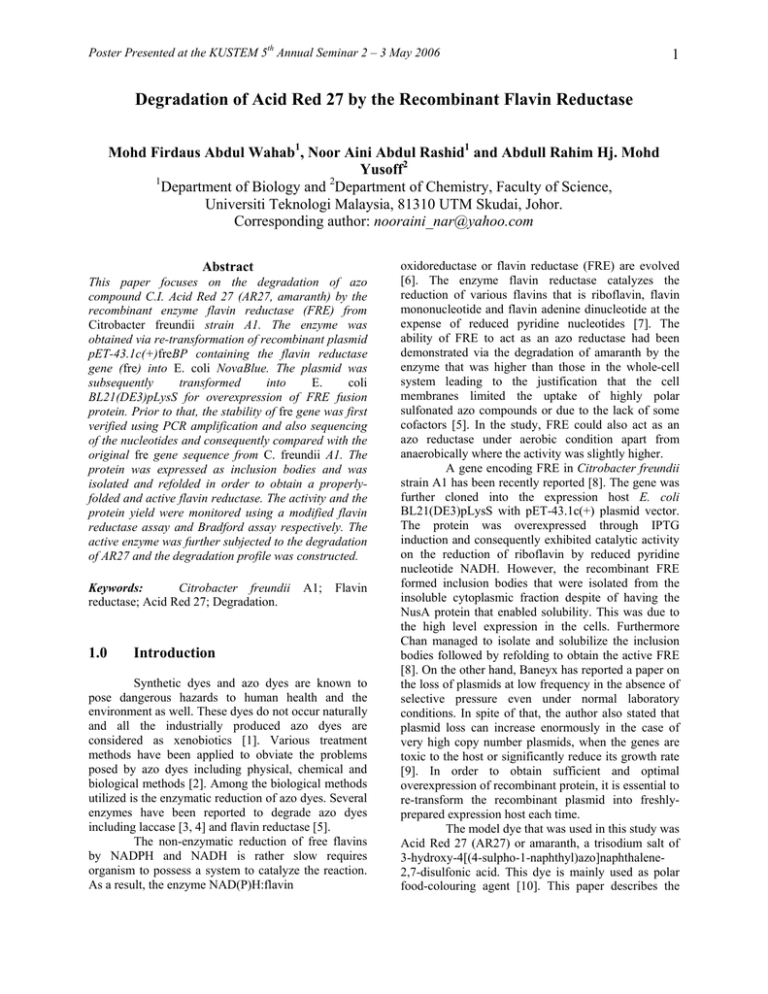
Poster Presented at the KUSTEM 5th Annual Seminar 2 – 3 May 2006 1 Degradation of Acid Red 27 by the Recombinant Flavin Reductase Mohd Firdaus Abdul Wahab1, Noor Aini Abdul Rashid1 and Abdull Rahim Hj. Mohd Yusoff2 1 2 Department of Biology and Department of Chemistry, Faculty of Science, Universiti Teknologi Malaysia, 81310 UTM Skudai, Johor. Corresponding author: nooraini_nar@yahoo.com Abstract This paper focuses on the degradation of azo compound C.I. Acid Red 27 (AR27, amaranth) by the recombinant enzyme flavin reductase (FRE) from Citrobacter freundii strain A1. The enzyme was obtained via re-transformation of recombinant plasmid pET-43.1c(+)freBP containing the flavin reductase gene (fre) into E. coli NovaBlue. The plasmid was subsequently transformed into E. coli BL21(DE3)pLysS for overexpression of FRE fusion protein. Prior to that, the stability of fre gene was first verified using PCR amplification and also sequencing of the nucleotides and consequently compared with the original fre gene sequence from C. freundii A1. The protein was expressed as inclusion bodies and was isolated and refolded in order to obtain a properlyfolded and active flavin reductase. The activity and the protein yield were monitored using a modified flavin reductase assay and Bradford assay respectively. The active enzyme was further subjected to the degradation of AR27 and the degradation profile was constructed. Keywords: Citrobacter freundii reductase; Acid Red 27; Degradation. 1.0 A1; Flavin Introduction Synthetic dyes and azo dyes are known to pose dangerous hazards to human health and the environment as well. These dyes do not occur naturally and all the industrially produced azo dyes are considered as xenobiotics [1]. Various treatment methods have been applied to obviate the problems posed by azo dyes including physical, chemical and biological methods [2]. Among the biological methods utilized is the enzymatic reduction of azo dyes. Several enzymes have been reported to degrade azo dyes including laccase [3, 4] and flavin reductase [5]. The non-enzymatic reduction of free flavins by NADPH and NADH is rather slow requires organism to possess a system to catalyze the reaction. As a result, the enzyme NAD(P)H:flavin oxidoreductase or flavin reductase (FRE) are evolved [6]. The enzyme flavin reductase catalyzes the reduction of various flavins that is riboflavin, flavin mononucleotide and flavin adenine dinucleotide at the expense of reduced pyridine nucleotides [7]. The ability of FRE to act as an azo reductase had been demonstrated via the degradation of amaranth by the enzyme that was higher than those in the whole-cell system leading to the justification that the cell membranes limited the uptake of highly polar sulfonated azo compounds or due to the lack of some cofactors [5]. In the study, FRE could also act as an azo reductase under aerobic condition apart from anaerobically where the activity was slightly higher. A gene encoding FRE in Citrobacter freundii strain A1 has been recently reported [8]. The gene was further cloned into the expression host E. coli BL21(DE3)pLysS with pET-43.1c(+) plasmid vector. The protein was overexpressed through IPTG induction and consequently exhibited catalytic activity on the reduction of riboflavin by reduced pyridine nucleotide NADH. However, the recombinant FRE formed inclusion bodies that were isolated from the insoluble cytoplasmic fraction despite of having the NusA protein that enabled solubility. This was due to the high level expression in the cells. Furthermore Chan managed to isolate and solubilize the inclusion bodies followed by refolding to obtain the active FRE [8]. On the other hand, Baneyx has reported a paper on the loss of plasmids at low frequency in the absence of selective pressure even under normal laboratory conditions. In spite of that, the author also stated that plasmid loss can increase enormously in the case of very high copy number plasmids, when the genes are toxic to the host or significantly reduce its growth rate [9]. In order to obtain sufficient and optimal overexpression of recombinant protein, it is essential to re-transform the recombinant plasmid into freshlyprepared expression host each time. The model dye that was used in this study was Acid Red 27 (AR27) or amaranth, a trisodium salt of 3-hydroxy-4[(4-sulpho-1-naphthyl)azo]naphthalene2,7-disulfonic acid. This dye is mainly used as polar food-colouring agent [10]. This paper describes the Poster Presented at the KUSTEM 5th Annual Seminar 2 – 3 May 2006 degradation of Acid Red 27 using this recombinant flavin reductase enzyme. 2.0 Materials and Methods 2.1 Microorganism and Culture Conditions transformants. The transformants that grew on the plate was screened for the presence of fre gene insert with PCR method using primers as in Section 2.3. Transformation into BL21(DE3)pLysS followed the above protocol replaced with the antibiotics carbenicillin (100 µg/mL) and chloramphenicol (34 µg/mL). 2.5 The clone BL21(DE3)pLysS:pET43.1c(+)freBP, an E. coli host cloned with flavin reductase gene (fre) from C. freundii A1 was used courtesy of previous research [8]. E. coli strain NovaBlue was used as the maintenance host for the recombinant plasmid and the recombinant plasmid was transformed into expression host BL21(DE3)pLysS for overexpression. 2.2 Plasmid DNA Purification Plasmid DNA isolation was performed using Wizard™ DNA Purification Kit (Promega) according to the manufacturer’s instruction. 2.3 fre Gene Detection and Sequencing PCR amplification was done in a 50 µL mixture containing template DNA 0.5 µL, forward and reverse primers 2 µL each, PCR Mastermix (Promega) 25 µL and topped-up with nuclease-free water to 50 µL. The mixture was subjected to 30 cycles in a thermocycler - GeneAmp® PCR System 9700 (PE Applied Biosystems). Each cycle consisting of denaturation at 94°C for 1 minute, primer annealing at 51°C for 1 minute and chain extension at 72°C for 2 minutes; with 8 minutes of initial denaturation and 8 minutes of final chain extension. The PCR and sequencing primers used were S•Tag18F (5’– GAACGCCAGCACATGGAC) and COLIDOWN (5’– TTCACTTCTGAGTTCGGCATG). The product was analyzed using 1% agarose gel electrophoresis. DNA sequencing was performed by First Base Laboratories Sdn. Bhd (Malaysia). 2.4 Transformation of Recombinant Plasmid into E. coli Novablue and BL21(DE3)pLysS Recombinant plasmid (1-100 µl) was added to the NovaBlue competent cells (0.1 ml) which were melted on ice. The mixture was incubated on ice for one hour, followed by heat shock at 42°C for 45-90 seconds with consequent chilling on ice for another 2 minutes. LB broth was added and the cells were incubated for one hour at 37°C with shaking. The cells were then plated out on LB agar with ampicillin (100 µg/mL) and tetracycline (12.5 µg/mL) for screening of 2 Isolation of Recombinant Flavin Reductase A single colony of BL21(DE3)pLysS:pET43.1c(+)freBPnew was inoculated into LB broth (10 mL) with carbenicillin (100 µg/mL) and chloramphenicol (34 µg/mL) and incubated overnight at 37°C until OD600 reached 0.4 – 0.6. Isopropyl-β-Dthiogalactopyranoside (IPTG) was added to the final concentration of 1 mM and further incubated for 5 hours. The induced cells will then be used for inclusion bodies solubilization and refolding using Protein Refolding Kit (Novagen) according to the manufacturer’s protocol. 2.6.1 Flavin Reductase Assay and Bradford Protein Assay The flavin reductase activity was detected using a modified flavin reductase assay as reported in [5] and [8]. 150 µL of the solubilized inclusion bodies was added to the reaction mixture with Tris-HCl (15 mM, pH 7.5), NADH (0.2 mM) and riboflavin (15 µM). The activity of flavin reductase was determined at 30°C from the decrease of the absorbance at 340 nm (ε340 = 6.22 mM-1 cm-1) due to the oxidation of NADH, using Cary 100 UV-Visible Spectrophotometer (Varian). One unit (U) of enzyme activity is defined as the amount of enzyme that converts 1 nmol of substrate NADH per minute. Protein concentration was determined using Bradford method with bovine serum albumin (BSA) as standard [11]. Bradford reagent (Sigma, USA) was utilized in this assay and conducted according to the manufacturer’s instruction based on the standard 3.1 mL assay. Initially, a standard curve was constructed before the actual assay of the unknown protein content was made. The unknown protein concentration was determined from the standard curve. 2.7 Degradation of Acid Red 27 by the Solubilized and Refolded Flavin Reductase AR27 were subjected to degradation by FRE based on the modified method developed by Chan [8] to obtain the degradation profile for the azo compound. 150 µL of the solubilized and refolded inclusion bodies was added to the reaction mixture with Tris-HCl (15 mM, pH 7.5), NADH (0.2 mM), riboflavin (15 µM) Poster Presented at the KUSTEM 5th Annual Seminar 2 – 3 May 2006 and AR27 in the final concentration of 1 × 10⁻⁴ M. Spectrophotochemical monitoring was performed at 521 nm that corresponds to the λmax of Acid Red 27 and at 340 nm that corresponds to the λmax of NADH simultaneously at 30°C for 30 minutes. 3.0 3 in the conformational change allowing the expression of high level of T7 RNA polymerase for transcription of target gene in the vector by activating the T7 promoter [12]. The induction of pET-43.1c(+)freBP recombinant plasmid led to protein expressed as inclusion bodies attributed to the high level of expression in the E. coli host [8]. The problem of inclusion bodies was overcome by isolating the inclusion bodies using the Protein Refolding Kit (Novagen), solubilizing it and further refolded into active protein. The solubilization involved CAPS buffer at alkaline pH and N-lauroylsarcosine with DTT followed by dialysis in Tris-HCl buffer in the presence of DTT. The final product was a refolded and active flavin reductase with the total activity of 106.44 U from the flavin reductase assay conducted and a total protein of 2.45 mg/mL assayed using the Bradford method giving the FRE specific activity of 1.44 U/mg. The recombinant FRE was further subjected to the degradation of AR27 to investigate the direct reduction of azo compound by this enzyme. The experiment was performed in the presence of NADH and riboflavin as demonstrated in [8] that FAD or riboflavin and NADH were required for the azo reduction to occur. Figure 1 shows the AR27 removal by FRE as a function of contact time together with the reduction in NADH concentration. . Results and Discussion From the PCR performed, it can be loosely confirmed that fre gene was still stable in the recombinant plasmid from the observation of a 1 kb band representing the region that was amplified by the primers. This is based on the finding that the sequenced gene matched that of the fre gene published by Chan [8]. After the transformation of recombinant plasmid into NovaBlue, the plasmid was then subsequently transformed into BL21(DE3)pLysS. Following transformation, PCR re-amplification was done to confirm the presence of the recombinant plasmid in the expression host. A band at around 1 kb symbolizing fre gene was observed. Hence, the recombinant plasmid was successfully re-transformed into BL21(DE3)pLysS. Induction of E. coli transformed with pET43.1c(+) plasmid with IPTG leads to the binding of IPTG to lac repressor in the plasmid system resulting 0.400 0.107 0.350 Conc. AR27 Conc. NADH 0.097 0.300 0.092 0.087 0.250 0.082 0.200 Concentration of NADH (mM) Concentration of AR27 (mM) 0.102 0.077 0.072 0.150 0 5 10 15 20 25 30 Time (minutes) Figure 1: The reduction of AR27 by FRE and concomitant reduction in NADH concentration during the 30 minutes incubation time. Poster Presented at the KUSTEM 5th Annual Seminar 2 – 3 May 2006 NADH 4 Reduced Riboflavin Aromatic Amines SO3-Na+ FRE NAD+ Oxidized Riboflavin N N OH +- Na O3S SO3-Na+ Acid Red 27 Figure 2: The reduction of azo dye AR27 by flavin reductase mediated by NADH and riboflavin. From Figure 1, maximum dye removal was obtained after 25 minutes of incubation. The direct reduction rate of AR27 in the linear region was calculated as 1.05 × 10⁻³ mM min-1. Simultaneously, the reduction in NADH concentration was also recorded in order to investigate the relation between NADH and the reduction of the azo compound. Interestingly NADH was also utilized in the reaction depicted from the decrease in absorbance at 340 nm. The similar trend indicates that reduction of AR27 was halted once the reduction in NADH concentration reached a plateau. The figure obviously supports the fact that azo reduction by FRE is made possible only in the presence of NADH and riboflavin as cofactors. Flavin reductase acts by reducing free flavins (riboflavin) where the H+ is obtained from reduced pyridine nucleotides (NADH) [8]. The reduced flavins subsequently transfer the electrons to the dye for the reduction to occur chemically. Figure 2 is a simplified diagram showing the transfer of electrons from NADH to azo dyes via the acts of flavin reductase based on the work in [13]. The utilization of NADH by crude azoreductase from Rhodobacter sphaeroides in the reduction of Methyl Orange had also been reported [14]. The reaction was performed under aerobic condition although it has been reported that reduced flavins react rapidly with molecular oxygen [5]. This is due to the further report that some decrease in the concentration of Acid Red 27 or amaranth (their model dye) was observed even though the reaction rate was lower to those of under anaerobic condition [5]. This means that significant reduction of amaranth by the enzyme could take place even under aerobic condition as reported in this paper. BL21(DE3)pLysS for the expression of flavin reductase enzyme (FRE) and the activity was analyzed using flavin reductase assay and Bradford protein assay. The enzyme was expressed as inclusion bodies and further solubilized and refolded yielding the active FRE with the specific activity of 1.44 U/mg. The enzyme was able to reduce Acid Red 27 directly at the rate of 1.05 × 10⁻³ mM min-1 in the linear region until maximum reduction time (25 minutes) with a concomitant reduction in NADH concentration. It was concluded that the recombinant flavin reductase was able to degrade Acid Red 27 directly via reduction of riboflavin at the expense of reduced pyridine nucleotide NADH. Acknowledgements The authors would like to acknowledge Universiti Teknologi Malaysia and MOSTI for a UTM-PTP scholarship granted to Mohd Firdaus Abdul Wahab and for a research grant under VOT 74199. References [1] [2] [3] Conclusions [4] The recombinant plasmid pET-43.1c(+)freBP was successfully re-transformed into E. coli Stolz, A. (2001). “Basic and Applied Aspects in the Microbial Degradation of Azo Dyes”. Appl. Microbiol. Biotechnol. 56. 69 – 80. Nam, S. (1998). “Azo Dye Transformation by Enzymatic and Chemical Systems”. Oregon Graduate Institute of Science and Technology: PhD Thesis. Liu, W., Chao, Y., Yang, X.., Bao, H., Qian, S. (2004). “Biodecolorization of Azo, Anthraquinonic and Triphenylmethane Dyes by White-rot Fungi and a Laccase-Secreting Engineered Strain”. J. Ind. Microbiol. Biotechnol. 31. 127 – 132. Ünyayar, A., Mazmanci, A. M., Erkurta, A., Atacaga, H., Gizir, A. M., (2005). “Decolorization Kinetics of the Azo Dye Drimaren Blue X3LR by Poster Presented at the KUSTEM 5th Annual Seminar 2 – 3 May 2006 [5] [6] [7] [8] [9] Laccase”. React. Kinet. Catal. Lett. 86. 99 – 107. Russ, R., Rau, J., Stolz, A. (2000). “The Function of Cytoplasmic Flavin Reductases in the Reduction of of Azo Dyes by Bacteria”. Appl. Environ. Microbiol. 66. 1429 – 1434. Ingelman, M., Ramaswamy, S., Niviére, V., Fontecave, M., Eklund, H. (1999). “Crystal Structure of NAD(P)H:Flavin Oxidoreductase from Escherichia coli”. Biochemistry. 38. 7040 – 7049. Spyrou, G., Haggård-Ljungquist, E., Krook, M., Jörnvall, H., Nilsson, E., Reichard, P. (1991). “Characterization of the Flavin Reductase Gene (fre) of Escherichia coli and Construction of a Plasmid for Overproduction of the Enzyme”. J. Bact. 173. 3673 – 3679. Chan, G. F. (2004). “Molecular and Enzymatic Studies on the Decolourization Mechanism of Azo Dyes by Citrobacter freundii A1”. Universiti Teknologi Malaysia: PhD Thesis. Baneyx, F. (1999). “Recombinant Protein Expression in Escherichia coli”. Current Opinion in Biotechnology. 10. 411 – 421. [10] [11] [12] [13] [14] 5 Mallett, A. K., King, L. J., Walker, R. (1982). “A Continuous Spectrophotometric Determination of Hepatic Microsomal Azo Reductase Activity and Its Dependence on Cytochrome P-450”. Biochem. J. 201. 589 – 595. Bradford, M. M. (1976). “A Rapid and Sensitive Method for the Quantitation of Microgram Quantities of Protein Utilizing the Principle of Protein-Dye Binding”. Analytical Biochemistry. 72. 248 – 254. Mierendorf, R., Yeager, K. and Novy, R. (1994). “The pET System: Your Choice for Expression”. inNovations. 1. 1 – 3. Field, J. A. and Brady, J. (2003). “Riboflavin as a Redox Mediator Accelerating the Reduction of the Azo Dye Mordant Yellow 10 by Anaerobic Granular Sludge”. Water Science and Technology. 48. 187 – 193. Song, Z-Y., Zhou, J-T., Wang, J., Yan, B., Du, CH. (2003). “Decolorization of Azo Dyes by Rhodobacter sphaeroides”. Biotechnol. Lett. 25. 1815 – 1818.


