Clinical Physics Implementation of Proton Therapy
advertisement

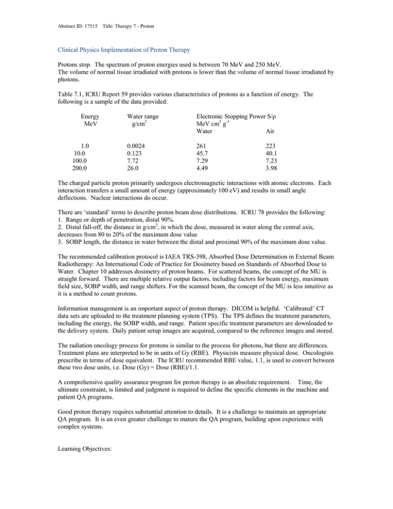
Abstract ID: 17515 Title: Therapy 7 - Proton Clinical Physics Implementation of Proton Therapy Protons stop. The spectrum of proton energies used is between 70 MeV and 250 MeV. The volume of normal tissue irradiated with protons is lower than the volume of normal tissue irradiated by photons. Table 7.1, ICRU Report 59 provides various characteristics of protons as a function of energy. The following is a sample of the data provided: Energy MeV 1.0 10.0 100.0 200.0 Water range g/cm2 Electronic Stopping Power S/ρ MeV cm2 g-1 Water Air 0.0024 0.123 7.72 26.0 261 45.7 7.29 4.49 223 40.1 7.23 3.98 The charged particle proton primarily undergoes electromagnetic interactions with atomic electrons. Each interaction transfers a small amount of energy (approximately 100 eV) and results in small angle deflections. Nuclear interactions do occur. There are ‘standard’ terms to describe proton beam dose distributions. ICRU 78 provides the following: 1. Range or depth of penetration, distal 90%. 2. Distal fall-off, the distance in g/cm2, in which the dose, measured in water along the central axis, decreases from 80 to 20% of the maximum dose value 3. SOBP length, the distance in water between the distal and proximal 90% of the maximum dose value. The recommended calibration protocol is IAEA TRS-398, Absorbed Dose Determination in External Beam Radiotherapy: An International Code of Practice for Dosimetry based on Standards of Absorbed Dose to Water. Chapter 10 addresses dosimetry of proton beams. For scattered beams, the concept of the MU is straight forward. There are multiple relative output factors, including factors for beam energy, maximum field size, SOBP width, and range shifters. For the scanned beam, the concept of the MU is less intuitive as it is a method to count protons. Information management is an important aspect of proton therapy. DICOM is helpful. ‘Calibrated’ CT data sets are uploaded to the treatment planning system (TPS). The TPS defines the treatment parameters, including the energy, the SOBP width, and range. Patient specific treatment parameters are downloaded to the delivery system. Daily patient setup images are acquired, compared to the reference images and stored. The radiation oncology process for protons is similar to the process for photons, but there are differences. Treatment plans are interpreted to be in units of Gy (RBE). Physicists measure physical dose. Oncologists prescribe in terms of dose equivalent. The ICRU recommended RBE value, 1.1, is used to convert between these two dose units, i.e. Dose (Gy) = Dose (RBE)/1.1. A comprehensive quality assurance program for proton therapy is an absolute requirement. Time, the ultimate constraint, is limited and judgment is required to define the specific elements in the machine and patient QA programs. Good proton therapy requires substantial attention to details. It is a challenge to maintain an appropriate QA program. It is an even greater challenge to mature the QA program, building upon experience with complex systems. Learning Objectives: Abstract ID: 17515 Title: Therapy 7 - Proton Review the following: 1. Interactions of protons with matter 2. Basic proton therapy terminology 3. Calibration protocol for protons 4. Physical dose and Dose Equivalent Abstract ID: 17515 Title: Therapy 7 - Proton 1.


