Automated Treatment Planning Using a Database of Prior Patient Treatment Plans
advertisement

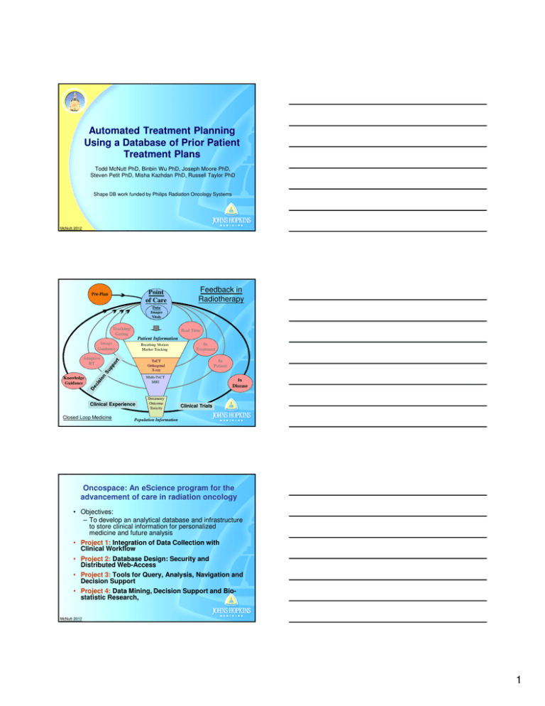
Automated Treatment Planning Using a Database of Prior Patient Treatment Plans Todd McNutt PhD, Binbin Wu PhD, Joseph Moore PhD, Steven Petit PhD, Misha Kazhdan PhD, Russell Taylor PhD Shape DB work funded by Philips Radiation Oncology Systems McNutt 2012 Point of Care Pre-Plan Feedback in Radiotherapy Data Images Vitals Tracking/ Gating Real Time Patient Information Image Guidance Breathing Motion Marker Tracking Adaptive RT In Treatment In Patient TxCT Orthogonal X-ray Multi-TxCT MRI Knowledge Guidance Clinical Experience Closed Loop Medicine Dosimetry Outcome Toxicity In Disease Clinical Trials Population Information Oncospace: An eScience program for the advancement of care in radiation oncology • Objectives: – To develop an analytical database and infrastructure to store clinical information for personalized medicine and future analysis • Project 1: Integration of Data Collection with Clinical Workflow • Project 2: Database Design: Security and Distributed Web-Access • Project 3: Tools for Query, Analysis, Navigation and Decision Support • Project 4: Data Mining, Decision Support and Biostatistic Research, McNutt 2012 1 Tumor information is stored including staging and image based RECIST assessment. Personal Health Information is stored in a single table to facilitate anonymization. Toxicity and Outcomes. Chemotherapy, medications, and surgeries Lab values Patient History Regions of interest are stored as run-length encoded masks associated with each Patient Representation. Shape and shape relationship descriptors are stored for fast query of patient shape similarities. Dose Volume Histograms are stored for each region of interest for fast query. DVH stored for both treatment summary and individual treatment sessions Dose distributions are stored for each radiotherapy session. Each radiotherapy session is associated with a single patient representation. The transformation table stores the transformation between multiple patient representations enabling dose accumulation. Oncospace Website Grade 2 & 3 Influence of Shape • Shape Characteristics – Volume – Positional relationships between structures – Separation of surfaces • • Shape Change in Time Influence – Plan quality (IMRT) – Ability to achieve Tx goal • Motion management – Toxicity • Simplification of information without loss of relevance? Patient Shape Clusters 2 Use of ROI Shapes in Oncospace Overlap Volume Histogram DB of prior patients far Shape Relationship Descriptor 10 OVH for similar patients to predict expected DVH 1 L: OVH58 L:OVH63 0.9 L:OVH70 0.8 R:OVH58 R:OVH63 0.7 R:OVH70 0.6 0.5 0.4 0.3 0.2 0.1 0 -2 -1 0 1 2 3 4 5 6 7 8 99.5 d3:OVH of PTVH norm overlap volume 8 6 4 2 0 -2 8 6 4 2 0 Expansion Distance (cm) 2 close 1 0 -1 -2 -2 d1:OVH of PTVL DVH Prediction 1 Right parotid Left parotid DVH predict toxicity? normalized volume 0.9 Decisions: • Plan Quality Assess • Automated IMRT • Expected Toxicities • Dosimetric Trade-offs 3 4 d2:OVH of PTVM 0.8 0.7 0.6 0.5 0.4 0.3 0.2 0.1 0 0 5 10 15 20 25 30 35 40 45 50 55 60 65 70 Dose (Gy) McNutt 2009 Overlap Histogram (OVH) ThatVolume was descriptive only OVH maps the shape of an OAR to a volume-distance plane target expanding and contracting Actualthrough computation is with a Euclidean Distance Organ Transform Algorithm Target which is more efficient than amm Volume the process described. amm amm Michael Kazhdan, Patricio Simari, Todd McNutt, Binbin Wu, Robert Jacques, Ming Chuang, and Russell Taylor, “A Shape Relationship Descriptor for RadiationTherapy Planning” Medical Image Computing and Computer-Assisted Intervention 5762/2009(12), 100–108 (2009) amm -2a mm -a mm 0 a mm 2a mm Distance 3 From a point to an organ Which dose? Maximum dose? Dose to e.g. 50% DVH OAR 1 OAR2 PTV Overlap Volume Histogram describes distance from a (sub)volume to the PTV 50% volume at 1.5 cm from PTV An example of OVH OAR2 OAR1 For parallel organs, OAR2 (red) is more easily spared. For serial organs, OAR1 (blue) is more easily spared. Comparison between 1L and 5L: 1L is an outlier Patient 1 Patient 5 1 1L:OVH58 1L:OVH63 1L:OVH70 5L:OVH58 5L:OVH63 5L:OVH70 0.9 0.8 0.7 0.6 0.5 0.4 3-D shapes of the left parotid and PTV58.1. 0.3 0.2 0.1 0 -1 0 1 2 3 4 Distance (cm) 5 6 7 8 OVH of 1L and 5L 1 1L:re-plan 1L:original 5L:original 0.9 0.8 3-D shapes of the left parotid and PTV63. 0.7 0.6 0.5 0.4 0.3 0.2 0.1 0 0 3-D shapes of the left parotid and PTV70. 10 20 30 40 Dose(Gy) 50 60 70 80 DVH of 1L and 5L 4 Re-plan results of patient 1 Original plan Re-plan Treatment Plan Quality Control (outlier detection): parotids OVH M OVH L 1 DVH of parotid OVH H 1 0.8 0.8 0.6 0.5 0.4 0.6 0.5 0.4 0.2 0.2 0 -2 0 2 40 2 4 82 6 Distance (cm) Distance (cm) 4 6 8 0 0 20 Distance (cm) 40 Dose (Gy) 60 80 Clinical goal: Parotid: V(30Gy)<50% of volume D V H = T [O V H L , O V H M , O V H H ] Dose corresponding corresponding ⇓ Distance 50% of volume: ⇓ 50% of volume: D G y = T [ d L cm , d M cm , d H cm ] Detection rule: For covering the same percentage volume of the OAR, the larger the expanded distance is, the easier to spare the OAR. Treatment Plan Quality Control (outlier detection): parotids 10 d3:OVH of PTVH 8 6 4 2 0 -2 8 6 4 3 4 2 2 1 0 0 d2:OVH o f PTVM -1 -2 -2 Dose corresponding 50% of volume: : 30—35Gy +:40—45Gy • : 0—25Gy × : 25—30Gy : 35—40Gy :45—50Gy ∗ d1:OVH of PTVL :50—55Gy :55—60Gy : >60Gy 5 Re-plan results of the 17 outlier patients Clinical goal (parotid): V(30Gy)<0.5 0.7 1 original: V(30Gy) re-plan: V(30Gy) 0.65 0.9 original: V(30Gy) re-plan: V(30Gy) 0.8 0.55 V(30Gy) V(30Gy) 0.6 0.7 0.5 0.6 0.45 0.4 0.5 0.35 0.4 0.3 1 2 3 4 5 6 7 8 9 10 11 12 13 14 15 16 17 0.3 1 2 17 outlier parotids 3 4 5 6 7 8 9 9 non-outlier parotids • 26 re-plan patients, 17 are outliers, 9 non-outlier indicated sby the OVH. • V(30Gy) of 8 parotids among the 17 outlier parotids are reduced to below 50%! • Non-outliers were not significantly improved • All 26 re-plans are reviewed by physician. Predict dose minimal dose to e.g. 50% of organ • • • • • Lookup distance PTV to 50% of the organ Read OVHs of all prior patients Select all patients for which the 50% was closer to the PTV Lookup dose at 50% DVH for selected patients Select the lowest achieved dose = prediction of what is achievable New pt More difficult Less difficult OVH-based Prediction 5 1 brain 34 10 ….. L parotid cord+4 Achievable DVHs for a new patient IMRT Optimization brainstem 13 …… mandible 4 21 Comparing OVH Predicting DVH IMRT optimization 6 Use of SQL DB reduces search to an SQL query H&N Retrospective Planning Demonstration 15 random pts from a DB of 91 H&N pts for OVHassisted planning demonstration IMRT-SIB: 58.1 Gy, 63 Gy and 70 Gy • DVH objectives of 13 OARs queried from the DB as initial planning goals in a leave-one-out manner • Dosimetry of 3 sets of plans were compared: • CP - Clinical plans • OP1 - OVH-assisted plans after 1 optimization • OP2 - Final OVH-assisted plans • 15 pts: PTV comparisons among CP,OP1 and OP2 PTV coverage and homogeneity were slightly better in both OPs; conformity was similar. 7 15 pts: OAR Sparing among CP,OP1 and OP2 Significantly lower in both OPs: cord4mm (~6 Gy), brainstem (~7.4 Gy) and contra-lateral parotid (~7%) Re-plan results of patient 1 1 0.9 0.8 0.7 Red: re-plan Blue: original Dot: right parotid No-dot: left parotid 0.6 0.5 0.4 0.3 0.2 0.1 0 0 10 20 30 40 Dose(Gy) 50 60 70 80 Re-plan results for other OARs Patient 1 brain (Gy) Brainstem (Gy) Cord4mm (Gy) L inner ear (Gy) original 61.25 54.58 41.75 57.18 re-plan 56.33 46.48 37.89 43.72 Patient 1 R inner ear (Gy) mandible (Gy) larynx for edema esophagus (Gy) original 40.57 66.58 61% 63.74 Re-plan 38.38 63.78 59% 61 Brain, brainstem, cord4mm, esophagus and mandible: maximal dose Inner ear: mean dose Larynx for edema: V(50Gy) Plan comparison: efficiency (15 plans) Average number of optimization rounds per OP is 1.9; that number for a CP is 27.6; 3 OPs finished in a single round. 8 Prospective clinical trial study Binbin Wu PhD, Giuseppe Sanguineti MD - IRB Approved Purpose: Explore the feasibility of the automated OVH planning tool in clinic. Pt accrual: 40 Pts accrued from 7/10 – 12/10 (26 oropharynx; 9 larynx; 5 nasopharynx) Protocol: Definitive IMRT to 70 Gy in 35 Fractions to GTV and 63 Gy and 58.1 Gy to high and low risk CTVs. Three PTVs for each pt: PTV58.1, PTV63 and PTV70 Volume in cc Volume distribution of 40 pts 1500 1400 1300 1200 1100 1000 900 800 700 600 500 400 300 200 100 0 1 3 PTV58.1 63 PTV 70 PTV 5 7 9 11 13 15 17 19 21 23 25 27 29 31 33 35 37 39 Patient No. PTV70: mean (173.4cc); median (130.78 cc); SD (153 cc). PTV63: mean (363.5 cc); median (309.4 cc); SD (210.6 cc). PTV58.1: mean (914.14 cc); median (922.72cc); SD (253.17cc) Study work flow 2 plans generated for each Patient New patient: contours of the OARs and CTVs CP- Clinical Plan manually created by dosimetrists (unaware of the study). AP- Automated Plan plans are automatically generated by the proposed TPS. Clinical planning is guided by Dr. Sanguineti and in-house dosimetric guidelines. 1 week of post-approval of CP, both AP and CP are blindly reviewed by Dr. Sanguineti. One of the plans is chosen as the better one. 9 Dosimetric Results: CP vs. AP Primary OARs (optic nerve, chiasm, brainstem, brain, cord and mandible) • AP: reduced by 1.14 Gy (p=0.004) overall PTV coverage (V95 in %) • AP: increased by 0.26% (p=0.02) overall Secondary OARs (parotid, brachial plexus, larynx, inner ear, oral mucosa, esophagus • AP: reduced by 1.16 Gy (p=0.04) overall PTV homogeneity and conformity • AP: significant better homogeneity in PTV63 (p=0.002) and PTV70 (p < 0.0001) • AP: significant better conformity in PTV58.1 (p=0.009). AP: fully automated plans CP: clinical plans manually created by dosimetrists in their regular way Planning efficiency AP: 2 optimization runs per plan (~23 minutes) CP: ~40 (SD: 29) optimization runs per plan oropharynx larynx nasopharynx No. of clicks in CPs 140 130 120 110 100 90 80 70 60 50 40 30 20 10 0 1 3 5 7 9 11 13 15 17 19 21 23 25 27 29 31 33 35 37 3940 Patient No. Physician Preference Dr. Sanguineti reviewed the isodose distributions and DVH curves without knowing the origins of the plans. Based on his opinion, – All APs (40/40) are clinically acceptable and can be used to treat patients – 27/40 APs are clinically superior to the CPs 10 73.5 Gy 70 Gy 63 Gy AP completed in 22.1 minutes 58.1 Gy 45 Gy Both clinically acceptable; physician preferred CP due to less hot spots inside PTV70 although better organ sparing in AP Dash curve: AP Solid curve: CP Pancreas example Steven Petit Mean kidney dose decrease 6 Gy! Results: liver x Original plan – predicted • Replanned – predicted • 25% constraint: 91% within 1 Gy 96% within 2 Gy • 50% constraint: 86% within 1 Gy 100% within 2 Gy • 65% constraint: 93% within 1 Gy 100% within 2 Gy • After replanning: decrease in mean dose = 10% [0 – 27%] 11 Results: Kidneys • 25% constraint: 89% within 1 Gy 100% within 2 Gy • 50% constraint: 96% within 1 Gy 96% within 2 Gy • 75% constraint: 65% within 1 Gy 87% within 2 Gy • After replanning: decrease in mean dose = 17% [0 – 76%] Does this really work? One modification • Consider part of organ within beams-eye-view of the beams (+ margin) head Liver PTV Field borders (Beams-eye-view + margin) toes Clinical Release Joseph Moore • • • • • • Tool is easily adapted to any site Release for Pancreas first Standardization of ROI Names Standardization of technique to some extent Query DB for predicted dose level for each objective function Completed new plans push to DB ROI Name Mapping IMRT Objective Function Query Green boxes queried from DB Red if failed Developed by Joe Moore 12 Autoplanned Joseph Moore + Dosimetry Summary • Automated TPS without user intervention OVH: retrieve geometrically “similar” pts DB of prior plans: control plan quality of future plans • Quality of new plans is independent of experience of planners; consistent with quality of prior plans in DB • Clinical trade-offs made by physician are captured in the database • Easily implemented to other disease sites (pancreas and prostate) • Easily implemented to VMAT modality (used current DB for VMAT) • Easily applied with any commercial TPS Acknowledgments • JHU - CS – Russ Taylor PhD – Misha Kazhdan PhD – Patricio Simari PhD • JHU - Physics – Alex Szalay PhD • Philips PROS – Karl Bzdusek • Erasmus/MAASTRO – Steven Petit PhD • JHU-RO – – – – – – – – – Binbin Wu PhD Kim Evans MS Joseph Moore PhD Wuyang Yang MS, MD Giuseppe Sanguinetti MD Russell Hales MD Joseph Herman MD John Wong PhD Theodore DeWeese MD 13

